|
|
 |
| Mitochondria are essential and multifunctional components of cells in the nervous system. Their failure is associated with neurodegeneration, which is why a new study by Jadiya et al. investigated the link between mitochondrial calcium regulation and Alzheimer’s disease progression. The study reports that loss of a calcium exchanger in mitochondria advances Alzheimer’s pathology, making it a potential therapeutic target. Use our tools for studying mitochondrial function, detecting protein aggregates, and visualizing neurodegeneration to find new disease mechanisms that may be targeted by future treatments. |
 |
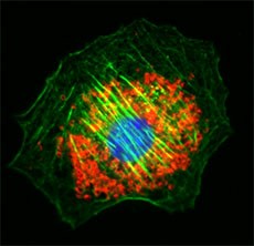
Uncover novel links between mitochondrial function and Alzheimer’s disease with MitoSpy™ probes for tracking mitochondrial health and localization, and antibodies specific for mitochondrial markers like VDAC1, TOMM40, and PINK1. |
 NIH3T3 cells co-stained with MitoSpy™ Red (red), Phalloidin (green) and DAPI (blue). NIH3T3 cells co-stained with MitoSpy™ Red (red), Phalloidin (green) and DAPI (blue). |
 |
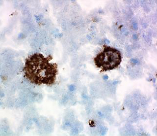
 IHC staining of diseased human brain tissue with anti-β-Amyloid, 1-15 antibody (3A1). |
Aggregation of Aβ and phosphorylation of Tau are hallmarks of Alzheimer’s disease. Accurately characterize disease severity and pathogenesis with antibodies that prefer aggregated Aβ (3A1, A17171C), detect both Aβ and APP (6E10, 4G8), or are specific for phosphorylated Tau. |
 |
Visualize mitochondria, protein aggregates, and other disease markers like ubiquitin, with our tools for microscopy. Utilize web resources, including protocols for IHC/IF, and specialized reagents like Ultra Streptavidin HRP Detection Kits to generate clear, detailed microscope images.
 |
 View the Immunofluorescence Tissue Staining video protocol, or browse our full video library. View the Immunofluorescence Tissue Staining video protocol, or browse our full video library. |
 |
|
|
*Any references to promotions on this page may not be valid at this time. View our promotions page for the most up-to-date promotions.

 Login / Register


Follow Us