 |
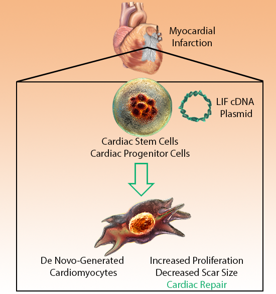
| Heart failure is one of the leading causes of death in the world, with an estimated 26 million people at risk1. While treatment has improved over the years, it remains one of the leading causes of mortality. As heart failure is mainly caused by myocyte loss, many labs are focused on cardiomyocyte regeneration, particularly from dormant cardiac stem cells (CSCs) and progenitor cells (CPCs). Leukemia Inhibitory Factor (LIF) has been shown to stimulate the proliferation of skeletal muscle cells and induce myocyte growth. As such, Kanda et al. investigated the ability of LIF to stimulate de novo myocyte generation and improve chances of cardiac repair. BioLegend provides new reagents for LIF investigation, including quantitative ELISAs, recombinant LIF proteins, and highly specific antibodies. |
 |
Adapted from Kanda, M. et al. 2016. PLoS One. 11:e0156562. Pubmed
1Ambrosy, A.P., et al. 2014. J. Am. Coll. Cardiol. 63:1123. |
| Kanda et al. labeled existing cardiomyocytes so that they expressed β-galactosidase and could react with X-gal to create an observable stain. Newly formed cardiomyocytes would thus be X-gal negative. Myocardial infarction (death of heart tissue or heart attack) led to an increase in newly generated myocytes, primarily from CPC or CSC populations, which can express c-kit, MDR1, or Sca-1, and cardiac transcription factors Nkx2.5 and GATA4. Cytokines can be instrumental following the aftermath of inflammation, and LIF administration was previously shown to increase cardiomyocyte generation. Administration of a plasmid containing LIF cDNA led to increased blood LIF levels, and proliferation and expansion of CSCs, leading to a decrease in the size of the myocardial infarction scar. This presents an interesting avenue for potential future treatments and clinical applications. |


 Login / Register


Follow Us