Watch your multicolor panels grow with Spark Dyes, a family of small, synthetic fluorophores. The addition of these expertly-crafted dyes to BioLegend’s portfolio provides researchers with the ability to fill spectral spaces between existing fluorophores, maximizing panel flexibility. Spark Dyes are advantageous due to their relatively narrow emission profile, stability, and solubility. In addition, their synthetic nature means they are not typically sensitive to standard fixatives, including organic solvents used for phospho-flow. Learn more about each of our Spark Dyes and how you can best incorporate them in multicolor applications.

Spark PLUS UV395™


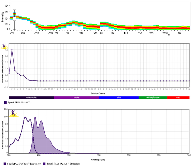
Spark PLUS UV395™ is a bright UV laser-excited dye that can be used on both conventional and spectral flow cytometers. It exhibits peak excitation/emission wavelengths at 355 nm and 385 nm respectively, most closely matching that of BD Horizon™ BUV395. On conventional cytometers, it can be detected with a 355 nm laser and 379/28 filter set (or equivalent). With its intense brightness, Spark PLUS UV395™ is a versatile option for detecting antigens at most expression levels and is stable towards exposure to heat or a variety of commonly used fixative solutions. Spark PLUS UV395™ is the first in the family of Spark PLUS™ Dyes, all formulated to provide brighter signals and superior performance.


Excitation and Emission Spectra of Spark PLUS UV395™


Emission spectra (top) and normalized emission spectra (middle) of Spark PLUS UV395™ run on a 5-laser Cytek™ Aurora Spectral Cytometer. To compare Spark PLUS UV395™ with other fluorophores on a spectral cytometer, use our Aurora Spectral Analyzer tool.


Normalized excitation and emission spectra (bottom) of Spark PLUS UV395™ obtained from a spectrophotometer. To compare Spark PLUS UV395™ with other fluorophores, use our Fluorescence Spectra Analyzer tool.


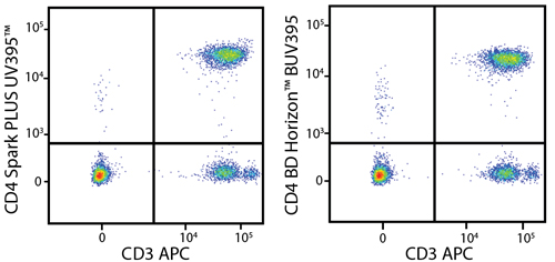
A New Alternative to BD Horizon™ BUV395


Human peripheral blood lymphocytes were stained with anti-human CD3 APC and anti-human CD4 (clone SK3) Spark PLUS UV395™ (left) or anti-human CD4 (clone SK3) BD Horizon™ BUV395 (right). Samples were acquired on a 5-laser Cytek® Aurora.


Multicolor Staining With Spark PLUS UV395™


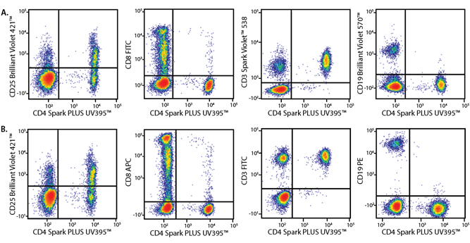
Panel A demonstrates how Spark PLUS UV395™ performs in a multicolor panel with fluorophores that may exhibit spectral overlap with it. Panel B is shown as a reference panel using pre-optimized fluorophores to ensure that staining patterns are similar.


Marker

Panel A Fluorophore

Panel B Fluorophore

CD3

Spark Violet™ 538

FITC

CD4

Spark PLUS UV395™

Spark PLUS UV395™

CD8

FITC

APC

CD19

Brilliant Violet 570™

PE

CD25

Brilliant Violet 421™

Brilliant Violet 421™


Human PBMCs were stained with the indicated antibodies and analyzed on a conventional flow cytometer. All plots are gated on lymphocytes.


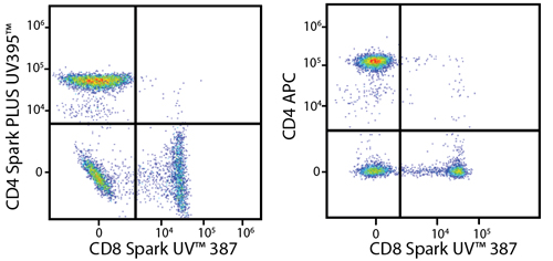
Unmix Spark PLUS UV395™ and Spark UV™ 387


Human PBMCs were stained with anti-human CD8 Spark UV 387 and anti-human CD4 (clone SK3) Spark PLUS UV395™ (left) or anti-human CD4 (clone SK3) APC (right). Samples were acquired on a 5-laser Cytek® Aurora cytometer.


Stability and Validation Testing


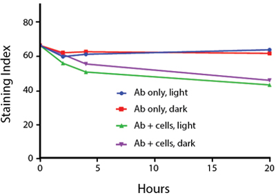
All BioLegend fluorophores undergo rigorous testing procedures to determine how light, heat, and fixation may affect the performance and ensure they will perform reliably. To compare the signal across different conditions and timepoints, we used the Stain Index (formula below) to measure the relative brightness of the antibody. 

Photostability Testing


The photostability of Spark PLUS UV395™ was tested in two ways that mimic how an antibody may be exposed to light over the course of an experiment.


  1. Antibodies were stored in the dark or exposed to fluorescent lighting. Then, the antibodies were used to stain freshly harvested cell samples and analyzed immediately.
  2. Cells were stained with antibody that had been kept under recommended storage conditions. Prior to analysis, the stained cells were stored in the dark or exposed to fluorescent lighting.


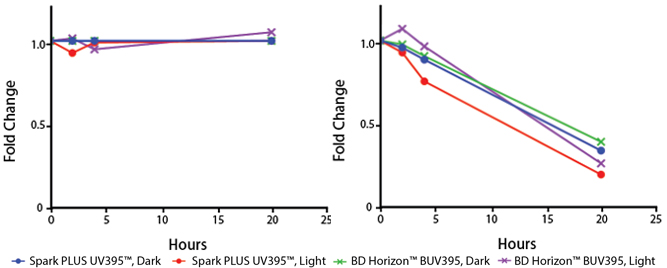
To assess the photostability of Spark PLUS UV395™-conjugated antibodies, anti-human CD4 (clone SK3) Spark PLUS UV395™ or BD Horizon™ BUV395 conjugates were exposed to light or protected in the dark (left). The samples were then used to stain human PBMCs and the fold change over the staining index at zero hours was calculated. To measure the photostability of Spark PLUS UV395™ stained cells, human PBMCs were stained with anti-human CD4 (clone SK3) Spark PLUS UV395™ or BD Horizon™ BUV395. The fold change in staining index of stained cells kept in the dark or exposed to light was calculated from the initial staining index at zero hours (right). Cells were gated on lymphocytes and data was acquired on a 5-laser Cytek® Aurora.


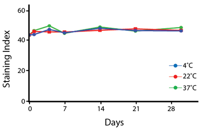
Heat Stability


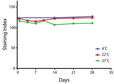
Anti-human CD4 (clone SK3) Spark PLUS UV395™ was aliquoted and incubated at the indicated temperatures over the course of 28 days, and then analyzed on a 5-laser Cytek™ Aurora cytometer. The antibodies were then used to stain human lysed whole blood from a single donor.


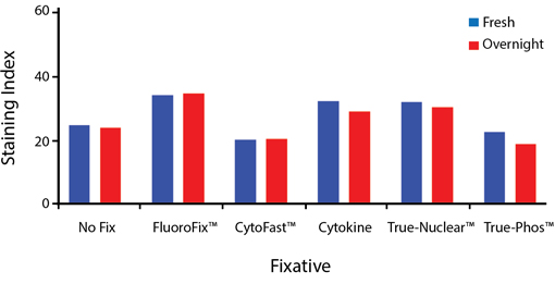
Fixative Stability


A guide to the fixatives used in this experiment:


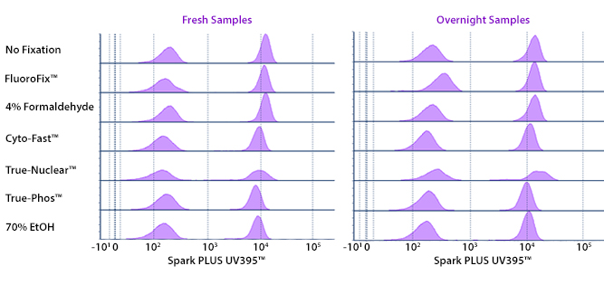
Human PBMCs were stained with anti-human CD4 (clone SK3) conjugated to Spark PLUS UV395™ and fixed using the respective protocols for each buffer set. Fresh samples were fixed and read on a 5-laser Cytek™ Aurora Cytometer immediately. Overnight samples were fixed and stored in Cyto-Last™ Buffer overnight before reading.


Spark Blue™ 550


Spark Blue™ 550 is excited off of the 488 nm laser and emits at 540 nm, filling the spectral gap between commonly used fluors like FITC and PE (R-Phycoerythrin). Emission off of the 405 nm violet laser further adds to the accuracy with which it can be unmixed from neighboring fluorophores and does not significantly impact the detection of any violet laser fluorophores. Because Spark Blue™ 550 is a relatively dim fluorophore, it is best used to detect a high abundance antigen.


Excitation and Emission Spectra of Spark Blue™ 550


Spark Blue 550 spectra signature

Spark Blue 550 aurora spectra

Spark Blue 550 spectra


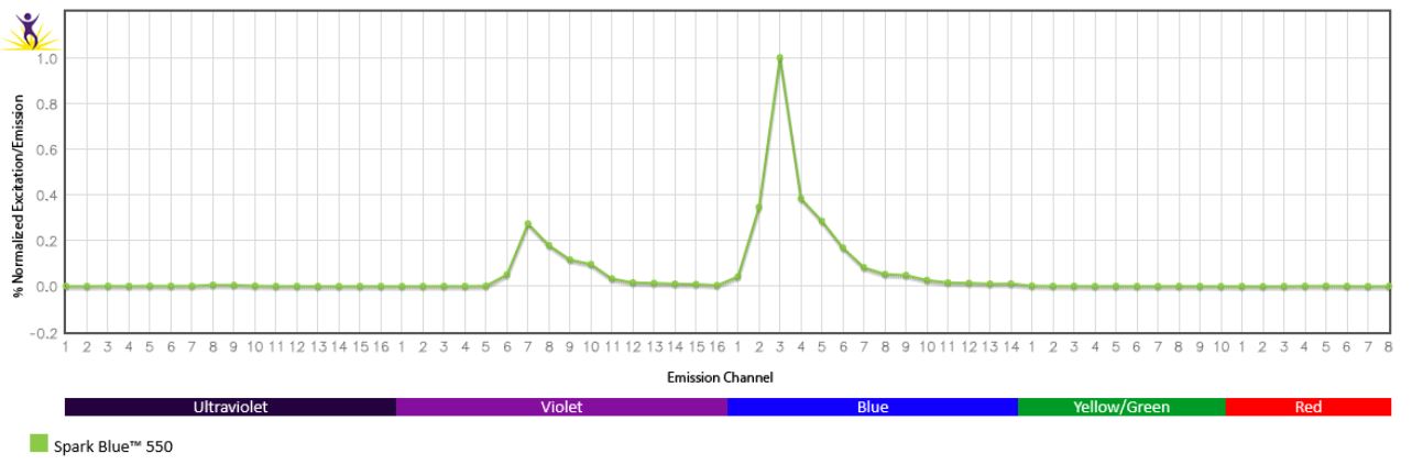
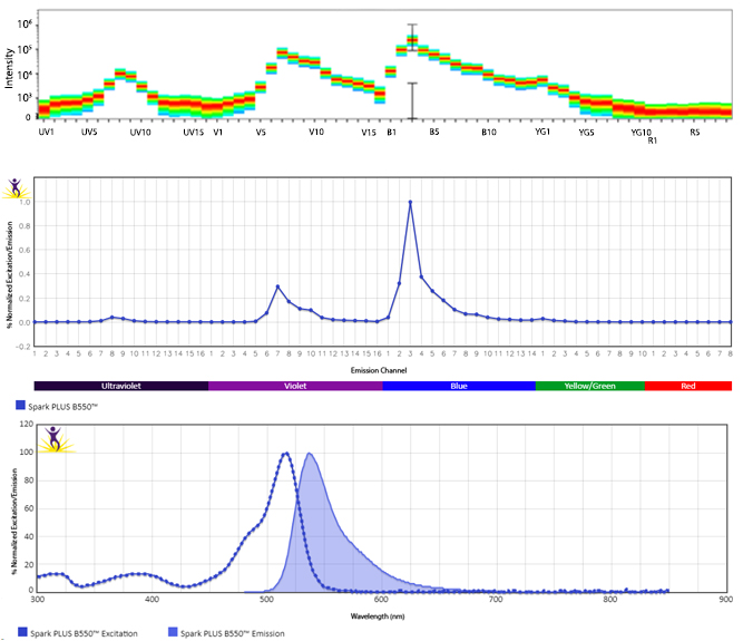
Emission spectra (top) and normalized emission spectra (middle) of Spark Blue™ 550 run on a 5-laser Cytek™ Aurora Spectral Cytometer. To compare Spark Blue™ 550 with other fluorophores on a spectral cytometer, use our Aurora Spectral Analyzer tool.


Normalized excitation and emission spectra (bottom) of Spark Blue™ 550 obtained from a spectrophotometer. To compare Spark Blue™ 550 with other fluorophores, use our Fluorescence Spectra Analyzer tool.


Minimal Spillover Considerations


Spark Blue™ 550 does not exhibit high spillover with other fluorophores, making it easy to place into a large panel. It is maximally excited by the 488 nm laser with minimal emission off of the 561 nm laser. While emission off of the 405 nm violet laser allows it to be easily unmixed from neighboring fluors, it does not significantly impact spreading into the violet laser fluors. Spectrally, it falls between the emissions of FITC and PE, but can easily be used in the same panel as these fluors with minimal spillover considerations.


Spark Blue 550

Spreading impact of Spark Blue™ 550 into detection channels of a 5-laser Cytek™ Aurora Spectral Cytometer.


Human whole blood was stained with anti-CD19 Spark Blue™ 550, anti-CD4 BV570™, anti-CD38 PE, and anti-CD40 FITC antibodies. Samples were unmixed on a Cytek™ Aurora Cytometer using compensation beads and cells. All plots are gated on lymphocytes.


A Brighter Alternative to Alexa Fluor® 532


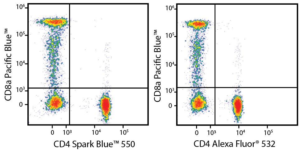
Spark Blue™ 550 is spectrally similar to Alexa Fluor® 532 with a peak emission in the B3 channel on the Cytek™ Aurora and offers a brighter signal when compared to Alexa Fluor® 532 in a side-by-side experiment. Due to its unique emission peak off of the violet laser, Spark Blue™ 550 is typically easier to unmix from neighboring fluors when compared to Alexa Fluor® 532.


Spark Blue 550 compared to Alexa Fluor 532

Human peripheral blood lymphocytes were stained with anti-CD8 Pacific Blue™, and either anti-CD4 (clone SK3) Spark Blue™ 550 (left) or anti-CD4 (clone SK3) Alexa Fluor® 532 (right).


Stability and Validation Testing


All BioLegend fluorophores undergo rigorous testing procedures to determine how light, heat, and fixation may affect the performance and ensure they will perform reliably. To compare the signal across different conditions and timepoints, we used the Stain Index (formula below) to measure the relative brightness of the antibody. 

Spark 550

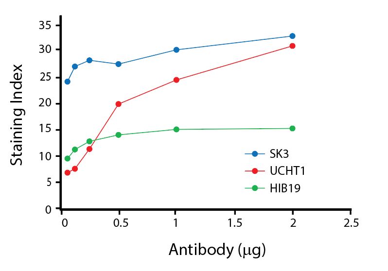
Titration of Spark Blue™ 550

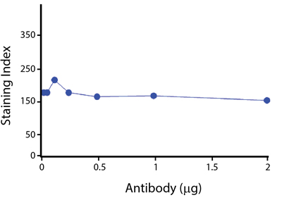
Titration curves generated by staining human lysed whole blood with Spark Blue™ 550 conjugated anti-CD4 (SK3), anti-CD3 (UCHT1), and anti-CD19 (HIB19) antibodies.


Photostability Testing


The photostability of Spark Blue™ 550 was tested in two ways that mimic how an antibody may be exposed to light over the course of an experiment.


  1. Antibodies were stored in the dark or exposed to fluorescent lighting. Then, the antibodies were used to stain freshly harvested cell samples and analyzed immediately.
  2. Cells were stained with antibody that had been kept under recommended storage conditions. Prior to analysis, the stained cells were stored in the dark or exposed to fluorescent lighting.


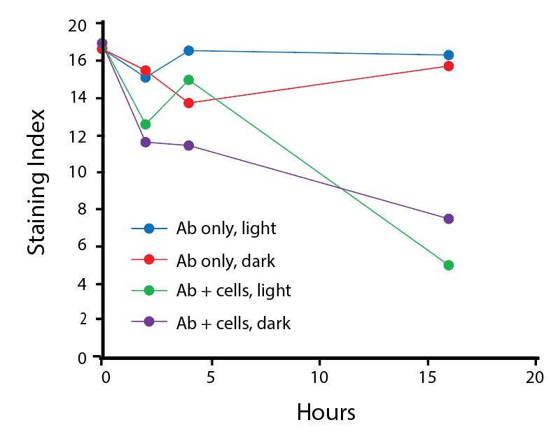
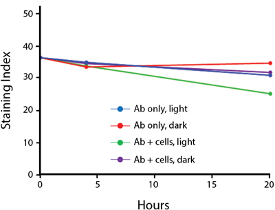
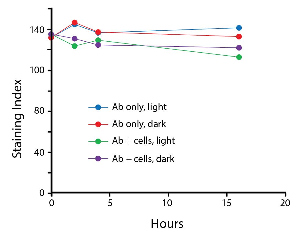
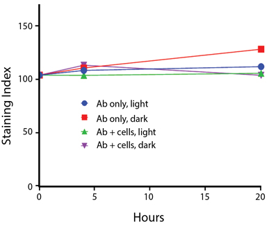
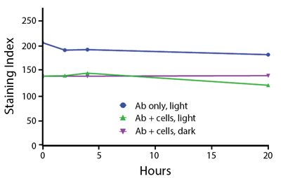
Spark Blue™ 550 antibodies are stable when left under fluorescent lighting overnight. When stained cells are stored overnight in either condition, there is an accelerated loss of signal due to the additive oxidative stress from the cells on the reagent.


Anti-human CD4 (clone SK3) Spark Blue™ 550 was stored in a clear vial (Ab only) and left exposed to light or protected in the dark, as indicated. Antibodies were stored for the indicated timepoints and then used to stain human lysed whole blood. Samples labeled Ab+ cells contain human lysed whole blood that was stained with anti-human CD4 Spark Blue™ 550. Stained cells were then left in the light or protected, as indicated.


Heat Stability


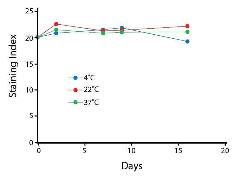
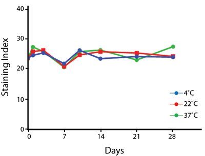
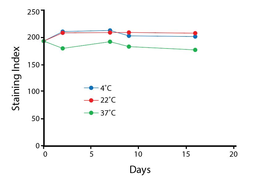
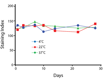
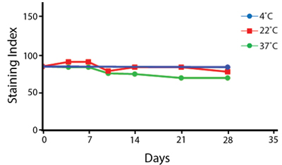
Anti-human CD4 (clone SK3) Spark Blue™ 550 was aliquoted and incubated at the indicated temperatures over the course of 17 days. The antibodies were then used to stain human lysed whole blood from a single donor.


Fixative Stability


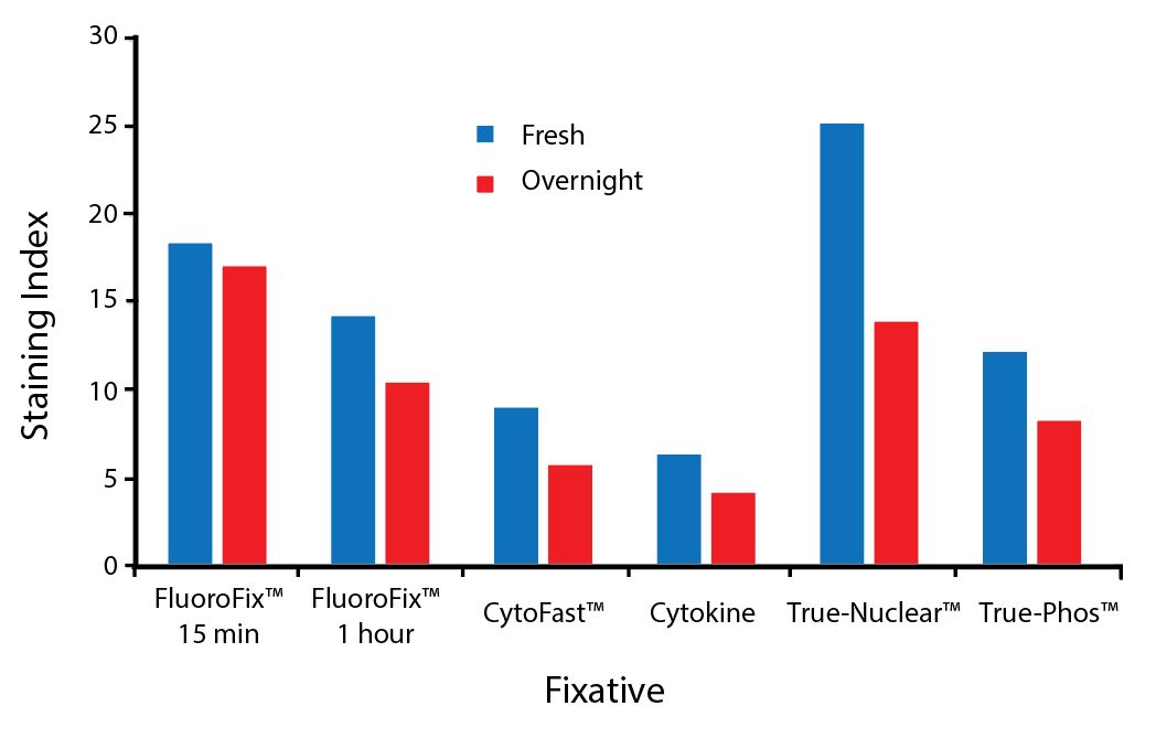
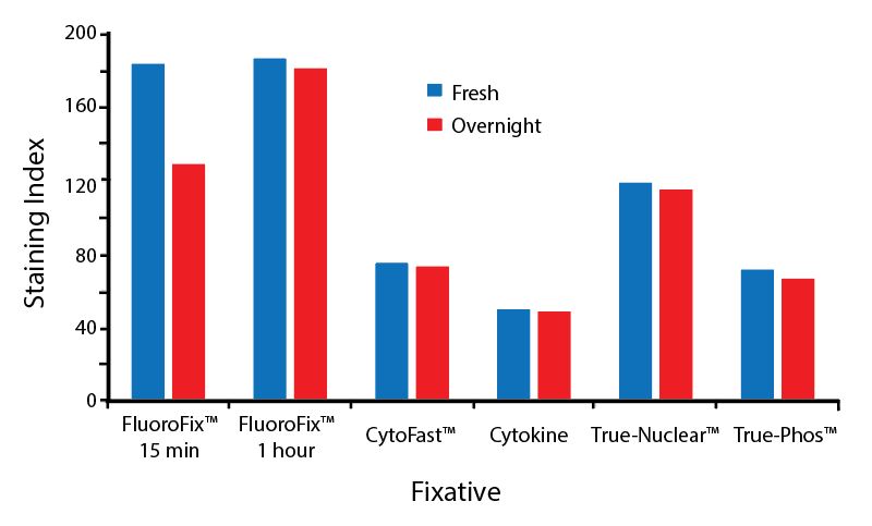
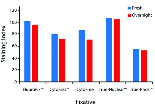
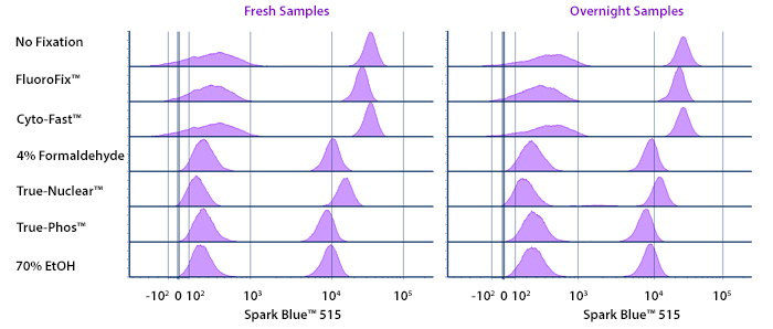
Spark Blue™ 550 is compatible with all BioLegend fixation buffers. For each buffer set, fresh fixed samples were tested immediately following staining or stored overnight in Cyto-Last™ Buffer before being read on a cytometer.


A guide to the fixatives used in this experiment:


Spark Blue 550

Human PBMCs were stained with anti-human CD4 (clone SK3) conjugated to Spark Blue™ 550 and fixed using the respective protocols for each buffer set. Fresh samples were fixed and read on a Cytek® Aurora immediately. Overnight samples were fixed and stored overnight in Cyto-Last™ Buffer before reading.

Spark Blue™ 574


Spark Blue™ 574 is mainly excited by the 488 nm blue laser. With minimal excitation from the 561 yellow/green laser, this allows it to be unmixed from PE. Spark Blue™ 574's peak emission is at 574 nm. Given the low brightness of this fluor, Spark Blue™ 574 is recommended for an antigens with high expression levels. 


Excitation and Emission Spectra of Spark Blue™ 574


Spark Blue 574 spectra signature


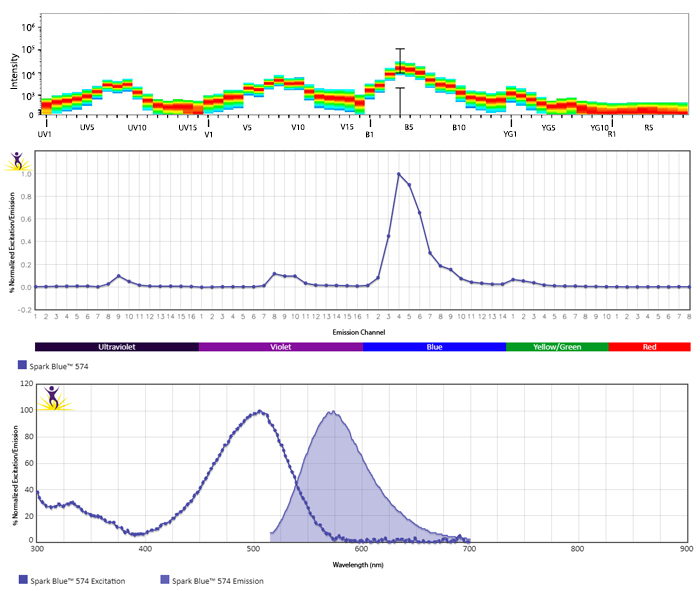
Emission spectra (top) and normalized emission spectra (middle) of Spark Blue™ 574 run on a 5-laser Cytek™ Aurora Spectral Cytometer. To compare Spark Blue™ 574 with other fluorophores on a spectral cytometer, use our Aurora Spectral Analyzer tool.


Normalized excitation and emission spectra (bottom) of Spark Blue™ 574 obtained from a spectrophotometer. To compare Spark Blue™ 574 with other fluorophores, use our Fluorescence Spectra Analyzer tool.


A Novel Blue Laser Fluorophore for Spectral Cytometry


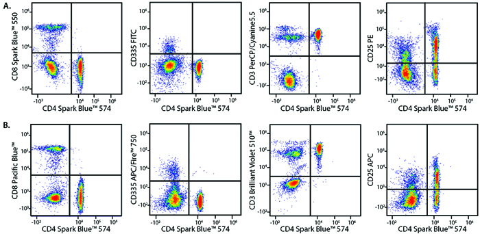
Panel A demonstrates how Spark Blue™ 574 performs in a multicolor panel with fluorophores that share significant spectra overlap with it. Panel B is shown as a reference panel using pre-optimized fluorophores to ensure that staining patterns are similar.


Spark Blue 574 Panel


Marker

Panel A Fluorophore

Panel B Fluorophore

CD3

PerCP/Cyanine5.5

Brilliant Violet 510™

CD4

Spark Blue™ 574

Spark Blue™ 574

CD8

Spark Blue™ 550

Pacific Blue™

CD25

PE

APC

CD335

FITC

APC/Fire™ 750


Human lysed whole blood was stained with the indicated antibodies and unmixed on a 5-laser Cytek™ Aurora Cytometer using cells. All plots are gated on lymphocytes.


Stability and Validation Testing


All BioLegend fluorophores undergo rigorous testing procedures to determine how light, heat, and fixation may affect the performance and ensure they will perform reliably. To compare the signal across different conditions and timepoints, we used the Stain Index (formula below) to measure the relative brightness of the antibody. 

Spark 550

Photostability Testing


The photostability of Spark Blue™ 574 was tested in two ways that mimic how an antibody may be exposed to light over the course of an experiment.


  1. Antibodies were stored in the dark or exposed to fluorescent lighting. Then, the antibodies were used to stain freshly harvested cell samples and analyzed immediately.
  2. Cells were stained with antibody that had been kept under recommended storage conditions. Prior to analysis, the stained cells were stored in the dark or exposed to fluorescent lighting.


Spark Blue 574 Photostability

Anti-human CD3 (clone UCHT1) Spark Blue™ 574 was stored in a clear vial (Ab only) and left exposed to light or protected in the dark, as indicated. Antibodies were stored for the indicated timepoints and then used to stain human lysed whole blood. Samples labeled Ab + cells contain human lysed whole blood that was stained with anti-human CD3 Spark Blue™ 574. Stained cells were then left in the light or protected, as indicated.


Heat Stability


Spark Blue 574 Heat Stability

Anti-human CD3 (clone UCHT1) Spark Blue™ 574 was aliquoted and incubated at the indicated temperatures over the course of 28 days. The antibodies were then used to stain human lysed whole blood from a single donor.


Fixative Stability


The compatibility of Spark Blue™ 574 with fixatives should be tested by the end user to ensure the antigen's expression is maintained post-fixation. 


A guide to the fixatives used in this experiment:


Spark Blue 574 Fixative Bar Graph


Human PBMCs were stained with anti-human CD3 (clone UCHT1) conjugated to Spark Blue™ 574 and fixed using the respective protocols for each buffer set. Fresh samples were fixed and read on a 5-laser Cytek™ Aurora Cytometer immediately. Overnight samples were fixed and stored overnight in Cyto-Last™ Buffer before reading.

Spark NIR™ 685


Spark NIR™ 685 expands your options off of the red laser to build larger multicolor panels. Traditionally, the 633 nm red laser has been limited to only 2-3 fluorophores due to the fluors’ wide emission spectra in the near infrared (NIR) range. With the help of spectral unmixing cytometers, the spectral gap between the emission peaks of APC and Alexa Fluor® 700 can now be used to detect an additional fluorophore. Spark NIR™ 685 fills that spectral space with a maximum emission at 685 nm.


Excitation and Emission Spectra of Spark NIR™ 685


Spark NIR685 Spectra Signature

Spark NIR685 Aurora Spectra

Spark NIR685 Spectra


Emission spectra (top) and normalized emission spectra (middle) of Spark NIR™ 685 run on a 5-laser Cytek™ Aurora Spectral Cytometer. To compare Spark NIR™ 685 with other fluorophores on a spectral cytometer, use our Aurora Spectral Analyzer tool.


Normalized excitation and emission spectra of Spark NIR™ 685 obtained from a spectrophotometer (bottom). To compare Spark NIR™ 685 with other fluorophores, use our Fluorescence Spectra Analyzer tool.


Utilize Spark NIR™ 685 With Other Red Laser Fluorophores


Spark NIR™ 685 can be used in the same panel as APC or Alexa Fluor® 647; however, spreading error with these fluors must be considered during panel design and panels should be built to match proper fluor brightness with antigen expression. For example, in an ideal panel, Spark NIR™ 685 and Alexa Fluor® 647 would not be used to detect two co-expressed markers.


Spark NIR 685

Spreading impact of Spark NIR™ 685 into detection channels of a 5-laser Cytek™ Aurora Spectral Cytometer.


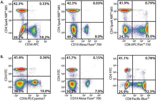
Below, Panel A demonstrates how Spark NIR™ 685 can be used in combination with other red laser fluorophores. Panel B is shown as a reference panel using pre-optimized fluorophores to ensure that staining patterns are similar.


Spark NIR 685 panel


Marker

Panel A Fluorophore

Panel B Fluorophore

CD4

Spark NIR™ 685

FITC

CD8

APC/Fire™ 750

Pacific Blue™

CD19

Alexa Fluor® 700

Alexa Fluor® 700

CD56

APC

PE/Cyanine7


Human whole blood from the same donor was stained with the indicated antibodies in either Panel A or Panel B. Cells were washed and fixed with FluoroFix™ prior to analysis.


A Brighter Alternative to Alexa Fluor® 660


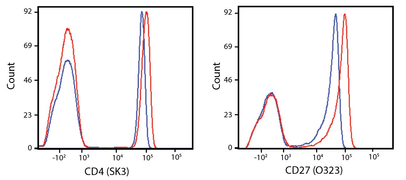
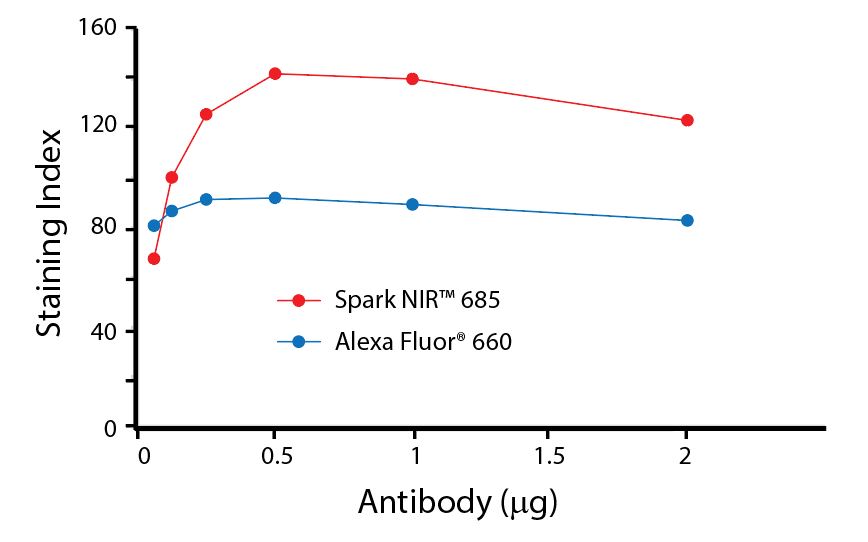
Spark NIR™ 685 is spectrally similar to Alexa Fluor® 660 with a peak emission in the R3 channel on the Cytek™ Aurora. Spark NIR™ 685 offers a brighter signal when compared to Alexa Fluor® 660 in a side-by-side experiment.


Spark NIR 685


Human lysed whole blood was stained with either anti-CD4 (clone SK3) or anti-CD27 (O323). Antibodies were conjugated to either Spark NIR™ 685 (red) or Alexa Fluor® 660 (blue).


Titration curves generated by staining human lysed whole blood with anti-CD4 (clone SK3) antibodies conjugated either to Spark NIR™ 685 or Alexa Fluor® 660 as indicated.


Stability and Validation Testing


All BioLegend fluorophores undergo rigorous testing procedures to determine how light, heat, and fixation may affect the performance and ensure they will perform reliably. To compare the signal across different conditions and timepoints, we used the Stain Index (formula below) to measure the relative brightness of the antibody

Spark NIR equation


Photostability Testing


The photostability of Spark NIR™ 685 was tested in two ways to mimic how an antibody may be exposed to light over the course of an experiment.


  1. Antibodies were stored in the dark or exposed to fluorescent lighting. Then, the antibodies were used to stain freshly harvested cell samples and analyzed immediately.
  2. Cells were stained with antibody that had been kept under recommended storage conditions. Prior to analysis, the stained cells were stored in the dark or exposed to fluorescent lighting.

Spark NIR

Anti-human CD4 (clone SK3) Spark NIR™ 685 was stored in a clear vial (Ab only) and left exposed to light or protected in the dark, as indicated. Antibodies were stored for the indicated timepoints and then used to stain human lysed whole blood. Samples labeled Ab+ cells contain human lysed whole blood that was stained with anti-human CD4 Spark NIR™ 550. Stained cells were then left in the light or protected, as indicated.


Heat Stability


Anti-human CD4 (clone SK3) antibody conjugated to Spark NIR™ 685 was aliquoted and incubated at the indicated temperatures over the course of 17 days. The antibodies were then used to stain human lysed whole blood from a single donor.


Fixative Stability


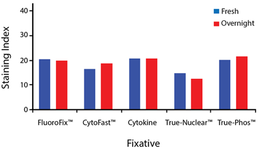
Spark NIR™ 685 is compatible with all BioLegend fixation buffers. For each buffer set, fresh fixed samples were tested immediately following staining or stored overnight in Cyto-Last™ Buffer before being read on a cytometer.


A guide to the fixatives used in this experiment:


Human PBMCs were stained with anti-human CD4 (clone SK3) Spark NIR™ 685 and fixed using the respective protocols for each buffer set. Fresh samples were fixed and read on a cytometer immediately following fixation. Overnight samples were fixed and then stored in Cyto-Last™ Buffer (overnight) before reading.

Spark YG™ 570


Ideal for building multicolor microscopy panels, Spark YG™ 570 provides a bright and photostable signal. With an excitation max of 555 nm and an emission max of 570 nm, it can be imaged using the filter sets commonly used for Alexa Fluor® 555, Cyanine3, or TRITC in either widefield or confocal microscopy. While Spark YG™ 570 can be detected on a spectral cytometer, it is difficult to accurately unmix from PE due to a high degree of spectral similarity.


Spark YG 570 spectra

Normalized excitation and emission spectra of Spark YG™ 570 obtained from a spectrophotometer. To compare Spark YG™ 570 with other fluorophores, use our Fluorescence Spectra Analyzer tool.


Build Multicolor Microscopy Panels with Spark YG™ 570


Depending on the filter sets and light source of your microscope, Spark YG™ 570 can be used in combination with a variety of other fluorophores including DAPI, Alexa Fluor® 488, Alexa Fluor® 647, and Alexa Fluor® 750.


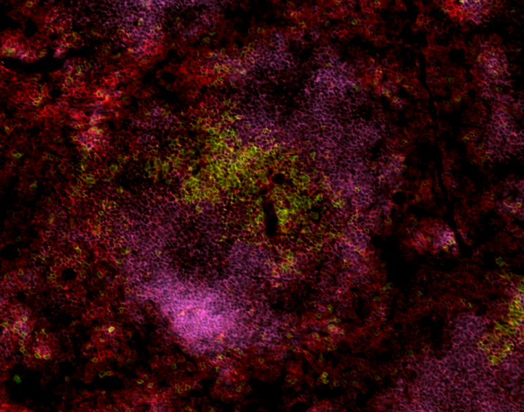
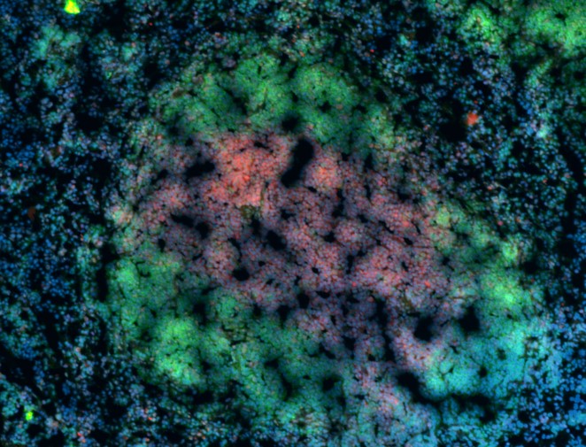
Immunohistochemistry on frozen mouse spleen tissue. Tissue on the left was stained with anti-CD3 Spark YG™ 570 (red), anti-B220 Alexa Fluor® 647 (green) and DAPI (blue). Tissue on the right was stained with anti-CD45 Spark YG™ 570 (red), anti-CD3 Alexa Fluor® 647 (green), and anti-B220 Alexa Fluor® 647 (blue).


Spark YG™ 570 Provides Similar Brightness to Alexa Fluor® 555


Spark YG 570 compared to Alexa Fluor 555

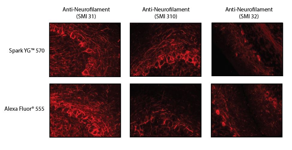
Rat (left and middle) or mouse (right) brain sections were stained with the indicated antibody conjugated to either Spark YG™ 570 or Alexa Fluor® 555. Across clones, tissues showed a similar brightness and staining pattern between the two antibodies.


Spark YG™ 570 Demonstrates Minimal Bleed-through into Nearby Channels


Spark YG 570 Bleedthrough

Human substantia nigra tissue was stained with anti-Tyrosine Hydrolase Spark YG™ 570 and imaged under a 40x objective. No bleed-through was observed in the 488 nm and 647 nm channels, even when using a five-fold longer exposure time.


Photostability of Spark YG™ 570


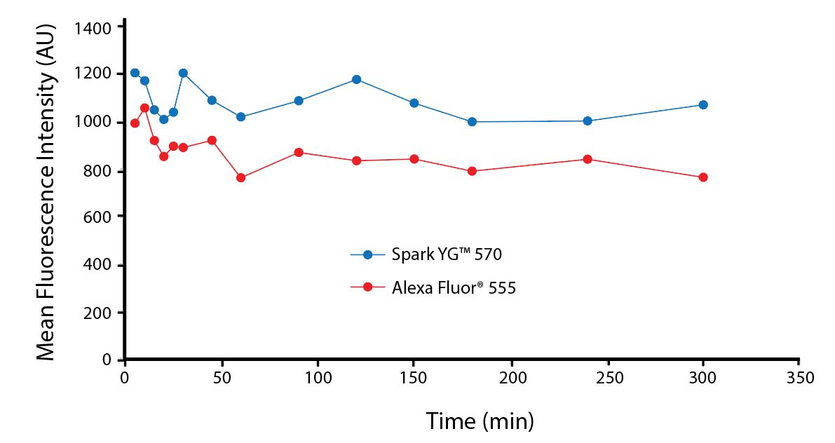
To test the photostability of Spark YG™ 570, we stained tissue sections with a purified antibody, followed by incubation with either an Alexa Fluor® 555 or Spark YG™ 570 conjugated secondary antibody. After staining, images were captured by exposing tissue sections to light at the specified time points.


Photostability Spark YG 570

FFPE human tonsil tissues were stained with purified anti-CD8a (Clone C8/144B) followed by incubation with Alexa Fluor® 555 or Spark YG™ 570 goat-anti-mouse IgG (Poly4053). Images were captured at the indicated time points and mean fluorescence intensity, expressed as Arbitrary Units (AU) was measured using ImageJ.

Spark Violet™ 538


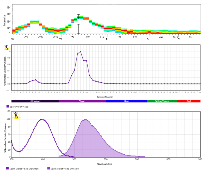
Spark Violet™ 538 slots in as an additional violet laser fluor option for multicolor flow cytometry users. This dye is optimally excited by the 405 nm violet laser and emits maximally at 538 nm, placing its peak emission between Brilliant Violet 510™ and Brilliant Violet 570™. When used on a spectral cytometer, Spark Violet™ 538 can be successfully unmixed from neighboring dyes, such as BV510™, BV570™, and BV605™. While it is brighter than Pacific Orange™, Spark Violet™ 538 is relatively dim and should be paired with antigens expressed at high levels.


Excitation and Emission Spectra of Spark Violet™ 538
 

Spark Violet 538 Spectra Signature


Emission spectra (top) and normalized emission spectra (middle) of Spark Violet™ 538 run on a 5-laser Cytek™ Aurora Spectral Cytometer. To compare Spark Violet™ 538 with other fluorophores on a spectral cytometer, use our Aurora Spectral Analyzer tool.


Normalized excitation and emission spectra of Spark NIR™ 685 obtained from a spectrophotometer (bottom). To compare Spark Violet™ 538 with other fluorophores, use our Fluorescence Spectra Analyzer tool.

A Perfect Complement to Brilliant Violet™ Dyes

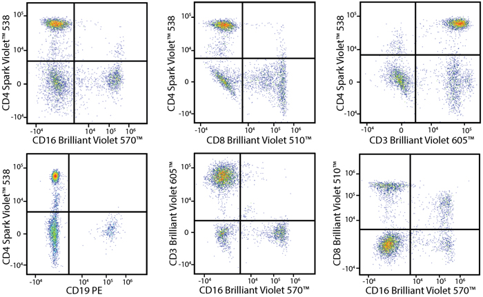
In the data below, Spark Violet™ 538 is utilized with several neighboring Brilliant Violet™ dyes, showcasing its ability to be spectrally unmixed from these dyes in a large, multicolor panel. 
 

Spark Violet 538 panel


Lysed human whole blood was stained with the indicated antibodies in the presence of Brilliant Stain Buffer. Samples were unmixed on a Cytek™ Aurora Cytometer using compensation beads and cells. All plots are gated on lymphocytes.


A New Alternative Pacific Orange™

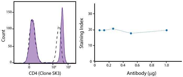
Spark Violet™ 538 is spectrally similar to Pacific Orange™ with a peak emission in the V8 channel on the Cytek™ Aurora. Spark Violet™ 538 offers a brighter signal when compared to Pacific Orange™ in a side-by-side experiment.
 

Spark Violet 538 Titration


(Left) Human lysed whole blood was stained with anti-human CD4, clone SK3, conjugated to either Spark Violet™ 538 (solid purple) or Pacific Orange™ (clone S3.5, clear dashed) and then analyzed on a Cytek™ Aurora cytometer. Plots are gated on lymphocytes. (Right) Titration curve generated by staining human lysed whole blood with Spark Violet™ 538 conjugated to anti-CD4, clone SK3, antibody.  


Stability and Validation Testing


All BioLegend fluorophores undergo rigorous testing procedures to determine how light, heat, and fixation may affect the performance and ensure they will perform reliably. To compare the signal across different conditions and timepoints, we used the Stain Index (formula below) to measure the relative brightness of the antibody

Spark Violet 538 equation


Photostability Testing


The photostability of Spark Violet™ 538 was tested in two ways that mimic how an antibody may be exposed to light over the course of an experiment.


  1. Antibodies were stored in the dark or exposed to fluorescent lighting. Then, the antibodies were used to stain freshly harvested cell samples and analyzed immediately.
  2. Cells were stained with antibody that had been kept under recommended storage conditions. Prior to analysis, the stained cells were stored in the dark or exposed to fluorescent lighting.
Spark Violet 538

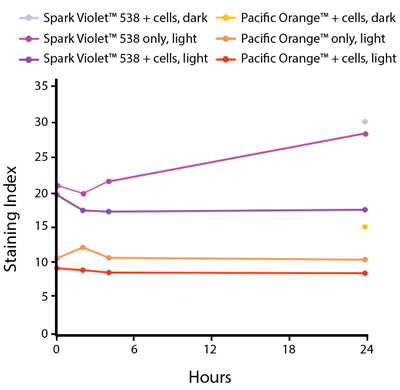
Anti-human CD4 Spark Violet™ 538 (clone SK3) or Pacific Orange™ (clone S3.5) antibodies were stored in a clear vial (dye only) and left exposed to light or protected in the dark, as indicated. Antibodies were stored for the indicated timepoints and then used to stain human lysed whole blood. “Dark” samples were only tested at the 24 hour time point as a control. Samples labeled "dye + cells" contain human lysed whole blood that was stained with the indicated antibody. Stained cells were then left in the light or protected, as indicated. 


Heat Stability
 

Spark Violet 538 Heat Stability

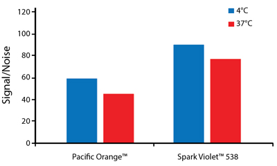
Anti-human CD4 antibodies, clone SK3 Spark Violet™ 538 or clone S3.5 Pacific Orange™, were kept at either 4°C or 37°C for seven weeks. The antibodies were then used to stain human lysed whole blood.


Fixative Stability


Spark Violet™ 538 is compatible with BioLegend buffers, but may be sensitive to alcohol-based buffers. The end user will need to test to ensure their antigen's signal is maintained post-fixation.


A guide to the fixatives used in this experiment:
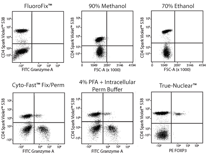


Human PBMCs were stained with anti-human CD4 (clone SK3) conjugated to Spark Violet™ 538 and fixed using the respective protocols for each buffer set. For intracellular staining buffers, additional antibody stains for Granzyme A or FOXP3 were performed. For the alcohols, 90% methanol and 70% ethanol were used. Samples were then transferred into FluoroFix™ Buffer and read on a 5-laser Cytek™ Aurora Cytometer immediately.

Spark YG™ 581


Spark YG™ 581 is excited primarily by the 561 nm yellow/green laser and has a peak emission at 581 nm. Although it emits in the same channel as PE, Spark YG™ 581 has minimal cross-beam excitation with the 488 nm laser, making Spark YG™ 581 distinguishable from PE and allowing these highly overlapping fluorophores to be accurately unmixed. Spark YG™ 581 is recommended for an intermediate density antigen. 


Excitation and Emission Spectra of Spark YG™ 581


Spark YG 581 spectra signature


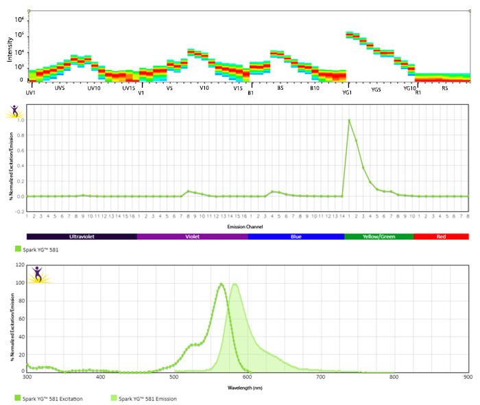
Emission spectra (top) and normalized emission spectra (middle) of Spark YG™ 581 run on a 5-laser Cytek™ Aurora Spectral Cytometer. To compare Spark YG™ 581 with other fluorophores on a spectral cytometer, use our Aurora Spectral Analyzer tool.


Normalized excitation and emission spectra (bottom) of Spark YG™ 581 obtained from a spectrophotometer. To compare Spark YG™ 581 with other fluorophores, use our Fluorescence Spectra Analyzer tool.


Unmix Spark YG™ 581 from PE and Yellow/Green Laser Options


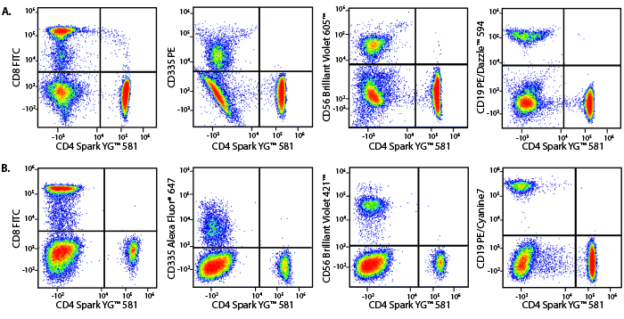
Panel A demonstrates how Spark YG™ 581 performs in a multicolor panel with fluorophores that share significant spectra overlap with it. Panel B is shown as a reference panel using pre-optimized fluorophores to ensure that staining patterns are similar.


Spark YG 581 Panel


Marker

Panel A Fluorophore

Panel B Fluorophore

CD4

Spark YG™ 581

Spark YG™ 581

CD8

FITC

FITC

CD19

PE/Dazzle™ 594

PE/Cyanine7

CD56

Brilliant Violet 605™

Brilliant Violet 421™

CD335

PE

Alexa Fluor® 647


Human lysed whole blood was stained with the indicated antibodies and unmixed on a 5-laser Cytek™ Aurora Cytometer using cells. All plots are gated on lymphocytes.


Stability and Validation Testing


All BioLegend fluorophores undergo rigorous testing procedures to determine how light, heat, and fixation may affect the performance and ensure they will perform reliably. To compare the signal across different conditions and timepoints, we used the Stain Index (formula below) to measure the relative brightness of the antibody. 

Spark 550

Titration of Spark YG™ 581

Spark YG 581 Titration

Titration curve generated by staining human lysed whole blood with Spark YG™ 581 conjugated anti-human CD4 (SK3) antibody.


Photostability Testing


The photostability of Spark YG™ 581 was tested in two ways that mimic how an antibody may be exposed to light over the course of an experiment.


  1. Antibodies were stored in the dark or exposed to fluorescent lighting. Then, the antibodies were used to stain freshly harvested cell samples and analyzed immediately.
  2. Cells were stained with antibody that had been kept under recommended storage conditions. Prior to analysis, the stained cells were stored in the dark or exposed to fluorescent lighting.


Spark YG 581 Photostability

Anti-human CD4 (clone SK3) Spark YG™ 581 was stored in a clear vial (Ab only) and left exposed to light or protected in the dark, as indicated. Antibodies were stored for the indicated timepoints and then used to stain human lysed whole blood. Samples labeled Ab + cells contain human lysed whole blood that was stained with anti-human CD4 Spark YG™ 581. Stained cells were then left in the light or protected, as indicated.


Heat Stability


Spark YG 581 Heat Stability

Anti-human CD4 (clone SK3) Spark YG™ 581 was aliquoted and incubated at the indicated temperatures over the course of 28 days. The antibodies were then used to stain human lysed whole blood from a single donor.


Fixative Stability


Spark YG™ 581 is compatible with all BioLegend fixation buffers. For each buffer set, fresh fixed samples were tested immediately following staining or stored overnight in Cyto-Last™ Buffer before being read on a cytometer.


A guide to the fixatives used in this experiment:


Spark YG 581 Fixative

Human PBMCs were stained with anti-human CD4 (clone SK3) conjugated to Spark YG™ 581 and fixed using the respective protocols for each buffer set. Fresh samples were fixed and read on a 5-laser Cytek™ Aurora Cytometer immediately. Overnight samples were fixed and stored overnight in Cyto-Last™ Buffer before reading.

Spark YG™ 593


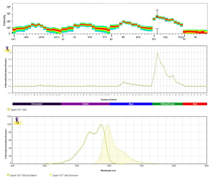
Spark YG™ 593 is excited primarily by the 561 nm yellow/green laser and has a peak emission at 593 nm, placing it between PE and PE/Dazzle™ 594. Spark YG™ 593 is primarily excited by the yellow/green laser and minimally excited by the blue laser, allowing it to be unmixed from fluorophores like PE. Spark YG™ 593 is recommended for an intermediate density antigen. 


Excitation and Emission Spectra of Spark YG™ 593


Spark YG 593 spectra signature


Emission spectra (top) and normalized emission spectra (middle) of Spark YG™ 593 run on a 5-laser Cytek™ Aurora Spectral Cytometer. To compare Spark YG™ 593 with other fluorophores on a spectral cytometer, use our Aurora Spectral Analyzer tool.


Normalized excitation and emission spectra (bottom) of Spark YG™ 593 obtained from a spectrophotometer. To compare Spark YG™ 593 with other fluorophores, use our Fluorescence Spectra Analyzer tool.


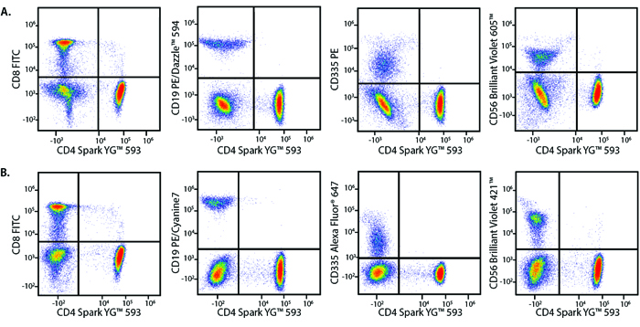
Unmix Spark YG™ 593 from PE and Yellow/Green Laser Options


Panel A demonstrates how Spark YG™ 593 performs in a multicolor panel with fluorophores that share significant spectra overlap with it. Panel B is shown as a reference panel using pre-optimized fluorophores to ensure that staining patterns are similar.


Spark YG 593 Panel


Marker

Panel A Fluorophore

Panel B Fluorophore

CD4

Spark YG™ 593

Spark YG™ 593

CD8

FITC

FITC

CD19

PE/Dazzle™ 594

PE/Cyanine7

CD56

Brilliant Violet 605™

Brilliant Violet 421™

CD335

PE

Alexa Fluor® 647


Human lysed whole blood was stained with the indicated antibodies and unmixed on a 5-laser Cytek™ Aurora Cytometer using cells. All plots are gated on lymphocytes.


Stability and Validation Testing


All BioLegend fluorophores undergo rigorous testing procedures to determine how light, heat, and fixation may affect the performance and ensure they will perform reliably. To compare the signal across different conditions and timepoints, we used the Stain Index (formula below) to measure the relative brightness of the antibody. 

Spark 550

Photostability Testing


The photostability of Spark YG™ 593 was tested in two ways that mimic how an antibody may be exposed to light over the course of an experiment.


  1. Antibodies were stored in the dark or exposed to fluorescent lighting. Then, the antibodies were used to stain freshly harvested cell samples and analyzed immediately.
  2. Cells were stained with antibody that had been kept under recommended storage conditions. Prior to analysis, the stained cells were stored in the dark or exposed to fluorescent lighting.


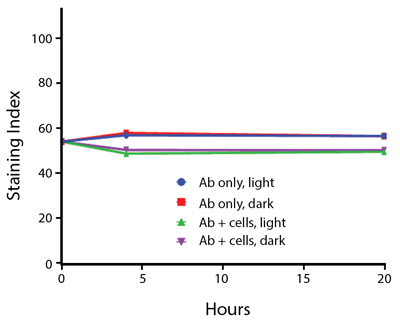
Spark YG 593 Photostability

Anti-human CD4 (clone SK3) Spark YG™ 593 was stored in a clear vial (Ab only) and left exposed to light or protected in the dark, as indicated. Antibodies were stored for the indicated timepoints and then used to stain human lysed whole blood. Samples labeled Ab + cells contain human lysed whole blood that was stained with anti-human CD4 Spark YG™ 593. Stained cells were then left in the light or protected, as indicated.


Heat Stability


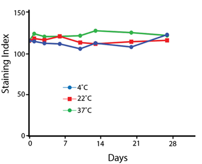
Spark YG 593 Heat Stability

Anti-human CD4 (clone SK3) Spark YG™ 593 was aliquoted and incubated at the indicated temperatures over the course of 28 days. The antibodies were then used to stain human lysed whole blood from a single donor.


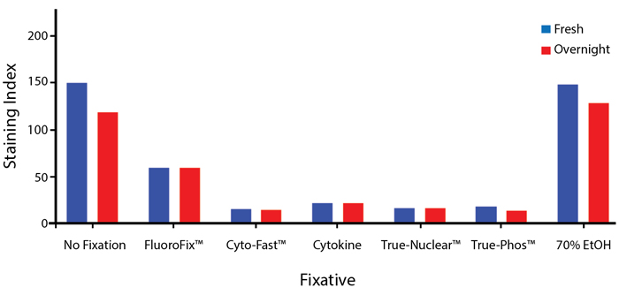
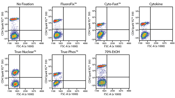
Fixative Stability


The compatibility of Spark YG™ 593 with fixatives should be tested by the end user to ensure the antigen's expression is maintained post-fixation. 


A guide to the fixatives used in this experiment:


Spark YG 593 Fixative Bar Graph

Spark YG 593 Fixative Flow

Human PBMCs were stained with anti-human CD4 (clone SK3) conjugated to Spark YG™ 593 and fixed using the respective protocols for each buffer set. Fresh samples were fixed and read on a 5-laser Cytek™ Aurora Cytometer immediately. Overnight samples were fixed and stored overnight in Cyto-Last™ Buffer before reading.

Spark Violet™ 423


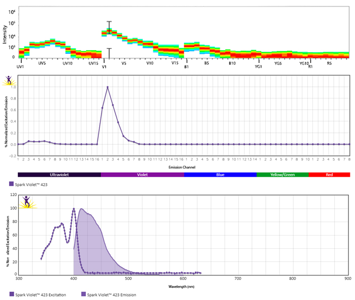
Spark Violet™ 423, is excited by the 405 nm violet laser and can easily be unmixed from Pacific Blue™ and Brilliant Violet 421™ on spectral cytometers. On conventional filters, this fluorophore can be detected with a 450/50 filter. With a moderate brightness, Spark Violet™ 423 is suitable for antigens of a variety of expression levels.


Excitation and Emission Spectra of Spark Violet™ 423


Spark Violet 423 spectra signature


Emission spectra (top) and normalized emission spectra (middle) of Spark Violet™ 423 run on a 5-laser Cytek™ Aurora Spectral Cytometer. To compare Spark Violet™ 423 with other fluorophores on a spectral cytometer, use our Aurora Spectral Analyzer tool.


Normalized excitation and emission spectra (bottom) of Spark Blue™ 574 obtained from a spectrophotometer. To compare Spark Violet™ 423 with other fluorophores, use our Fluorescence Spectra Analyzer tool.


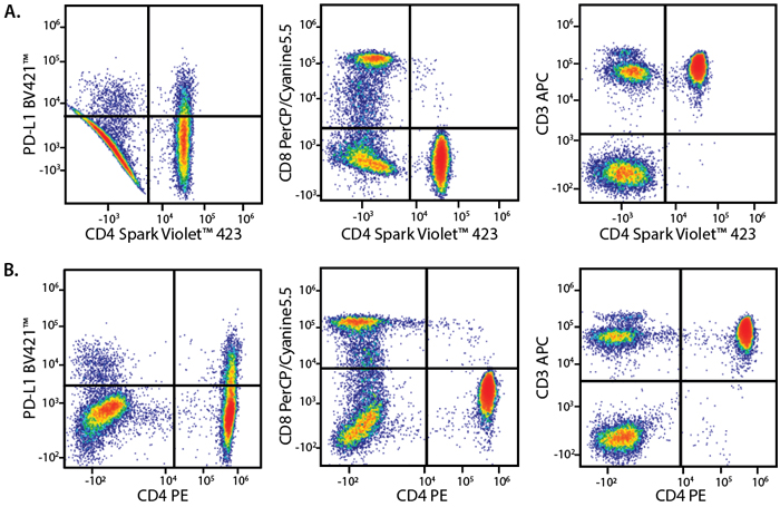
Unmix Spark Violet™ 423 from Brilliant Violet 421™


Panel A demonstrates how Spark Violet™ 423 performs in a multicolor panel with fluorophores that share significant spectra overlap with it. Panel B is shown as a reference panel using pre-optimized fluorophores to ensure that staining patterns are similar.


Spark Violet 423 Panel


Marker

Panel A Fluorophore

Panel B Fluorophore

CD3

APC

APC

CD4

Spark Violet™ 423

PE

CD8

PerCP/Cyanine5.5

PerCP/Cyanine5.5

PD-L1

BV421™

BV421™


Human lysed whole blood was stained with the indicated antibodies and unmixed on a 5-laser Cytek™ Aurora Cytometer using cells. All plots are gated on lymphocytes.


Stability and Validation Testing


All BioLegend fluorophores undergo rigorous testing procedures to determine how light, heat, and fixation may affect the performance and ensure they will perform reliably. To compare the signal across different conditions and timepoints, we used the Stain Index (formula below) to measure the relative brightness of the antibody. 

Spark 550

Photostability Testing


The photostability of Spark Violet™ 423 was tested in two ways that mimic how an antibody may be exposed to light over the course of an experiment.


  1. Antibodies were stored in the dark or exposed to fluorescent lighting. Then, the antibodies were used to stain freshly harvested cell samples and analyzed immediately.
  2. Cells were stained with antibody that had been kept under recommended storage conditions. Prior to analysis, the stained cells were stored in the dark or exposed to fluorescent lighting.


Spark Violet 423 Photostability

Anti-human CD3 (clone UCHT1) Spark Violet™ 423 was stored in a clear vial (Ab only) and left exposed to light or protected in the dark, as indicated. Antibodies were stored for the indicated timepoints and then used to stain human lysed whole blood. Samples labeled Ab + cells contain human lysed whole blood that was stained with anti-human CD3 Spark Violet™ 423. Stained cells were then left in the light or protected, as indicated.


Heat Stability


Spark Violet 423 Heat Stability

Anti-human CD3 (clone UCHT1) Spark Violet™ 423 was aliquoted and incubated at the indicated temperatures over the course of 28 days. The antibodies were then used to stain human lysed whole blood from a single donor.


Fixative Stability


The compatibility of Spark Violet™ 423 with fixatives should be tested by the end user to ensure the antigen's expression is maintained post-fixation. 


A guide to the fixatives used in this experiment:


Spark Violet 423 Fixative Bar Graph


Human PBMCs were stained with anti-human CD3 (clone UCHT1) conjugated to Spark Violet™ 423 and fixed using the respective protocols for each buffer set. Fresh samples were fixed and read on a 5-laser Cytek™ Aurora Cytometer immediately. Overnight samples were fixed and stored overnight in Cyto-Last™ Buffer before reading.

Spark UV™ 387


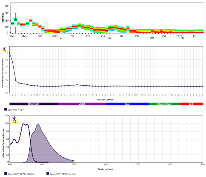
Spark UV™ 387 is selectively excited by the 355 nm ultraviolet laser and can easily be used in a multicolor panel with BUV496™ on both conventional (as an alternative for BUV395™) and spectral cytometers with appropriate instrument settings. Unmixing and simultaneous use with BUV395™ in a multicolor panel on a UV laser-equipped spectral cytometer is technically feasible. However, as these two fluorophores share significant spectral overlap, this requires careful panel building and is ideally reserved for analyzing two mutually-expressed markers on different cell types. On conventional flow cytometers, this fluorophore can be detected with a 379/28 filter, or similar (detector typically associated for BUV395™ detection). With a lower brightness, Spark UV™ 387 is best suited for antigens with high expression levels.


Excitation and Emission Spectra of Spark UV™ 387


Spark UV 387 spectra signature


Emission spectra (top) and normalized emission spectra (middle) of Spark UV™ 387 run on a 5-laser Cytek™ Aurora Spectral Cytometer. To compare Spark UV™ 387 with other fluorophores on a spectral cytometer, use our Aurora Spectral Analyzer tool.


Normalized excitation and emission spectra (bottom) of Spark UV™ 387 obtained from a spectrophotometer. To compare Spark Blue™ 574 with other fluorophores, use our Fluorescence Spectra Analyzer tool.


Multicolor Staining With Spark UV™ 387 


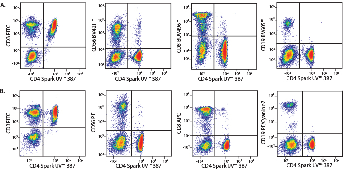
Panel A demonstrates how Spark UV™ 387 performs in a multicolor panel with fluorophores that share significant spectra overlap with it. Panel B is shown as a reference panel using pre-optimized fluorophores to ensure that staining patterns are similar.


Spark UV 387 Panel


Marker

Panel A Fluorophore

Panel B Fluorophore

CD3

FITC

FITC

CD4

Spark UV™ 387

Spark UV™ 387

CD8

BUV496™

APC

CD19

BV605™

PE/Cyanine7

CD56

BV421™

PE


Human lysed whole blood was stained with the indicated antibodies and unmixed on a 5-laser Cytek™ Aurora Cytometer using cells. All plots are gated on lymphocytes.


Stability and Validation Testing


All BioLegend fluorophores undergo rigorous testing procedures to determine how light, heat, and fixation may affect the performance and ensure they will perform reliably. To compare the signal across different conditions and timepoints, we used the Stain Index (formula below) to measure the relative brightness of the antibody. 

Spark 550

Photostability Testing


The photostability of Spark UV™ 387 was tested in two ways that mimic how an antibody may be exposed to light over the course of an experiment.


  1. Antibodies were stored in the dark or exposed to fluorescent lighting. Then, the antibodies were used to stain freshly harvested cell samples and analyzed immediately.
  2. Cells were stained with antibody that had been kept under recommended storage conditions. Prior to analysis, the stained cells were stored in the dark or exposed to fluorescent lighting.


Spark UV 387 Photostability

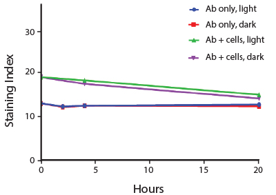
Anti-human CD4 (clone SK3) Spark UV™ 387 was stored in a clear vial (Ab only) and left exposed to light or protected in the dark, as indicated. Antibodies were stored for the indicated timepoints and then used to stain human PBMCs. Samples labeled Ab + cells contain human PBMCs that were stained with anti-human CD4 Spark UV™ 387. Stained cells were then left in the light or protected, as indicated.


Heat Stability


Spark UV 387 Heat Stability

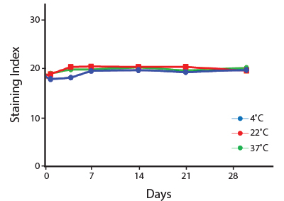
Anti-human CD4 (clone SK3) Spark UV™ 387 was aliquoted and incubated at the indicated temperatures over the course of 28 days. The antibodies were then used to stain human lysed whole blood from a single donor.


Fixative Stability


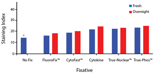
The compatibility of Spark UV™ 387 with fixatives should be tested by the end user to ensure the antigen's expression is maintained post-fixation. 


A guide to the fixatives used in this experiment:


Spark UV 387 Fixative Bar Graph


Human PBMCs were stained with anti-human CD4 (clone SK3) conjugated to Spark UV™ 387 and fixed using the respective protocols for each buffer set. Fresh samples were fixed and read on a 5-laser Cytek™ Aurora Cytometer immediately. Overnight samples were fixed and stored overnight before reading.
*No reading was taken for an unfixed sample kept overnight, as this would not follow typical lab protocols.

Spark Red™ 718


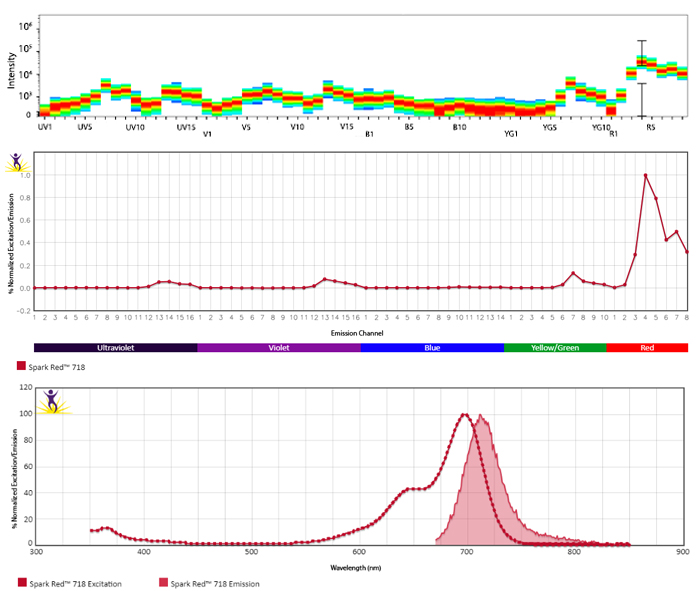
Spark Red™ 718 is a brighter alternative to Alexa Fluor® 700, adding to the fluorophore options for the red laser that can detect antigens with lower expression levels. It can be used on a conventional cytometer with a 633 nm red laser and 720/45 filter set (or equivalent filter used to detect Alexa Fluor® 700) spectral cytometers with appropriate instrument settings. On conventional cytometers, Spark Red™ 718 can be utilized with both APC and APC/Cyanine7. On spectral cytometers, it can be unmixed from APC, Spark NIR™ 685, Alexa Fluor® 660, APC/Cyanine7, and APC/Fire™ 810. As spectral properties of Spark Red™ 718 and Alexa Fluor® 700 are nearly identical, using them together in a multicolor panel is not recommended. With a medium brightness, Spark Red™ 718 is best suited for detecting antigens with low to moderate expression levels.


Excitation and Emission Spectra of Spark Red™ 718


Spark Red 718 spectra signature


Emission spectra (top) and normalized emission spectra (middle) of Spark Red™ 718 run on a 5-laser Cytek™ Aurora Spectral Cytometer. To compare Spark Red™ 718 with other fluorophores on a spectral cytometer, use our Aurora Spectral Analyzer tool.


Normalized excitation and emission spectra (bottom) of Spark Red™ 718 obtained from a spectrophotometer. To compare Spark Red™ 718 with other fluorophores, use our Fluorescence Spectra Analyzer tool.


Multicolor Staining With Spark Red™ 718 


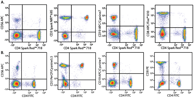
Panel A demonstrates how Spark Red™ 718 performs in a multicolor panel with fluorophores that share significant spectra overlap with it. Panel B is shown as a reference panel using pre-optimized fluorophores to ensure that staining patterns are similar.


Spark Red 718 Panel


Marker

Panel A Fluorophore

Panel B Fluorophore

CD3

Spark NIR™ 685

PerCP/Cyanine5.5

CD4

Spark Red™ 718

FITC

CD8

APC/Fire™ 810

PE

CD19

APC/Cyanine7

APC/Cyanine7

CD56

APC

APC


Human PBMCs were stained with the indicated antibodies and unmixed on a 3-laser Cytek™ Aurora Cytometer using single color-stained cells. All plots are gated on lymphocytes.


Stability and Validation Testing


All BioLegend fluorophores undergo rigorous testing procedures to determine how light, heat, and fixation may affect the performance and ensure they will perform reliably. To compare the signal across different conditions and timepoints, we used the Stain Index (formula below) to measure the relative brightness of the antibody. 

Spark 550

Photostability Testing


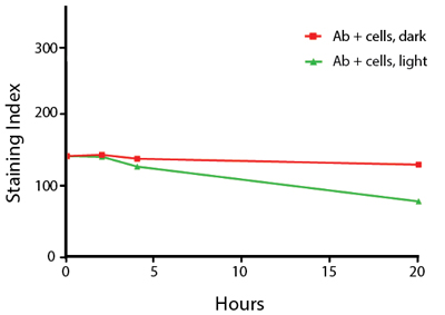
The photostability of Spark Red™ 718 was tested to mimic how an antibody may be exposed to light over the course of an experiment. Cells were stained with antibody that had been kept under recommended storage conditions. Prior to analysis, the stained cells were stored in the dark or exposed to fluorescent lighting.


Spark Red 718 Photostability

Human PBMCs were stained with anti-human CD4 (clone SK3) Spark Red™ 718, exposed to ambient light (Ab + cells, light) or kept in the dark (Ab + cells, dark) for the indicated times, and then analyzed on a 3-laser Cytek™ Aurora cytometer.


Heat Stability


Spark Red 718 Heat Stability

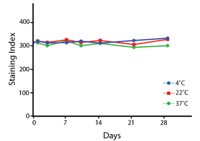
Anti-human CD4 (clone SK3) Spark Red™ 718 was aliquoted and incubated at the indicated temperatures over the course of 28 days, and then analyzed on a 3-laser Cytek™ Aurora cytometer. The antibodies were then used to stain human lysed whole blood from a single donor.


Fixative Stability


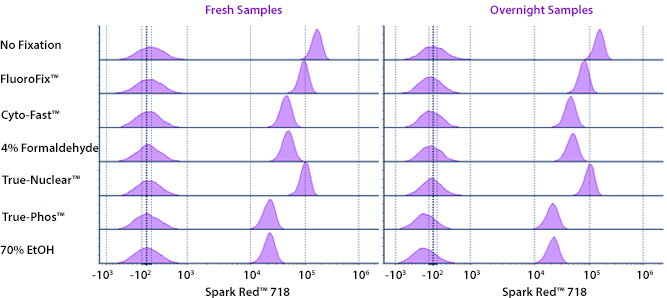
The compatibility of Spark Red™ 718 with fixatives should be tested by the end user to ensure the antigen's expression is maintained post-fixation. 


A guide to the fixatives used in this experiment:


Spark Red 718 Fixation


Human PBMCs were stained with anti-human CD4 (clone SK3) conjugated to Spark Red™ 718 and fixed using the respective protocols for each buffer set. Fresh samples were fixed and read on a 3-laser Cytek™ Aurora Cytometer immediately. Overnight samples were fixed and stored in Cyto-Last™ Buffer overnight before reading.

Spark Violet™ 500


Spark Violet™ 500 exhibits peak excitation/emission wavelengths at 393 nm and 500 nm respectively, most closely matching that of BD Horizon™ V500 (V500-C) and AmCyan. On conventional cytometers, it can be detected with a 405 nm laser and 510/50 filter set (or equivalent). On spectral cytometers with appropriate instrument settings, it can be unmixed from Spark Violet™ 538, Pacific Blue™, and Brilliant Violet 570™. With a medium brightness, Spark Violet™ 500 is best suited for detecting antigens with low to moderate expression levels. In addition, Spark Violet™ 500 can be cocktailed with other fluorophore conjugated antibodies, either in liquid or dry-down formats, without exhibiting inter-dye interactions. It is also stable towards exposure to heat or a variety of commonly used fixative solutions.


Excitation and Emission Spectra of Spark Violet™ 500


Spark Violet 500 spectra signature


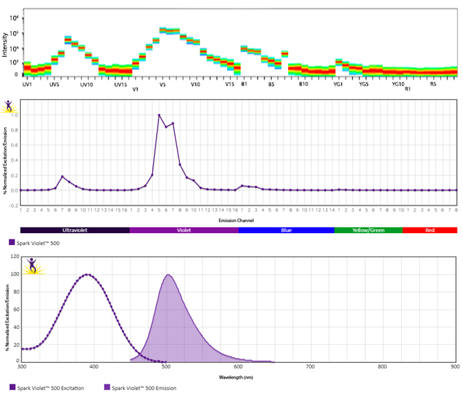
Emission spectra (top) and normalized emission spectra (middle) of Spark Violet™ 500 run on a 5-laser Cytek™ Aurora Spectral Cytometer. To compare Spark Violet™ 500 with other fluorophores on a spectral cytometer, use our Aurora Spectral Analyzer tool.


Normalized excitation and emission spectra (bottom) of Spark Violet™ 500 obtained from a spectrophotometer. To compare Spark Violet™ 500 with other fluorophores, use our Fluorescence Spectra Analyzer tool.


Multicolor Staining With Spark Violet™ 500


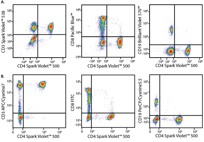
Panel A demonstrates how Spark Violet™ 500 performs in a multicolor panel with fluorophores that share significant spectra overlap with it. Panel B is shown as a reference panel using pre-optimized fluorophores to ensure that staining patterns are similar.


Spark Violet 500 Panel


Marker

Panel A Fluorophore

Panel B Fluorophore

CD3

Spark Violet™ 538

APC/Cyanine7

CD4

Spark Violet™ 500

Spark Violet™ 500

CD8

Pacific Blue™

FITC

CD19

Brilliant Violet 570™

PerCP/Cyanine5.5


Human PBMCs were stained with the indicated antibodies and unmixed on a 5-laser Cytek™ Aurora Cytometer using compensation beads. All plots are gated on lymphocytes.


Stability and Validation Testing


All BioLegend fluorophores undergo rigorous testing procedures to determine how light, heat, and fixation may affect the performance and ensure they will perform reliably. To compare the signal across different conditions and timepoints, we used the Stain Index (formula below) to measure the relative brightness of the antibody. 

Spark 550

Photostability Testing


The photostability of Spark Violet™ 500 was tested in two ways that mimic how an antibody may be exposed to light over the course of an experiment.


  1. Antibodies were stored in the dark or exposed to fluorescent lighting. Then, the antibodies were used to stain freshly harvested cell samples and analyzed immediately.
  2. Cells were stained with antibody that had been kept under recommended storage conditions. Prior to analysis, the stained cells were stored in the dark or exposed to fluorescent lighting.


Spark Violet 500 Photostability


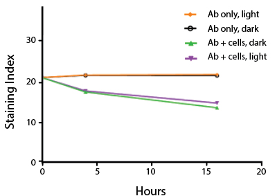
Anti-human CD4 (clone SK3) Spark Violet™ 500 was stored in a clear vial (Ab only) and left exposed to light or protected in the dark, as indicated. Antibodies were stored for the indicated timepoints and then used to stain human lysed whole blood. Samples labeled Ab + cells contain human lysed whole blood that was stained with anti-human CD4 Spark Violet™ 500. Stained cells were then left in the light or protected, as indicated.


Heat Stability


Spark Violet 500 Heat Stability

Anti-human CD4 (clone SK3) Spark Violet™ 500 was aliquoted and incubated at the indicated temperatures over the course of 30 days, and then analyzed on a 5-laser Cytek™ Aurora cytometer. The antibodies were then used to stain human lysed whole blood from a single donor.


Fixative Stability


The compatibility of Spark Violet™ 500 with fixatives should be tested by the end user to ensure the antigen's expression is maintained post-fixation. 


A guide to the fixatives used in this experiment:


Spark Violet 500 Fixation


Human PBMCs were stained with anti-human CD4 (clone SK3) conjugated to Spark Violet™ 500 and fixed using the respective protocols for each buffer set. Fresh samples were fixed and read on a 5-laser Cytek™ Aurora Cytometer immediately. Overnight samples were fixed and stored in Cyto-Last™ Buffer overnight before reading.

*No reading was taken for an unfixed sample kept overnight, as this would not follow typical lab protocols.


Spark Blue™ 515


Spark Blue™ 515 exhibits peak excitation/emission wavelengths at 506 nm and 516 nm respectively, most closely matching the spectra of cFluor® B515. On conventional cytometers, it can be detected with a 488 nm blue laser and the 530/30 filter set typically used by FITC or Alexa Fluor® 488. On spectral instruments with the proper settings, it is possible to unmix Spark Blue™ 515 from dyes like FITC, Alexa Fluor® 488, and KIRAVIA Blue 520™ as its peak emission is shifted over 10 nm. With a dim brightness, Spark Blue™ 515 is best suited for detecting antigens with medium to high expression levels. It is also stable towards exposure to heat or a variety of commonly used fixative solutions.


Excitation and Emission Spectra of Spark Blue™ 515


Spark Blue 515 spectra signature


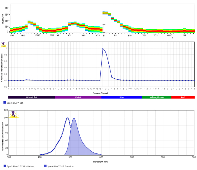
Emission spectra (top) and normalized emission spectra (middle) of Spark Blue™ 515 run on a 5-laser Cytek™ Aurora Spectral Cytometer. To compare Spark Blue™ 515 with other fluorophores on a spectral cytometer, use our Aurora Spectral Analyzer tool.


Normalized excitation and emission spectra (bottom) of Spark Blue™ 515 obtained from a spectrophotometer. To compare Spark Blue™ 515 with other fluorophores, use our Fluorescence Spectra Analyzer tool.


Multicolor Staining With Spark Blue™ 515


The panel below demonstrates how Spark Blue™ 515 performs in a multicolor panel with fluorophores that can share significant spectra overlap with it.


Spark Blue 515 Panel


Marker

Fluorophore

Marker

Fluorophore

Marker

Fluorophore

CCR7

BV421™ 

CD11c

PE/Cyanine7

CD45RA

Alexa Fluor® 488

CD1c

Alexa Fluor® 647

CD14

Spark Blue™ 550

CD56

BV750™

CD3

BV570™

CD16

BV605™

CD123

Alexa Fluor® 700

CD4

Spark Blue™ 574 

CD19

Spark NIR™ 685

CD141

Spark Blue™ 515

CD8

PerCP/Cyanine5.5

CD20

Pacific Blue™

HLA-DR

APC/Fire™ 810


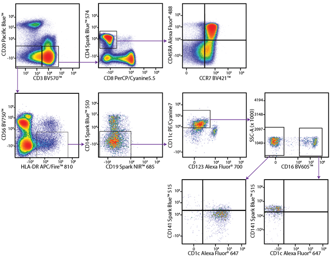
Representative multicolor staining that includes Spark Blue™ 515 anti-human CD141 and Alexa Fluor® 488 anti-human CD45RA. Hierarchical gating methods to analyze CD45RA and CD141 expression on CD4+ T cells and CD11c+ dendritic cells, respectively, are represented by the indicated arrows. Staining was performed on human peripheral blood mononuclear cells, and analyzed on a 5-laser Cytek™ Aurora.


Stability and Validation Testing


All BioLegend fluorophores undergo rigorous testing procedures to determine how light, heat, and fixation may affect the performance and ensure they will perform reliably. To compare the signal across different conditions and timepoints, we used the Stain Index (formula below) to measure the relative brightness of the antibody. 

Spark 550

Photostability Testing


The photostability of Spark Blue™ 515 was tested in two ways that mimic how an antibody may be exposed to light over the course of an experiment.


  1. Antibodies were stored in the dark or exposed to fluorescent lighting. Then, the antibodies were used to stain freshly harvested cell samples and analyzed immediately.
  2. Cells were stained with antibody that had been kept under recommended storage conditions. Prior to analysis, the stained cells were stored in the dark or exposed to fluorescent lighting.


Spark Blue 515 Photostability


Anti-human CD4 (clone SK3) Spark Blue™ 515 was stored in a clear vial (Ab only) and left exposed to light or protected in the dark, as indicated. Antibodies were stored for the indicated timepoints and then used to stain human lysed whole blood. Samples labeled Ab + cells contain human lysed whole blood that was stained with anti-human CD4 Spark Blue™ 515. Stained cells were then left in the light or protected, as indicated.


Heat Stability


Spark Blue 515 Heat Stability

Anti-human CD4 (clone SK3) Spark Blue™ 515 was aliquoted and incubated at the indicated temperatures over the course of 30 days, and then analyzed on a 5-laser Cytek™ Aurora cytometer. The antibodies were then used to stain human lysed whole blood from a single donor.


Fixative Stability


The compatibility of Spark Blue™ 515 with fixatives should be tested by the end user to ensure the antigen's expression is maintained post-fixation. 


A guide to the fixatives used in this experiment:


Spark Blue 515 Fixation


Human PBMCs were stained with anti-human CD4 (clone SK3) conjugated to Spark Blue™ 515 and fixed using the respective protocols for each buffer set. Fresh samples were fixed and read on a 5-laser Cytek™ Aurora Cytometer immediately. Overnight samples were fixed and stored in Cyto-Last™ Buffer overnight before reading.


Spark PLUS UV395™


Spark PLUS UV395™ is a bright UV laser-excited dye that can be used on both conventional and spectral flow cytometers. It exhibits peak excitation/emission wavelengths at 355 nm and 385 nm respectively, most closely matching that of BD Horizon™ BUV395. On conventional cytometers, it can be detected with a 355 nm laser and 379/28 filter set (or equivalent). With its intense brightness, Spark PLUS UV395™ is a versatile option for detecting antigens at most expression levels and is stable towards exposure to heat or a variety of commonly used fixative solutions. Spark PLUS UV395™ is the first in the family of Spark PLUS™ Dyes, all formulated to provide brighter signals and superior performance.


Excitation and Emission Spectra of Spark PLUS UV395™


Emission spectra (top) and normalized emission spectra (middle) of Spark PLUS UV395™ run on a 5-laser Cytek™ Aurora Spectral Cytometer. To compare Spark PLUS UV395™ with other fluorophores on a spectral cytometer, use our Aurora Spectral Analyzer tool.


Normalized excitation and emission spectra (bottom) of Spark PLUS UV395™ obtained from a spectrophotometer. To compare Spark PLUS UV395™ with other fluorophores, use our Fluorescence Spectra Analyzer tool.


A New Alternative to BD Horizon™ BUV395


Human peripheral blood lymphocytes were stained with anti-human CD3 APC and anti-human CD4 (clone SK3) Spark PLUS UV395™ (left) or anti-human CD4 (clone SK3) BD Horizon™ BUV395 (right). Samples were acquired on a 5-laser Cytek® Aurora.


Multicolor Staining With Spark PLUS UV395™


Panel A demonstrates how Spark PLUS UV395™ performs in a multicolor panel with fluorophores that may exhibit spectral overlap with it. Panel B is shown as a reference panel using pre-optimized fluorophores to ensure that staining patterns are similar.


Marker

Panel A Fluorophore

Panel B Fluorophore

CD3

Spark Violet™ 538

FITC

CD4

Spark PLUS UV395™

Spark PLUS UV395™

CD8

FITC

APC

CD19

Brilliant Violet 570™

PE

CD25

Brilliant Violet 421™

Brilliant Violet 421™


Human PBMCs were stained with the indicated antibodies and analyzed on a conventional flow cytometer. All plots are gated on lymphocytes.


Unmix Spark PLUS UV395™ and Spark UV™ 387


Human PBMCs were stained with anti-human CD8 Spark UV 387 and anti-human CD4 (clone SK3) Spark PLUS UV395™ (left) or anti-human CD4 (clone SK3) APC (right). Samples were acquired on a 5-laser Cytek® Aurora cytometer.


Stability and Validation Testing


All BioLegend fluorophores undergo rigorous testing procedures to determine how light, heat, and fixation may affect the performance and ensure they will perform reliably. To compare the signal across different conditions and timepoints, we used the Stain Index (formula below) to measure the relative brightness of the antibody. 

Photostability Testing


The photostability of Spark PLUS UV395™ was tested in two ways that mimic how an antibody may be exposed to light over the course of an experiment.


  1. Antibodies were stored in the dark or exposed to fluorescent lighting. Then, the antibodies were used to stain freshly harvested cell samples and analyzed immediately.
  2. Cells were stained with antibody that had been kept under recommended storage conditions. Prior to analysis, the stained cells were stored in the dark or exposed to fluorescent lighting.


To assess the photostability of Spark PLUS UV395™-conjugated antibodies, anti-human CD4 (clone SK3) Spark PLUS UV395™ or BD Horizon™ BUV395 conjugates were exposed to light or protected in the dark (left). The samples were then used to stain human PBMCs and the fold change over the staining index at zero hours was calculated. To measure the photostability of Spark PLUS UV395™ stained cells, human PBMCs were stained with anti-human CD4 (clone SK3) Spark PLUS UV395™ or BD Horizon™ BUV395. The fold change in staining index of stained cells kept in the dark or exposed to light was calculated from the initial staining index at zero hours (right). Cells were gated on lymphocytes and data was acquired on a 5-laser Cytek® Aurora.


Heat Stability


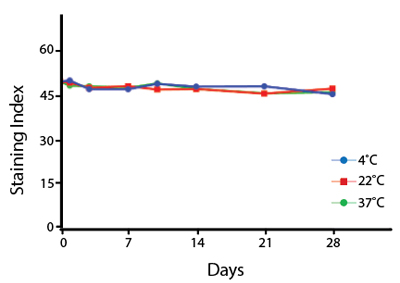
Anti-human CD4 (clone SK3) Spark PLUS UV395™ was aliquoted and incubated at the indicated temperatures over the course of 28 days, and then analyzed on a 5-laser Cytek™ Aurora cytometer. The antibodies were then used to stain human lysed whole blood from a single donor.


Fixative Stability


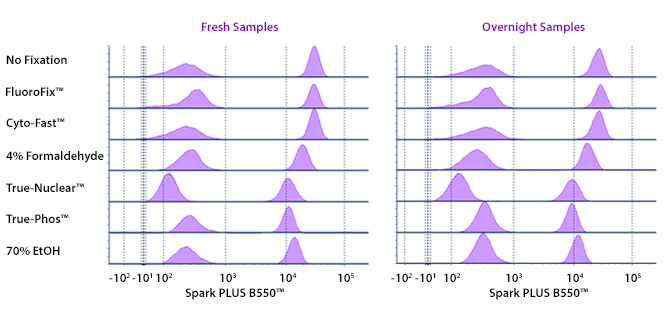
A guide to the fixatives used in this experiment:


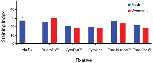
Human PBMCs were stained with anti-human CD4 (clone SK3) conjugated to Spark PLUS UV395™ and fixed using the respective protocols for each buffer set. Fresh samples were fixed and read on a 5-laser Cytek™ Aurora Cytometer immediately. Overnight samples were fixed and stored in Cyto-Last™ Buffer overnight before reading.


Spark PLUS B550™


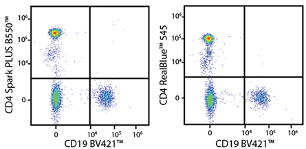
Spark PLUS B550™ is formulated for brighter signal and superior stability, maximizing signal from the blue laser with minimal yellow/green cross-excitation and spillover. This fluorophore serves as an alternative to BD Horizon™ RealBlue545 and can be used on conventional and spectral flow cytometers, and is ideal for combining with PE and FITC on spectral instruments.


Excitation and Emission Spectra of Spark PLUS B550™


Emission spectra (top) and normalized emission spectra (middle) of Spark PLUS B550™ run on a 5-laser Cytek™ Aurora Spectral Cytometer. To compare Spark PLUS B550™ with other fluorophores on a spectral cytometer, use our Aurora Spectral Analyzer tool.


Normalized excitation and emission spectra (bottom) of Spark PLUS B550™ obtained from a spectrophotometer. To compare Spark PLUS B550™ with other fluorophores, use our Fluorescence Spectra Analyzer tool.


A New Alternative to BD Horizon™ RealBlue 545


Human peripheral blood lymphocytes were stained with anti-human CD19 (clone HIB19) Brilliant Violet 421™ and either CD4 (clone SK3) Spark PLUS B550™ (left) or CD4 (clone SK3) BD Horizon RealBlue™ 545 (right). Samples were acquired on a 5-laser Cytek® Aurora.


Multicolor Staining with Spark PLUS B550™


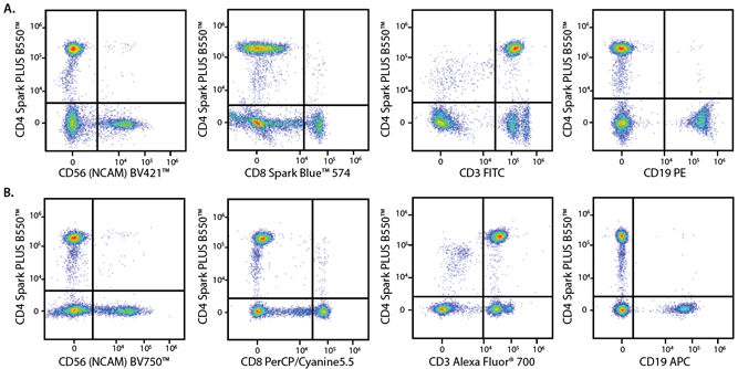
Panel A demonstrates how Spark PLUS B550™ performs in a multicolor panel with fluorophores that may exhibit spectral overlap with it. Panel B is shown as a reference panel using pre-optimized fluorophores to ensure that staining patterns are similar.


Marker

Panel A Fluorophore

Panel B Fluorophore

CD3

FITC

Alexa Fluor® 700

CD4

Spark PLUS B550™

Spark PLUS B550™

CD8

Spark Blue™ 574

PerCP/Cyanine5.5

CD19

PE

APC

CD56

Brilliant Violet 421™

Brilliant Violet 750™


Human PBMCs were stained with the indicated antibodies and analyzed on 5-laser Cytek® Aurora. All plots are gated on lymphocytes.


Stability and Validation Testing


All BioLegend fluorophores undergo rigorous testing procedures to determine how light, heat, and fixation may affect the performance and ensure they will perform reliably. To compare the signal across different conditions and timepoints, we used the Stain Index (formula below) to measure the relative brightness of the antibody. 

Photostability Testing


The photostability of Spark PLUS B550™ was tested in two ways that mimic how an antibody may be exposed to light over the course of an experiment.


  1. Antibodies were stored in the dark or exposed to fluorescent lighting. Then, the antibodies were used to stain freshly harvested cell samples and analyzed immediately.
  2. Cells were stained with antibody that had been kept under recommended storage conditions. Prior to analysis, the stained cells were stored in the dark or exposed to fluorescent lighting.


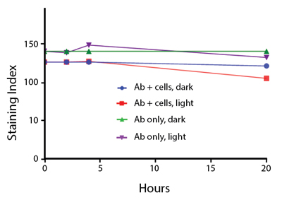
To assess the photostability of Spark PLUS B550™-conjugated antibodies, anti-human CD4 (clone SK3) Spark PLUS B550™ was exposed to light or protected in the dark for the indicated time points. The samples were then used to stain human lysed whole blood. To measure the photostability of Spark PLUS B550™ stained cells, human lysed whole blood was stained with anti-human CD4 (clone SK3) Spark PLUS B550™. Stained cells were then left in the light or protected in the dark as indicated. Cells in the lymphocyte gate were used for analysis.


Heat Stability


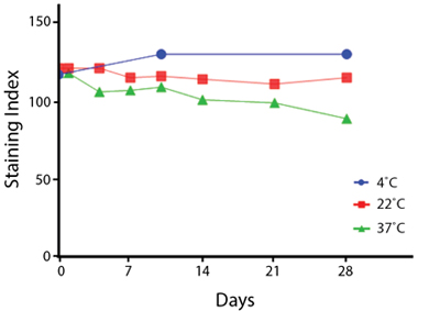
Anti-human CD4 (clone SK3) Spark PLUS B550™ was aliquoted and incubated at the indicated temperatures over the course of 28 days, and then analyzed on a 5-laser Cytek™ Aurora cytometer. The antibodies were then used to stain human lysed whole blood from a single donor.


Fixative Stability


A guide to the fixatives used in this experiment:


Human PBMCs were stained with anti-human CD4 (clone SK3) conjugated to Spark PLUS B550™ and fixed using the respective protocols for each buffer set. Fresh samples were fixed and read on a BD LSRFortessa™ cytometer immediately. Overnight samples were fixed and stored in Cyto-Last™ Buffer overnight before reading.


ProductsHere

Login / Register
Remember me
Forgot your password? Reset password?
Create an Account