Tolling the Dead with Zombie Dyes
For flow cytometry experiments, it is imperative to make sure samples are clean of debris and dying cells as they can non-specifically bind to dyes and antibodies meant to stain live cells. Dead cell exclusion is even more critical when working with small populations or rare events, as even small changes can have a big impact on your analysis. In addition, certain cell types (e.g., bone marrow resident plasma cells or mouse intestinal lamina propria cells) are more sensitive than others, and lengthy protocols for sample isolation can affect the viability of your samples. In these cases, it is particularly important to exclude dead cells from your analysis.
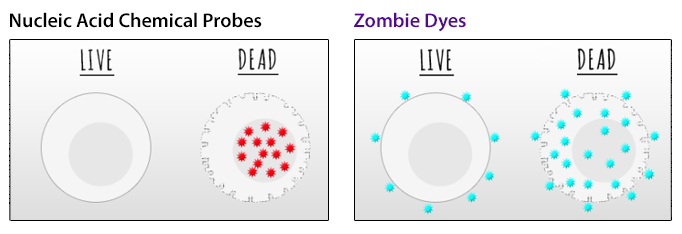
Chemical probes that target nucleic acids, such as 7-AAD or propidium iodide, are commonly used to differentiate live and dead cells. These dyes cannot penetrate the intact membranes of healthy cells but can access and stain nucleic acids through the disrupted membranes of dying cells. However, nucleic acid stains are not perfect for every occasion.
First, they cannot be used for viability staining on cells that will be fixed or permeabilized, as the live/dead staining pattern is not preserved through treatments that disrupt cell membranes. Second, nucleic acid probes can be limited in their excitation and emission properties, restricting options for their use in multicolor panel design.
In contrast, our amine-reactive Zombie Dyes are compatible with fixation and intracellular staining techniques and are offered in a wide range of colors – from our traditional offerings that can be used in the most common conventional flow cytometry channels to dyes designed to emit at unique wavelengths for large, optimized panel design, such as Zombie B550™, Zombie YG581™, Zombie R685™, and Zombie R718™.
|
Zombie Dye |
Excitation Laser |
Emission Max |
|
UV 355 nm |
459 nm |
|
|
Violet 405 nm |
423 nm |
|
|
Violet 405 nm |
516 nm |
|
|
Violet 405 nm |
572 nm |
|
|
Blue 488 nm |
515 nm |
|
|
Blue 488 nm |
540 nm |
|
|
Yellow/Green 561 nm |
581 nm |
|
|
Yellow/Green 561 nm |
624 nm |
|
|
Red 633 nm |
685 nm |
|
|
Red 633 nm |
718 nm |
|
|
Red 633 nm |
746 nm |

Mouse splenocytes were stored overnight to allow dead cells to accumulate. Cells were then stained with Zombie Dyes as indicated and analyzed before fixation (purple) or after fixation and permeabilization (red). Cells alone, without Zombie staining, are indicated in black.
Zombie Dyes detect amines on the surface of all cells at a low level but intensely stain dead and dying cells by accessing the abundant intracellular amines through disrupted membranes. As this covalent interaction is irreversible, cells can be washed, fixed, and permeabilized after viability staining without altering the initial live/dead staining pattern.

(Left) Impermeant nucleic acid stains can only stain DNA when the membrane is compromised. (Right) Zombie dyes bind to primary amines on proteins. Live cells receive low-level surface staining, while dead cells stain at a much brighter fluorescence level due to the exposure of internal proteins.
Whether you are interested in Zombie Dyes or any of our other chemical probes, you can always talk with our experts to determine which reagent is best for your experimental setup. To probe beyond live/dead discrimination, explore our growing portfolio of antibody and non-antibody based methods of assessing cell health and proliferation. With our products and guidance, we will ensure your data is consistent, trustworthy, and groundbreaking.
 Login / Register


Follow Us