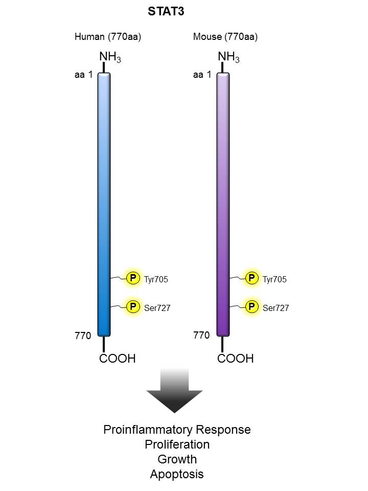
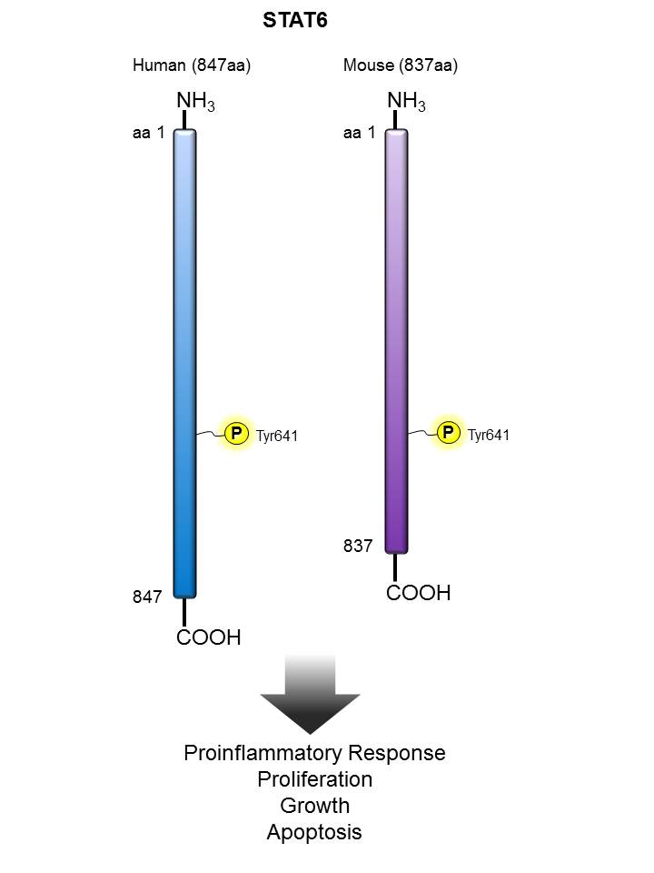
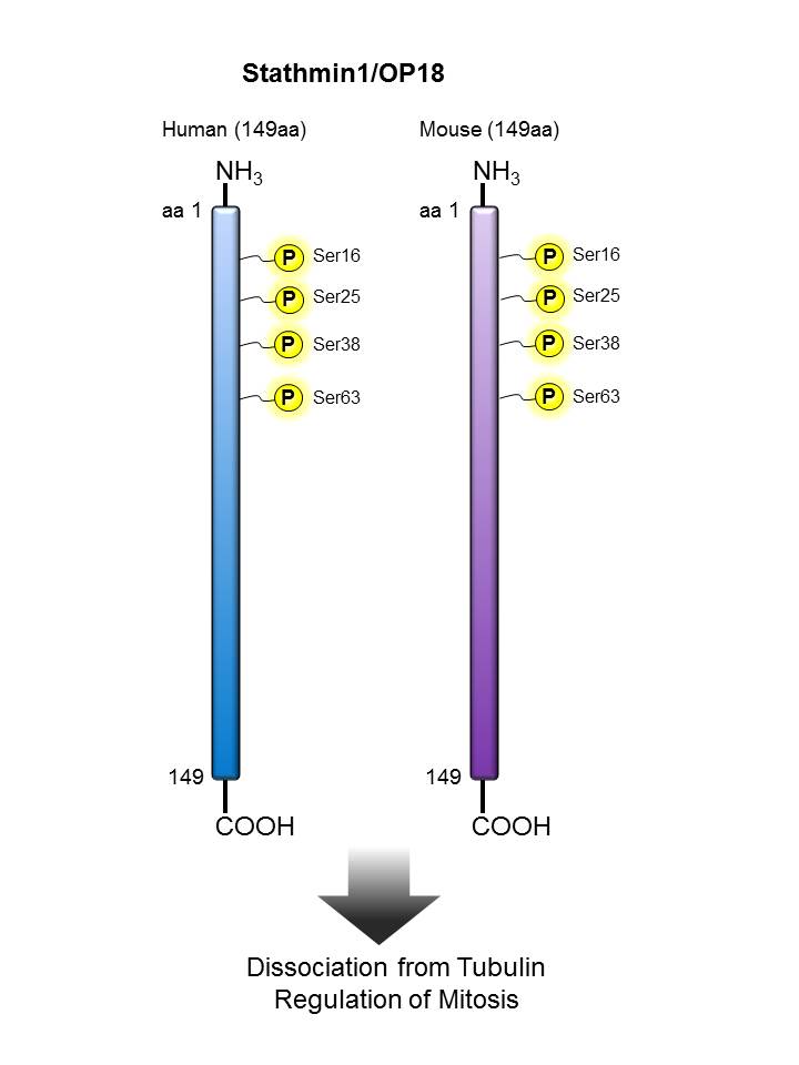
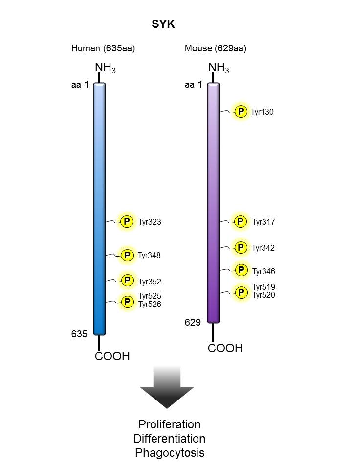
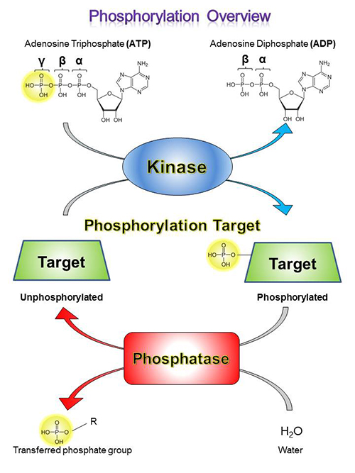
Phosphorylation and de-phosphorylation play critical roles as a mode of signal transfer in biological processes. This is defined as the transfer of phosphoryl groups [(PO3)2-] from one molecule to another, and serves as a transfer of energy that results in the activation, or deactivation of downstream proteins.

At BioLegend, we offer a wide array of phospho protein-related products for use in flow cytometry, western blotting, and microscopy applications. Whenever possible, the phospho protein-specific antibodies we offer are optimized to work for multiple applications, allowing for clone and product consistency across different experimental systems.

PhosphoPair Antibody Sets


PhosphoPair Antibody Sets bundle a pair of antibodies together for cost-savings and convenience. One antibody is specific for a phosphorylated form of a protein, while the other detects total or unphosphorylated levels of the protein. PhosphoPair Antibody Sets are quality tested in western blotting to ensure high quality results in each experiment.


View all PhosphoPair Antibody Sets...


Phosphorylation Targets


The table below lists a number of well known phosphorylated proteins. Learn more about each protein and view the important phosphorylation sites. Also find upstream kinases and phosphatases that interact with the protein.


AKT α-Synuclein ATM Aurora A BCL-2
B-Raf BTK CAK/CDK7 CD195 (CCR5) DNA-PKcs
EG5 EGFR ERK1/2 GluR1 Histone H2AX
Histone H3.1 ITK JNK LCK mTOR
Neurofilament
H
Neurofilament
L
Neurofilament
M
NPM/B23 Nucleolin
p38 MAPK PERK PKCα PLCγ PLK1
RPS6 STAT1 STAT3 STAT4 STAT5
STAT6 Stathmin/
OP18
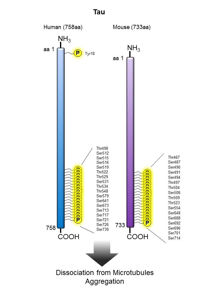
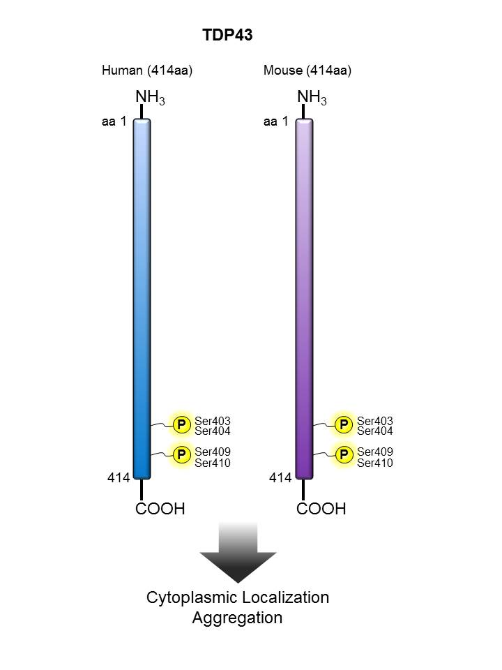
SYK Tau TDP43
TIF1β/
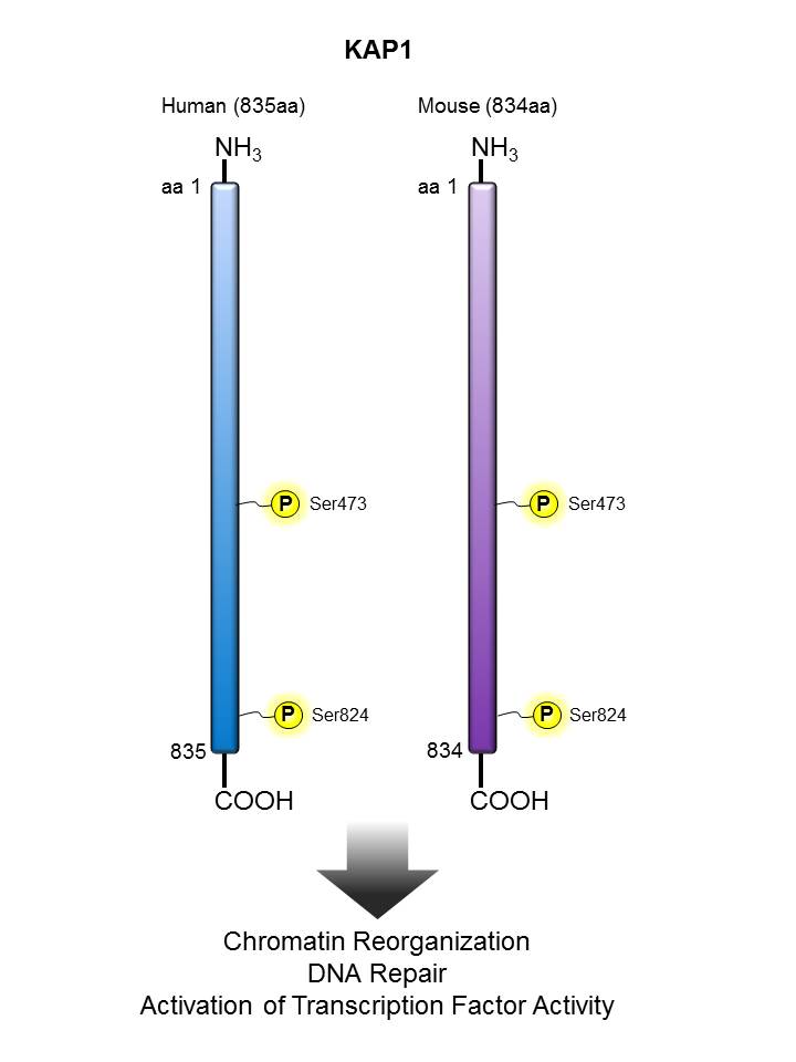
KAP1
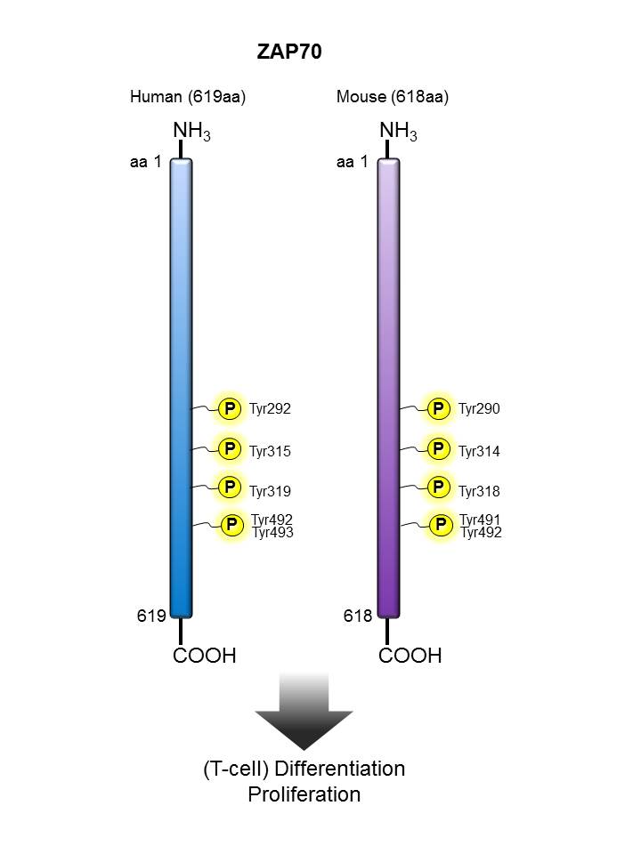
Zap70
  • Relevant phosphorylation site information obtained from PhosphoSitePlus®
  • Sequence homology determined via alignment of sequences available on UniProt
  • Notable Kinases/Phosphatases and listed associated functions for each target may not be entirely comprehensive. Please contact Technical Service if you have any feedback regarding the information listed on the page.

AKT


Description:

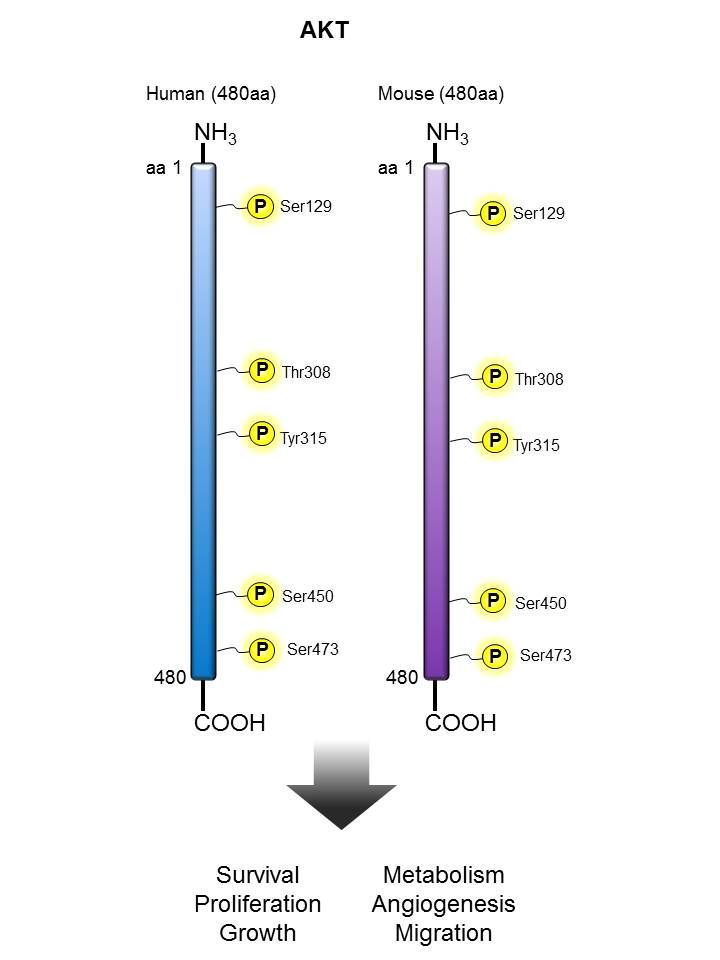
Akt (also known as protein kinase B alpha) is a 60 kD serine/threonine specific kinase containing a pleckstrin domain. Akt plays a critical role in controlling survival and apoptosis. This kinase is ubiquitously expressed and translocates to the membrane upon activation. This protein kinase is activated by insulin and various growth and survival factors to function in a wortmannin-sensitive pathway involving PI 3-kinase.  Akt is activated by phospholipid binding and activation loop phosphorylation at Thr308 by PDK1 (4) and by phosphorylation within the carboxy terminus at Ser473. The previously elusive PDK2, responsible for phosphorylation of Akt at Ser473, has been identified as a mammalian target of rapamycin (mTOR) in a rapamycin-insensitive complex with rictor and Sin1. Activated Akt can then phosphorylate a wide range of substrates including transcription factors (e.g. FOXO1), kinases (GSK-3,Raf-1, ASK, Chk1) and other proteins with important signaling roles (e.g. Bad, MDM2).  Akt promotes cell survival by inhibiting apoptosis through phosphorylation and by inactivation of several targets, including Bad, forkhead transcription factor, c-Raf and caspase-9. Akt also plays a critical role in cell growth by directly phosphorylating mTOR in a rapamycin-sensitive complex containing raptor. More importantly, Akt phosphorylates and inactivates tuberin (TSC2), an inhibitor of mTOR within the mTOR-raptor complex. The phosphor-Akt (S473) antibody specifically detects phosphorlated Akt at the Ser473 site.

Other Names:

Protein kinase B alpha (PRK-BA, PKB), Serine/Threonine specific kinase RAC alpha, Protein kinase Akt

Human to Mouse Sequence Homology:

98.30%

Notable Upstream Kinase(s):

PIP3, PDK1, mTOR/RICTOR

Notable Phosphatase(s):

PTEN, PP2A

Product Listing:

See products for AKT

Antigen References:

1. Jacinto E, et al. 2006. Cell 127:125.
2. Inoki K, et al. 2002. Nat. Cell. Biol. 4:648.
3. Franke TF, et al. 1995. Cell 81:727.
4. Brunet A, et al. 1999. Cell 96:857.
5. Yang WL, et al. 2010 Cell Cycle 9:487.

α-Synuclein


Description:

Alpha-synuclein is expressed principally in the central nervous system (brain) but is also expressed in low concentrations in a variety of tissues except liver. It is predominantly expressed in the neocortex, hippocampus, substantia nigra, thalamus, and cerebellum of the CNS. It is primarily a neuronal protein, but can also be found in the neuroglial cells. It is concentrated in presynaptic nerve terminals of neurons, as well as having reported nuclear and mitochondrial localization. Alpha-synuclein interacts with plasma membrane phospholipids. Alpha-synuclein in solution is considered to be an intrinsically disordered protein and thus lacks a stable secondary or tertiary structure. However, data suggests the presence of partial alpha helical as well as beta sheet structures as well as mostly structured tetrameric states in solution, the equilibrium of which may be altered by binding partners.

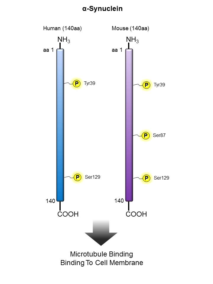
The human alpha-synuclein protein is made of 140 amino acids, encoded by the SNCA gene. The primary structure is divided in three distinct domains: (1-60) - An amphipathic N-terminal region dominated by four 11-residue repeats including the consensus sequence KTKEGV. This sequence has a structural alpha helix propensity similar to apolipoproteins-binding domains. (61-95) - a central hydrophobic region which includes the non-amyloid-β component (NAC) region, involved in protein aggregation. (96-140) - a highly acidic and proline-rich region. At least three isoforms of synuclein are produced through alternative splicing. The most common form of the protein, is the full 140 amino acid-long transcript. Other isoforms are alpha-synuclein-126, lacking residues 41-54; and alpha-synuclein-112, which lacks residues 103-130. a-synuclein may be involved in the regulation of dopamine release and transport and also may function to induce fibrillization of microtubule-associated protein tau. alpha-synuclein functions as a molecular chaperone in the formation of SNARE complexes. In particular, it can bind to phospholipids of the plasma membrane and to synaptobrevin-2 via its C-terminus domain to influence synaptic activity. alpha-synuclein is essential for normal development of the cognitive functions and that it significantly interacts with tubulin. It also reduces neuronal responsiveness to various apoptotic stimuli, leading to decreased caspase-3 activation. Alpha-synuclein fibrils are major substituent of the intracellular Lewy bodies seen in Parkinson’s disease.

Phosphorylation by CK1 appears to occur on residues distinct from the residue phosphorylated by other kinases. Phosphorylation of Ser-129 is selective and extensive in synucleinopathy lesions. In vitro, phosphorylation at Ser-129 promoted insoluble fibril formation. Phosphorylated on Tyr-125 by a PTK2B-dependent pathway upon osmotic stress.


Other Names:

Synuclein alpha-140, non-A4 component of amyloid, alpha-synuclein, isoform NACP140, non-A beta component of AD amyloid Parkinson disease (autosomal dominant, Lewy body) 4

Human to Mouse Sequence Homology:

95.00%

Notable Upstream Kinase(s):

SRC, FYN, LYN, FGR, SYK, c-ABL, PLK, CK, GRK

Notable Phosphatase(s):

PP2A, PP2B, PP5

Product Listing:

See products for α-Synuclein


ATM


Description:

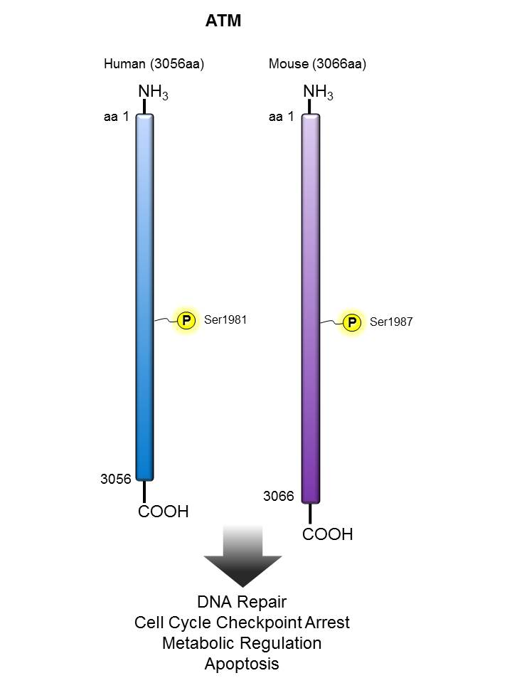
Ataxia telangiectasia mutated kinase (ATM) is a serine/threonine kinase that regulates cell cycle checkpoints and DNA repair. ATM is held inactive in unirradiated cells as a dimer or higher-order multimer. Cellular irradiation induces rapid intermolecular autophosphorylation of serine 1981 that causes dimer dissociation and initiates cellular ATM kinase activity. ATM kinase regulates a number of proteins involved in cell cycle checkpoint control, apoptosis, and DNA repair, including tumor suppressor proteins p53 and BRCA1, checkpoint kinase CHK2, checkpoint proteins RAD17 and RAD9, and DNA repair protein NBS1. Mutations in the corresponding ATM gene result in ataxia telangiectasia (AT), an autosomal recessive disease characterized by uncoordinated muscle movement and neurodegeneration. Cells from AT patients display defective DNA damage-induced checkpoint activation, sensitivity to radiation, and a higher frequency of chromosome breakage.

Other Names:

Ataxia telangiectasia mutated (ATM) kinase, serine-protein kinase, AT mutated

Human to Mouse Sequence Homology:

83.60%

Notable Upstream Kinase(s):

ATM (autophosphorylation)

Notable Phosphatase(s):

WIP1

Product Listing:

See products for ATM

Antigen References:

1. Lee JH and Paull TT 2007. Oncogene 26:7741.
2. Bakkenist CJ and Kastan MB 2003. Nature 421:499.
3. McConville CM, et al. 1996. Am. J. Hum. Genet. 59:320.
4. Tang X, et al. 2008. Mol. Cell Biol. 28:2559.


Aurora A


Description:

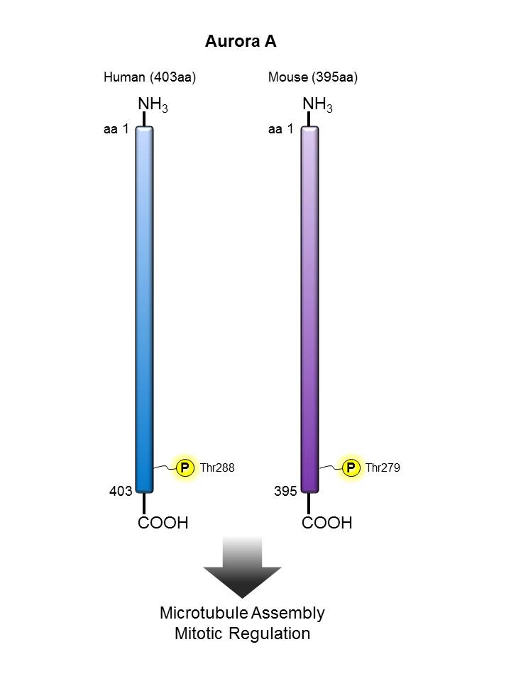
Aurora A (also known as Aurora 2) is a serine/threonine kinase with a molecular weight of approximately 46 kD. This kinase is highly expressed in the thymus and some tumors and is also expressed in other tissues including the lung, testis, colon, placenta, and fetal liver. Aurora A localizes in the midzone or central spindle in late anaphase and is concentrated in the midbody in telophase and during cytokinesis. This kinase is believed to act in cell cycle regulation during anaphase and/or telophase at centrosome/spindle pole during chromosome segregation. Aurora A has been shown to regulate cleavage of polar spindle microtubules at the onset of cytokinesis during mitosis. Defects in Aurora A cause numerous centrosome aberrations including aneuploidy (genetic variant with amino acid substitution F31I). Aurora A expression is cell cycle regulated, low in G1/S, and accumulating in G2/M. Expression is upregulated in cancer cells during M phase. Phosphorylation by PKA has been shown to regulate function. Aurora A phosphorylation has been reported on Thr 288. This kinase associates with the centrosome and mitotic spindles, NM23-H1, protein phosphatase type I, and co-localizes with γ-tubulin. The Phe 31 variant has been shown to interact with the E2 ubiquitin-conjugating enzyme, UBE2N. The Poly6187 antibody has been shown to react with phosphorylated human Aurora A. This antibody has weak avidity for Aurora B in immunofluorescence. To minimize Aurora B cross-reactivity, it is recommended that the reagent be titrated and more stringent wash conditions be employed.

Other Names:

Aurora 2, Aurora- and IPll-like kinase (AIK), Serine/threonine kinase 15, serine/threonine kinase 6

Human to Mouse Sequence Homology:

81.30%

Notable Upstream Kinase(s):

AuroraA (autophosphorylation), PKA, PAK1

Notable Phosphatase(s):

PP1, PPP1CA

Product Listing:

See products for Aurora A

Antigen References:

1. Shindo M, et al. 1998. Biochem. Biophys. Res. Commun. 244:285.
2. Tatsuka M, et al. 1998. Cancer Res. 58:4811.
3. Nigg EA. 2001. Nat. Rev. Mol. Cell. Biol. 2:21.

BCL-2


Description:

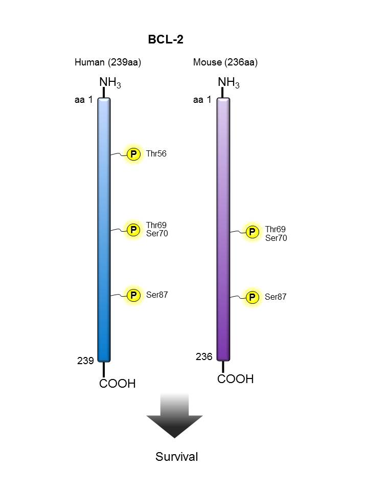
Bcl-2 (B-cell leukemia 2) is an apoptotic protein and a member of the Bcl-2 family containing BH1-4 domains. Two reported isoforms exist α=25 kD; β=22 kD. The Bcl-2 protein forms homo- or hetero-dimers with other Bcl-2 family members. Bcl-2 is distributed in the outer mitochondrial membrane, the nuclear envelope, and the endoplasmic reticulum. This protein blocks apoptotic death by controlling mitochondrial membrane permeability. Cleavage of Bcl-2 can convert to pro-apoptotic (by cleavage of BH4 domain). Bcl-2 has been reported to regulate cell cycle progression via ROS. This protein is modified by ASK1/JNK1, PKC, ERKs, and stress-activated kinase phosphorylation and can be ubiquitinated. Bcl-2 has been shown to interact with Apaf-1, Raf-1, TP53BP2, caspase-3, and form heterodimers with Bax, Bad, Bak, Bcl-xL, and Bag-1. Clone BCL/10C4 has been shown to be useful for Western blotting, immunoprecipitation, and immunofluorescence of the mouse and rat Bcl-2 protein.

Other Names:

B-cell leukemia 2

Human to Mouse Sequence Homology:

90.40%

Notable Upstream Kinase(s):

JNK1, ERK1/2, PKCA

Notable Phosphatase(s):

PP2A

Product Listing:

See products for BCL-2

Antigen References:

1. Tsujimoto Y, et al. 1986 P. Natl. Acad. Sci. USA 83:5214.
2. Yang E, et al. 1995. Cell 80:285.
3. Huang Z, et al. 2000. Oncogene 19:6627.
4. Deng X, et al. 2003. Blood. 102:3179.

B-Raf


Description:

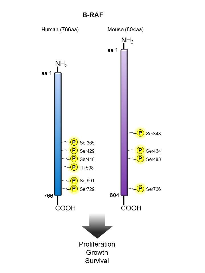
The Raf proteins are a family of serine/threonine-specific kinases that serve as a central intermediate in transmitting extracellular signals to the mitogen-activated protein kinase cascade, which controls cell growth, differentiation and survival. Three isoforms of Raf proteins have been found in mammalian cells: Raf-1, A-Raf and B-Raf. The B-raf involved in signal transduction from the membrane to the nucleus. It has been reported that T598 and S601 are the major phosphorylation sites of B-Raf in response to oncogenic Ras, and phosphorylation of these two residues is required for full activation of B-Raf. Mutations in the B-Raf gene have been reported in a number of human cancers, including malignant melanoma, thyroid cancer, and colorectal carcinoma. The poly6327 antibody recognizes human phosphorylated B-Raf (Thr598/Ser601) and has been shown to be useful for immunoflorence staining.

Other Names:

B-Raf proto-oncogene serine/threonine-protein kinase, BRAF, p94

Human to Mouse Sequence Homology:

84.70%

Notable Upstream Kinase(s):

RAS, PAK1

Antigen References:

1. Avruch J, et al. 1994. Trends Biochem Sci. 19:279.
2. Brose MS, et al. 2002. Cancer Res. 62:6997.
3. Zhang B-H, et al. 2000. EMBO 19:5429.

BTK


Description:

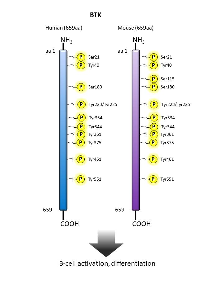
Bruton’s tyrosine kinase (BTK) is a Tec family kinase that plays a critical role in B cell development. Upon B cell receptor engagement, BTK translocates to the plasma membrane, where it is transphosphorylated by LYN and SYK kinases at Tyr 551. This initial phosphorylation event is followed by autophosphorylation at Tyr 223. Phosphorylated Tyr 223 is proposed to serve as a docking site for other proteins containing an SH2 domain. Downregulation of BTK activity is achieved through phosphorylation at Ser 180 by PKCβ, which results in reduced membrane recruitment and concomitant transphosphorylation. BTK-mediated B cell receptor signaling is required for B cell survival in the bone marrow and is a therapeutic target for certain B cell malignancies. Mutations in the BTK gene results in X-linked agammaglobulinemia (XLA), a primary immunodeficiency disease characterized by a failure in B cell maturation. Additionally, BTK plays a role in Toll-like receptor signaling in innate immune cells, and has been shown to play a direct role in NLRP3 inflammasome activation, thus contributing to ischaemic brain injury.

Other Names:

Bruton’s tyrosine kinase, Bruton agammaglobulinemia tyrosine kinase, AGMX1, ATK, IMD1, PSCTK1, XLA, BPK

Human to Mouse Sequence Homology:

98.33%

Notable Upstream Kinase(s):

BTK (autophosphorylation), PKCβ

Notable Phosphatase(s):

IBTK (not a phosphatase, but inhibits BTK activity), PTEN (via PIP3 level regulation)

Product Listing:

See products for BTK

Antigen References:


  1. Krysiak K, et al. 2017. Blood. 129:473.
  2. Burger JA, et al. 2016. Nat Commun. 7:11589.
  3. Wang Q, et al. 2015. Elife. 4:e06074.
  4. Ito M, et al. 2015. Nat. Commun. 6:7360.
  5. Pillinger G, et al. 2015. Sci. Rep. 5:12949.
  6. Cinar M, et al. 2013. Leuk. Res. 37:1271.
  7. Doyle SL, et al. 2007 J. Biol. Chem. 282:36953.
  8. Nore BF, et al. 2003. Biochem. Biophys. Acta. 1645:123.
  9. Li Z, et al. 1997. Proc. Natl. Acad. Sci. 94:13820.

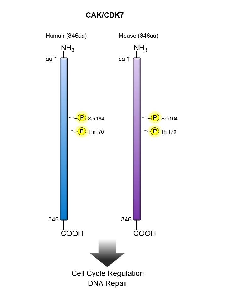
CAK/CDK7


Description:

CAK (also known as CDK-activating kinase, CDK7 and cell division protein kinase 7 is a 40 kD nuclear protein that is a member of the serine/threonine family of protein kinases, CDC2/CDKX subfamily. CAK activates cyclin-associated kinases CDC2/CDK1, CDK2, CDK4, CDK6 by threonine phosphorylation and controls cell cycle progression, DNA repair, and RNA polymerase II transcription. CAK is activated by phosphorylation at Thr170 and is inactivated by phosphorylation at Ser164. CAK has been reported to interact with cyclin H and MAT1 to form the CAK complex, the CAK complex then interacts with core-TFIIH to form the TFIIH basal transcription factor. The Poly6324 antibody is useful for immunofluorescence microscopy of human CAK protein.

Other Names:

Cyclin-dependent kinase 7, CDK-activating kinase (CAK)

Human to Mouse Sequence Homology:

95.10%

Notable Upstream Kinase(s):

PDK1, CDK1, CDK2, PKC1

Notable Phosphatase(s):

CDC25, KAP1, NMPP1

Antigen References:

1. Tassan J, et al. 1994. J. Cell. Biol. 127:467.
2. Tirode F, et al. 1999. Mol Cell. 3:87.
3. Akoulitchev S, et al. 1998. Genes Dev. 12:3541.
4. Chen J, et al. 2003. Nature. 424:228.

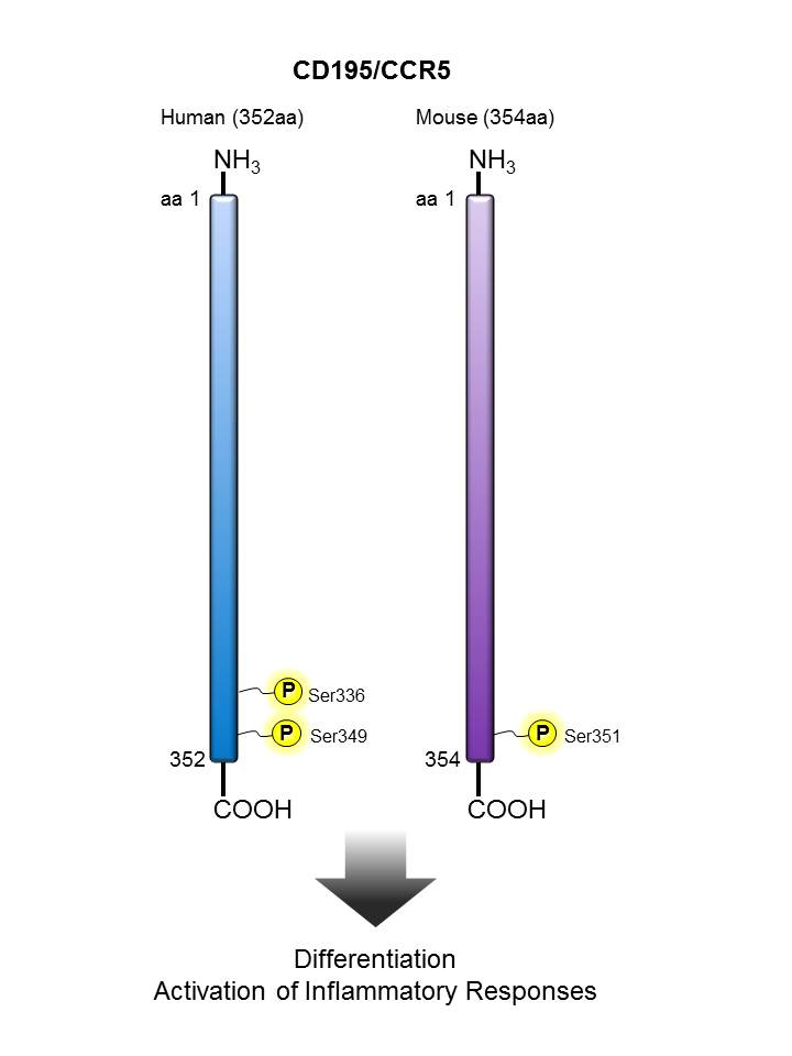
CD195 (CCR5)


Description:

CD195, also known as CCR5, is a 45 kD G protein-coupled seven transmembrane CC-chemokine receptor. It binds to MIP-1α, MIP-1β, and RANTES and is expressed on a subset of T cells and monocytes. CD195 mediates an intracellular signal thought to induce cell differentiation and proliferation. CCR5 has also been shown to act as a co-receptor for R5 HIV-1 cell entry; modification of CCR5 by sulfation contributes to the efficiency of HIV-1 entry.

Other Names:

CD195, CCR5, C-C chemokine receptor type 5, HIV-1 fusion co-receptor

Human to Mouse Sequence Homology:

81.90%

Notable Upstream Kinase(s):

GRK3

Product Listing:

See products for CD195 (CCR5)

Antigen References:

1. Samson M, et al. 1996. Biochemistry 35:3362.
2. Raport CJ, et al. 1996. J. Biol. Chem. 271:17161.
3. Combadiere C, et al. 1996. J. Leukoc. Biol. 60:147.
4. Deng H, et al. 1996. Nature 381:661.

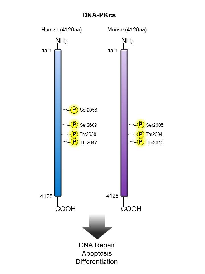
DNA-PKcs


Description:

DNA-protein kinase catalytic subunit (DNA-PKcs) also known as DNA-activated kinase is a nuclear 460-470 kD serine threonine kinase involved in double-stranded DNA break repair, VDJ recombination, and transcriptional modulation. DNA-PKcs must bind DNA ends to become active. DNA-PKcs is modified by phosphorylation and has been shown to interact with Ku70/Ku80, KIP, DNA-ligase IV, and XRCC4 proteins. Phosphorylated DNA-PKcs is upregulated after DNA damaage. The 10B1 monoclonal antibody recognizes phosphorylated human DNA-PKcs (Thr2609) and has been shown to be useful for immunofluorescence staining and Western blotting.

Other Names:

DNA-protein kinase catalytic subunit, DNA-activated protein kinase, DNA-dependent protein kinase

Human to Mouse Sequence Homology:

79.10%

Notable Upstream Kinase(s):

DNA-PK (autophosphorylation), ATM, ATR

Notable Phosphatase(s):

PP2A

Product Listing:

See products for DNA-PKcs

Antigen References:

1. Hartley K, et al. 1995. Cell. 82:849.
2. Connelly M, et al. 1996. Gene. 175:271.
3. Douglas P, et al. 2002. Biochem. J. 368:243
4. Calsou P, et al. 2003. J. Mol. Biol. 326:93.

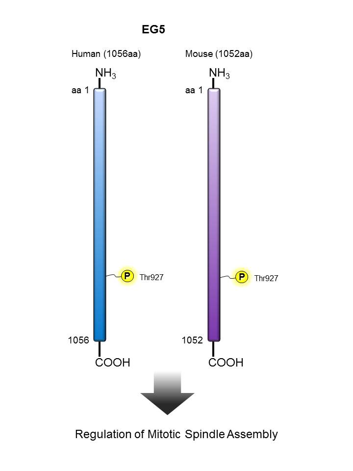
EG5


Description:

hEg5 (also known as kinesin-like protein KIF11, kinesin-related motor protein Eg5, kinesin-like spindle protein HKSP, and thyroid receptor interacting protein 5 (TRIP5)) is a 119 kD kinesin-like protein family, BimC subfamily. This protein is a catalytic kinesin motor with coiled-coil and Smc domains. hEg5 is localized at the centrosomes, spindle microtubules, and intracellular bridge. This motor protein is required for establishing the bipolar spindle. hEg5 is modified by phosphorylation on Thr927 by Cdc2 to allow association with the spindle apparatus. hEg5 has been shown to interact with the thyroid hormone receptor in presence of thyroid hormone and Cdc2. The Poly6205 antibody has been shown to react with phosphorylated human Eg5 (Thr927) by Western blot.

Other Names:

Kinesin-like protein KIF11, kinesin-related motor protein Eg5, kinesin-like spindle protein HKSP, thyroid receptor interacting protein 5 (TRIP5)

Human to Mouse Sequence Homology:

79.30%

Notable Upstream Kinase(s):

CDC2

Product Listing:

See products for EG5

Antigen References:

1. Lee J, et al. 1995. Mol. Endocrinol. 9:243.
2. Blangy A, et al. 1995. Cell 83:1159.
3. Whitehead C, et al. 1998. J. Cell Sci. 111:2551.
4. DeBonis S, et al. 2003. Biochemistry. 42:338.

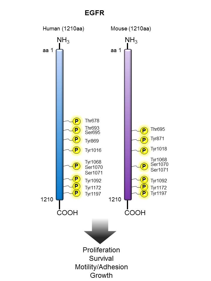
EGFR


Description:

EGFR is a transmembrane glycoprotein that belongs to protein kinase superfamily. It is expressed in tissues of epithelial, mesenchymal and neuronal origin and plays major roles in proliferation, differentiation and development. Binding of the protein to its ligand induces receptor dimerization and tyrosine autophosphorylation. Mutations or over-expression of this gene are associated with certain forms of cancer. Tyrosine 1068 is the primary autophosphorylation site in the C-terminal domain of the receptor.

Other Names:

Epidermal growth factor receptor, HER1

Human to Mouse Sequence Homology:

90.30%

Notable Upstream Kinase(s):

EGFR/HER2 (autophosphorylation), SRC, ERK1/2, PKCA

Notable Phosphatase(s):

PTP

Product Listing:

See products for EGFR

Antigen References:

1. Alameddine RS, et al. 2013. Curr. Opin. Oncol. 25:313.
2. Downward J, et al. 2003. Anticancer Res. 23:3639.
3. Hrustanovic G, et al. 2013. Cancer Biol. Ther. 14:304.
4. Koland JG, et al. 1990. Biochem. Biophys. Res. Commun. 166:90.
5. Wheeler SE, et al. 2012. Clin. Cancer Res. 18:2278.
6. Yano S, et al. 2003. Anticancer Res. 23:3639.

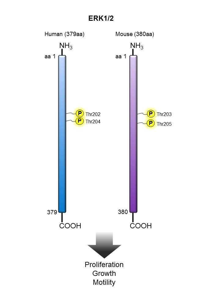
ERK1/2


Description:

ERK1/2 are members of mitogen-activated kinases (MAPKs) of serine/threonine protein kinases. ERK1/2 can be activated by a range of extracellular stimuli, such as mitogen, growth factors, neurotransmitters, chemokines, and cytokines, through receptor tyrosine kinases (RTK), G protein-coupled receptors (GPCRs), or protein kinase C (PKC). Upon stimulation, ERK1/2 are phosphorylated by the upstream kinase MEK on residues Thr202 and Tyr204 and in turn phosphorylate many other downstream molecules that are involved in a range of cellular processes such as cell proliferation, differentiation, motility and cell death.

Other Names:

P44/42 MAP (motogen activated protein) Kinase, Extracelluar signal-regulated kinase (ERK)

Human to Mouse Sequence Homology:

96.60%

Notable Upstream Kinase(s):

MEK1

Notable Phosphatase(s):

DUSP6

Product Listing:

See products for ERK1/2

Antigen References:

1. Futran AS, et al. 2013. Curr. Biol. 23:R972.
2. Mendoza MC, et al. 2011. Trends Biochem. Sci. 36:320.
3. Chambard JC, et al. 2007. Biochim. Biophys. Acta. 1773:1299.
4. Roux PP, et al. 2004. Microbiol. Mol. Biol. Rev. 68:320.
5. Torii S, et al. 2004. J. Biochem. 136:557.
6. Clark EA, et al. 1996. J. Biol. Chem. 271:14814.

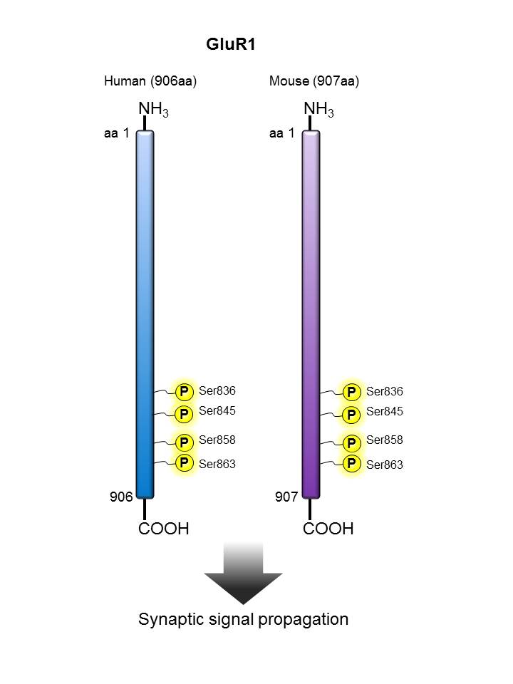
GluR1


Description:

GluR1 belongs to the family of Glutamate Receptors (GluRs) that are essential for regulating excitatory synaptic transmission by L-glutamate in the CNS of vertebrates. These are mostly expressed in granule and pyramidal cells in the hippocampus. GluRs can be categorized as ionotropic or metabotropic and subcategorized by their specific agonists (NMDA, AMPA or Kainic acid). There are four types of AMPA related GluR subunits (GluR1, GluR2, GluR3 and GluR4) that are generated by alternative RNA splicing. Tetrameric or pentameric combinations of different subunits contribute to the functional diversity of AMPA receptors.

Other Names:

Glutamate receptor 1, GluR1, glutamate receptor A, glutamate receptor subunit GluR1, AMPA-selective glutamate receptor 1, glutamate receptor, ionotropic, AMPA1 (alpha 1), GLUH1, GLUR1, GLURA, GluA1, HBGR1, gluR-1, gluR-A, gluR-K1

Human to Mouse Sequence Homology:

97.80%

Notable Upstream Kinase(s):

PKCA, CAMK2

Product Listing:

See products for GluR1


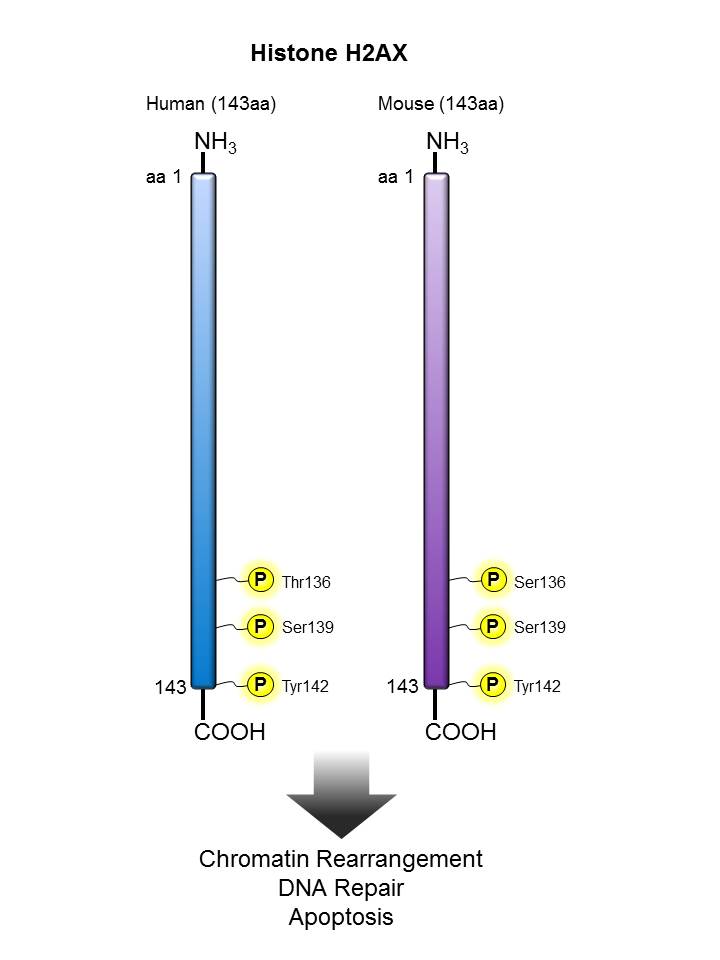
Histone H2AX


Description:
 

H2A.X is a 14 kD basal histone and a member of the H2 histone family. This nuclear protein is synthesized in the G1 and S phase of the cell cycle and is known to be important for DNA repair and maintaining genomic stability and for recombination between immunoglobulin switch regions. H2A.X becomes phosphorylated on serine 139 after double-stranded DNA breaks. Phosphorylated H2A.X promotes DNA repair and maintains genomic stability. The 2F3 monoclonal antibody reacts with phosphorylated human H2A.X (Ser139) and has been shown to be useful for Western blotting, immunofluorescence and flow cytometry.

Other Names:

H2A.x, H2a/x, Histone 2A, Histone 2A.X, Gamma-H2AX

Human to Mouse Sequence Homology:

97.20%

Notable Upstream Kinase(s):

ATM, DNA-PK

Notable Phosphatase(s):

PPM1D

Product Listing:

See products for Histone H2AX

Antigen References:

1. Mannironi C, et al.1989. Nucleic Acids Res. 17:9113.
2. Celeste A, et al. 2002. Science 296:922.
3. Bassing CH, et al. 2002. Proc. Natl. Acad. Sci. USA 99:8173.
4. Reina-San-Martin B, et al. 2003. J. Exp. Med. 197:1767.

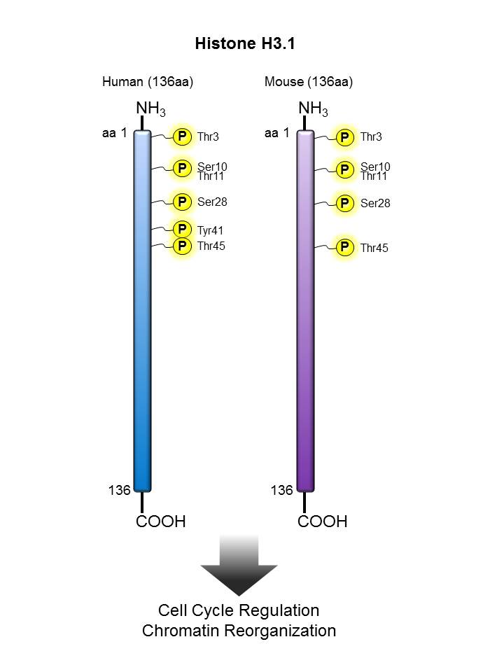
Histone H3.1


Description:

Histone proteins are classified into core histones (H2A, H2B, H3, H4) and linker histones (H1, H5). Core histones form an octamer, which contains two H2A-H2B dimers and one H3-H4 tetramer. Core histones are predominantly globular except for the unstructured N-terminal tails. Posttranslational modifications, such as acetylation, methylation, phosphorylation, ubiquitination, SUMOylation and ADP-ribosylation occur in histone tails.

Histone modifications induce changes of chromatin structure and thereby affect the accessibility of transcription factors, nuclear proteins and enzymes to genomic DNA, resulting in gene activation or repression. It is known that histone modifications play critical roles in DNA repair, DNA replication, transcription regulation, alternative splicing and chromosome condensation and some diseases including autoimmune diseases and cancers.

Other Names:

Histone-3

Human to Mouse Sequence Homology:

100%

Notable Upstream Kinase(s):

AuroraA, AuroraB, AKT2, MSK1, PIM1,

Notable Phosphatase(s):

CDC25A, PPP1CA, PPP2CA

Product Listing:

See products for Histone H3.1

Antigen References:

1. Choi HS, et al. 2005. J. Biol. Chem. 280:13545.
2. Goto H, et al. 2002. Genes Cells 7:11.
3. Garcia BA, et al. 2005. Biochemistry 44:13202.
4. Yoshimi T, et al. 2013. Monoclon. Immunodiagn. Immunother. 32:119.

ITK


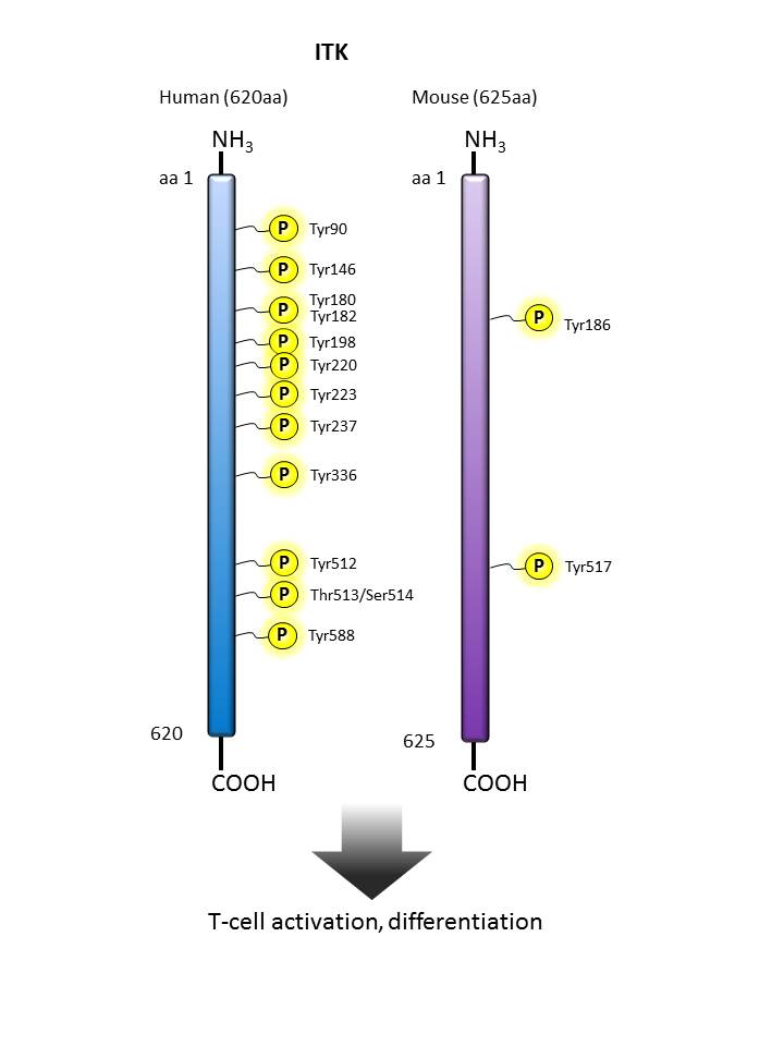
Description:

IL2-inducible T-cell kinase (ITK) is an intracellular tyrosine kinase that is expressed in T cells. It regulates the proliferation, differentiation and development of conventional T cells and NK T cells. This protein has 620 amino acids and contains one PH domain for cell membrane recruitment, one protein kinase domain, and both SH2 and SH3 domains which are commonly found in intracellular kinases. Once T cells are activated, a series of phosphorylation recruits and activates ITK through autophosphorylation. The activated ITK can activate downstream intracellular calcium mobilization and NFAT nucleus translocation, and in turn, activate T cells. The phenotypes of mice deficient in ITK show severely affected immune response and development.

Other Names:

Interleukin-2-inducible T-cell kinase, T-cell-specific kinase, EMT, LYK, PSCTK2

Human to Mouse Sequence Homology:

93.29%

Notable Upstream Kinase(s):

LCK, PKCβ

Notable Phosphatase(s):

PTEN (via PIP3 level regulation)

Product Listing:

See products for ITK

Antigen References:

1. Gomez-Rodriguez J, et al. 2016. Nat. Commun. 7:10857.
2. Cho HS, et al. 2015. J. Immunol. 195:4822.
3. Kannan A, et al. 2015. Eur. J. Immunol. 45:2276.
4. Ghosh S, et al. 2014. J. Clin. Immunol. 34:892.
5. Qi Q, et al. 2011. FEBS. J. 278:1970.
6. Prince AL, et al. 2009. Immunol. Rev. 228:115.

JNK


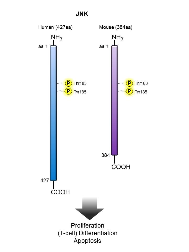
Description:

The protein encoded by this gene is a member of the MAP kinase family. MAP kinases act as an integration point for multiple biochemical signals and are involved in a wide variety of cellular processes such as proliferation, differentiation, transcription regulation, and development. This kinase is activated by various cell stimuli and targets specific transcription factors, thus mediating immediate-early gene expression in response to cell stimuli. As with the other MAPKs, the core signaling unit is composed of a MAPKKK, typically MEKK1-MEKK4, or by one of the mixed lineage kinases (MLKs), which phosphorylate and activate MKK4/7. Upon activation, MKKs phosphorylate and activate the SAPK/JNK kinase. SAPK/JNK, when active as a dimer, can translocate to the nucleus and regulate transcription through its effects on c-Jun, ATF-2, and other transcription factors. The activation of this kinase by tumor-necrosis factor alpha (TNF-alpha) is found to be required for TNF-alpha induced apoptosis. This kinase is also involved in UV radiation induced apoptosis, which is thought to be related to cytochrome c-mediated cell death pathway. Studies of the mouse counterpart of this gene suggested that this kinase plays a key role in T cell proliferation, apoptosis, and differentiation. Four alternatively spliced transcript variants encoding distinct isoforms have been reported.

Other Names:

Stress-activated protein kinase 1, Stress-activated protein kinase JNK1, mitogen-activated protein kinase 8 isoform JNK1 alpha1, mitogen-activated protein kinase 8 isoform JNK1 beta2

Human to Mouse Sequence Homology:

88.80%

Notable Upstream Kinase(s):

ASK1, MEKK6, MKK4, MKK7

Notable Phosphatase(s):

PPP5C

Antigen References:

1. Chen YR, et al. 1996. J. Biol. Chem. 271:631.
2. Su B, et al. 1994. Cell 77:727.
3. Brandt B, et al. 2010. Cell Death Dis. 1:e23.
4. Whitmarsh AJ, et al. 1998. Trends Biochem Sci. 23:481.

LCK


Description:

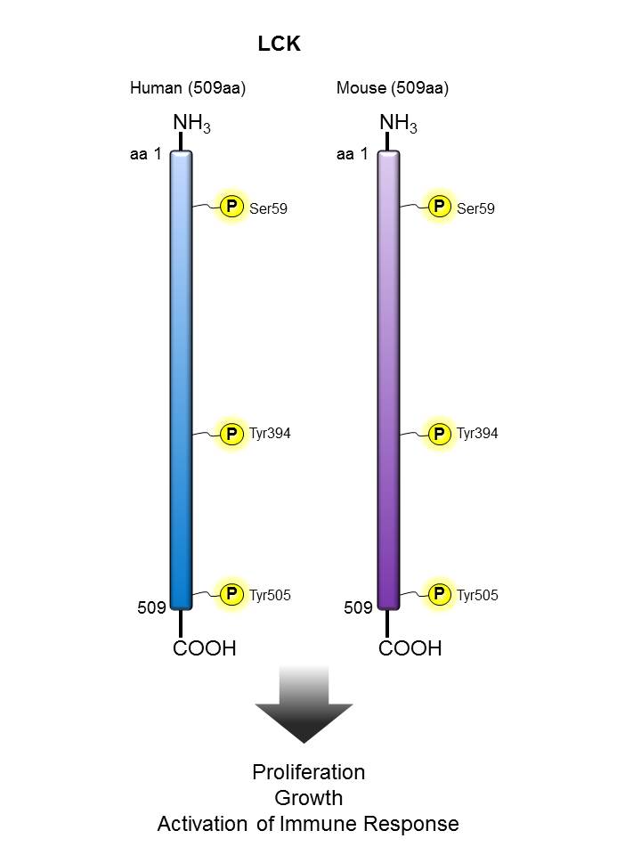
Lck is a 56 kD tyrosine protein kinase and a member of the src subfamily that contains both an SH2 and SH3 domain. Alternatively spliced isoforms of Lck have been reported. Lck is a cytoplasmic protein bound to CD4 and CD8 in T cells that participate in antigen-induced T cell activation through phosphorylation of the T cell receptor zeta chain. Lck is activated upon T cell receptor engagement and is involved in the onset of cell cycle progression induced by interleukin-2. Phosphorylation by Csk downregulates the activity of Lck. In addition to CD4 and CD8, Lck has been reported to interact with PI3K through its SH3 domain and tyrosine phosphorylated KHDRBS1/p70 through its SH2 domain. In addition, Lck has been shown to associate with the SAP transmembrane tyrosine phosphatase.

Other Names:

Proto-oncogene tyrosine kinase lck, p56-Lck, T-cell specific protein tyrosine kinase

Human to Mouse Sequence Homology:

96.90%

Notable Upstream Kinase(s):

LCK (autophosphorylation), CSK

Notable Phosphatase(s):

CD45/PTPRC, PTPN22

Product Listing:

See products for LCK

Antigen References:

1. Koga Y, et al. 1986. Eur. J. Immunol. 16:1643.
2. Vogel LB, et al. 1995. Biochem. Biophys. Acta 1264:168.
3. Vogel LB, et al. 1993. Mol. Cell. Biol. 13:7408.
4. Bergaman M, et al. 1992. EMBO J. 11:2919.

mTOR


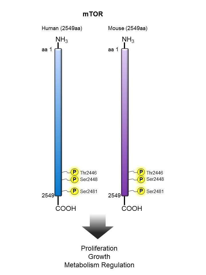
Description:

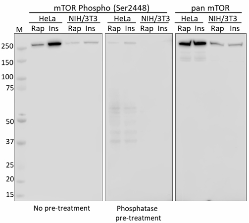
mTOR (mammalian target of rapamycin) is a Ser/Thr kinase acting as an ATP sensor and is reported to autophosphorylate Ser2481 in the presence of sufficient nutrients. Activated mTOR phosphorylates ribosomal protein S6 kinase 1 (S6K1) and the eukaryotic translation initiation factor 4E binding protein 1 (4EBP1) in response to PI3K/Akt signaling, in turn, promoting cell growth, survival, and proliferation. 

Other Names:

Mammalian target of rapamycin (mTOR), RAPT1, FK506 binding protein 12-rapamycin associated protein 1 (FRAP1)

Human to Mouse Sequence Homology:

98.90%

Notable Upstream Kinase(s):

AKT, P70S6K

Product Listing:

See products for mTOR

Antigen References:

1. Brown EJ, et al. 1994. Nature 369:756.
2. Dennis P, et al. 2001. Science 294:1102.
3. Fingar DC, et al. 2002. Genes Dev. 16:1472.
4. Nojima H, et al. 2003. J. Biol. Chem. 278:15461.
5. Soliman G, et al. 2010. J. Biol. Chem. 285:7866.
6. Vazquez-Martin A, et al. 2011. Cell Cycle 10:3140.

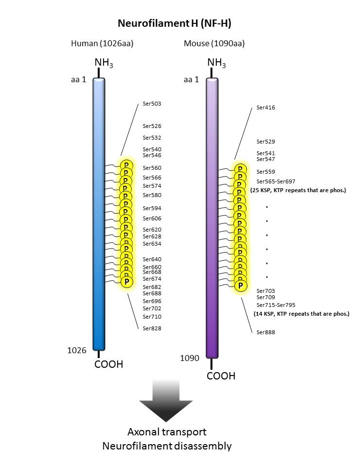
Neurofilament H


Description:

Neurofilaments (NFs) are ~10 nanometer, type IV intermediate filaments expressed in neurons. NFs are the major components of the neuronal cytoskeleton, and primarily function to provide structural support for axons and regulate axonal diameter. There are three major mammalian neurofilament subunits which are named based on their apparent molecular weight: 1) neurofilament light (NF-L, ~70 kD), 2) neurofilament medium (NF-M, ~145-160 kD), and 3) neurofilament heavy (NF-H, ~200-220 kD). Neurofilaments are extensively phosphorylated, and their phosphorylation status plays an important role in modulation of their function. Abnormal NF modifications, mutations, and accumulation have been associated with neurodegenerative diseases. NF immunostaining is commonly used as a diagnostic marker for neuropathology detection. NFs are also useful for differentiating neurons (positive for NF) from glia (negative for NF).

Other Names:

Neurofilament heavy polypeptide, NF-H, 200 kD neurofilament protein, neurofilament triplet H protein

Human to Mouse Sequence Homology:

73.34%

Notable Upstream Kinase(s):

PKA, PKC, ERK1/2, CDK5, GSK3β, CK1

Notable Phosphatase(s):

PP1, PP2A, PP2B

Product Listing:

See products for Neurofilament H


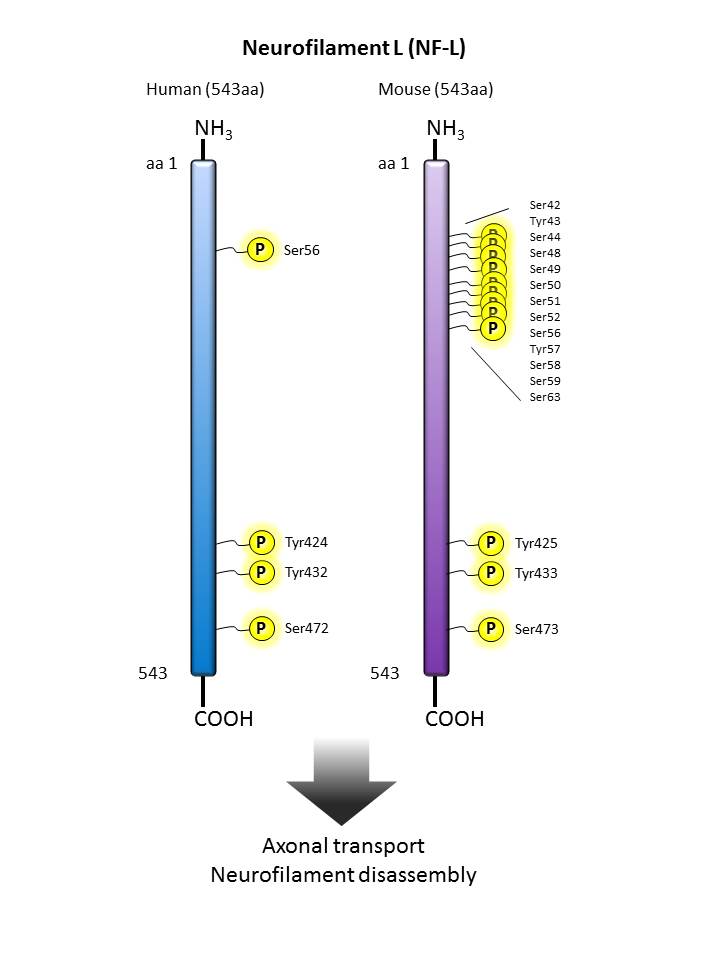
Neurofilament L


Description:

Neurofilaments (NF) are type IV intermediate filament heteropolymers composed of light, medium, and heavy chains. Detection of NF, and the subunits of NF, may serve as a biomarker for axonal degeneration. The degree of axonal degeneration is related to the amount of NF detected in the cerebrospinal fluid (CSF) and the blood. Neurofilament light (NF-L) levels are elevated in Alzheimer’s Disease.

Other Names:

Neurofilament Light Polypeptide 68kDa, Neurofilament Light Polypeptide, Neurofilament Subunit NF-L, CMT1F, NFL, NEFL, NF-L

Human to Mouse Sequence Homology:

95.22%

Notable Upstream Kinase(s):

PKA, PKC, ERK1/2, CDK5, GSK3β, CK1

Notable Phosphatase(s):

PP1, PP2A, PP2B

Product Listing:

See products for Neurofilament L

Antigen References:

1. Zetterberg H, Blennow K. 2015. JAMA Neurol. 11:1237-8.
2. Bäckström D, et al. 2015. JAMA Neurol. 72(10):1175.
3. Janelidze S, et al. 2015. Neurology 85:1834.
4. Magdalinou NK, et al. 2015. J. Neurol. Neurosurg. Psychiatry 86(11):1240.
5. Skillbäck T, et al. 2014. Neurology. 83:1945.

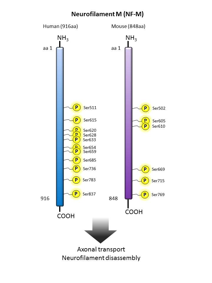
Neurofilament M


Description:

Neurofilaments can be defined as the intermediate or 10nm diameter filaments found in neuronal cells. They are composed a mixture of subunits which often includes the neurofilament triplet proteins, NF-L, NF-M and NF-H. Neurofilaments may also include peripherin, a-internexin, nestin and in some cases vimentin. Antibodies in the various neurofilament subunits are very useful cell type markers since the proteins are quite abundant, biochemically stable.

Other Names:

160 kD Neurofilament Protein, Neurofilament Triplet M Protein, Neurofilament-3 (150 Kd Medium), Neurofilament, Medium Polypeptide 150kD

Human to Mouse Sequence Homology:

82.29%

Notable Upstream Kinase(s):

PKA, PKC, ERK1/2, CDK5, GSK3β, CK1

Notable Phosphatase(s):

PP1, PP2A, PP2B

Product Listing:

See products for Neurofilament M


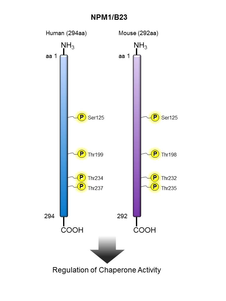
NPM/B23


Description:

NPM/B23 (also known as numatrin, nucleophosmin 1, nucleolar phosphoprotein B23) is a member of the nucleophosmin/nucleoplasmin family that shuttles between the nucleus and cytoplasm. This protein regulates the stability and transcriptional activity of p53 and acts as a molecular chaperone. NPM/B23 preferentially binds to denatured proteins and has been shown to stimulate DNA polymerase activity and control the duplication of centrosomes. The chaperone activity of NPM/B23 is regulated by protein kinase CK2 and promotes release of denatured substrates from NPM/B23. NPM/B23 is modified by phosphorylation onThr199. This protein has been shown to bind to RNA and DNA and interact with nucleolar proteins including nucleolin, protein P120, HIV-1 Rev protein, and hepatitis delta antigens. The Poly6191 antibody recognizes human phosphorylated NPM/B23 (Thr234/Thr237) and has been shown to be useful for Western blotting.

Other Names:

Numatrin, nucleophosmin 1, nucleolar phosphoprotein B23

Human to Mouse Sequence Homology:

93.90%

Notable Upstream Kinase(s):

CK2, CDK1, CDK2 CDK6, ATR

Product Listing:

See products for NPM/B23

Antigen References:

1. Szebeni A, et al. 2003. J. Biol. Chem. 278:9107.
2. Colombo E, et al. 2002. Nat. Cell Biol. 4:529.
3. Chan PK, et al. 1986. J. Biol. Chem. 261:14335.

Nucleolin


Description:

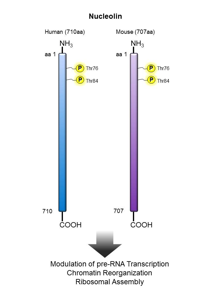
Nucleolin is a ubiquitously expressed 100 kD nuclear and nucleolar protein that contains RNA recognition motifs. Nucleoloin is the major nucleolar protein of eukaryotic cells in growth phase associated with intranucleolar chromatin and pre-ribosomal particles. This protein induces chromatin de-condensation by binding to histone H1 and plays a role in pre-rRNA transcription and ribosome assembly. Nucleolin exhibits self-cleaving, DNA helicase, RNA helicase and DNA-dependent ATPase activities; activity can be upregulated in response to ERK signaling, c-Myc expression, and genotoxic stress (UV radiation). This protein is modified by phosphorylation (Thr76/Thr84). This protein interacts with phosphatase 1δ, hepatitis C virus NS5B, part of B-cell specific transcription factor complex with LR1, and DNA Topoisomerase I. The 10C7 monoclonal antibody recognizes human phosphorylated nucleolin (Thr76/Thr84) and has been shown to be useful for Western blotting.

Other Names:

Nucleolin protein, C23

Human to Mouse Sequence Homology:

81.80%

Notable Upstream Kinase(s):

CDK2

Notable Phosphatase(s):

PP1γ

Antigen References:

1. Srivastava M, et al. 1989. FEBS Lett. 250:99.
2. Westmark C, et al. 2001. J. Biol. Chem. 276:1119.
3. Ying G, et al. 2000. J. Biol. Chem. 275:4152.
4. Hanakahi L, et al. 1997. P. Natl. Acad. Sci. USA 94:3605.

p38 MAPK


Description:

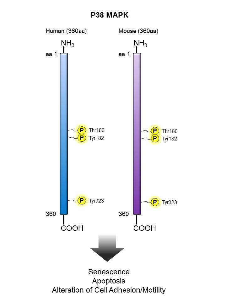
The p38 MAP kinase is a stress-activated protein kinase. The p38 MAP kinase is activated by treatment of cells with proinflammatory cytokines (e.g. TNF and IL-1) or by exposure of cells to environmental stress (e.g. UV radiation and osmotic shock). This activation results in the phosphorylation of residues Thr180 and Tyr182. This antibody is specific for the 39 kD p-38 protein phosphorylated at Thr180/Tyr182 It is purified by sequential chromatography on protein A and phospho- and dephospho- peptide affinity columns.

Other Names:

Mitogen-activated protein kinase 14, MAP kinase 14, p38alpha Exip, p38 MAP kinase, MAP kinase Mxi2, MAP kinase p38 alpha, CSAID-binding protein, MAX-interacting protein 2, stress-activated protein kinase 2A, p38 mitogen activated protein kinase, mitogen-a

Human to Mouse Sequence Homology:

99.40%

Notable Upstream Kinase(s):

ASK1, MEKK6, MKK3, MKK6

Notable Phosphatase(s):

MKP1, PPM1D

Product Listing:

See products for p38 MAPK


PERK


Description:

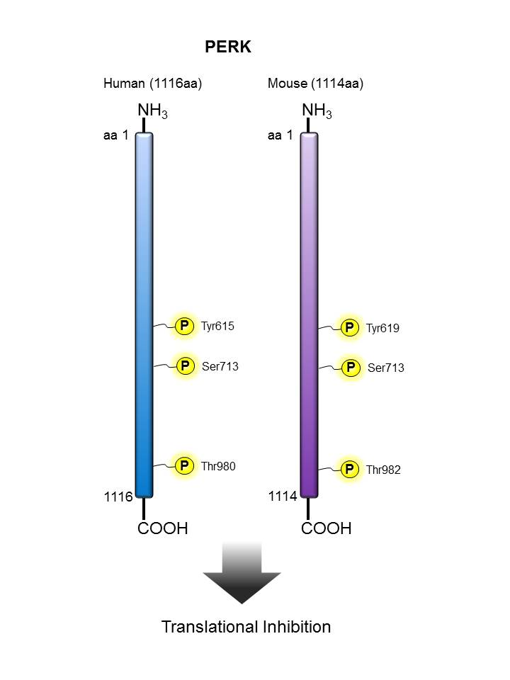
PERK (protein kinase R-like endoplasmic reticulum kinase) is a type I membrane protein located in the endoplasmic reticulum (ER). ER stress increases the activity of PERK, which then phosphorylates eIF2α to promote reduced translation, leading to its inactivation, and thus to a rapid reduction of translational initiation and repression of global protein synthesis. PERK-deficient mice have defects in pancreatic β cells several weeks after birth, suggesting a role for PERK-mediated translational control in protecting secretory cells from ER stress. PERK activation during ER stress correlates with autophosphorylation of its cytoplasmic kinase domain. Phosphorylation of PERK at Ser713 can be used as a marker for its activation status. The predicted phosphorylation site and surrounding residues of Ser713 are: HIEIIAPS*PQRSRSF.

Other Names:

Eukaryotic translation initiation factor 2 alpha kinase 3; PRKR-like endoplasmic reticulum kinase; heme-regulated EIF2-alpha kinase

Human to Mouse Sequence Homology:

87.00%

Notable Upstream Kinase(s):

PERK (autophosphorylation)

Antigen References:

1. Shi Y, et al. 1998. Mol. Cell. Biol. 18:7499.
2. Shi Y, et al. 1999. J. Biol. Chem. 274:5723.
3. Cybulsky AV, et al. 2005. J. Biol. Chem. 280:24396.
4. Kohno K. 2010. J. Biochem. 147:27.

PKCα


Description:

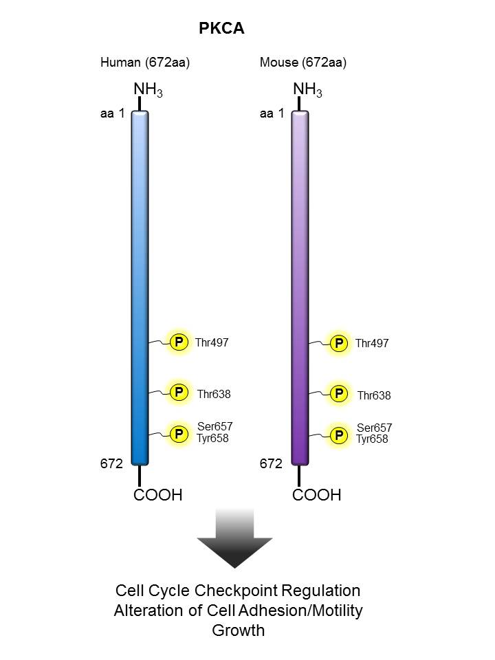
Protein kinase C alpha (PKCα), also known as PRKCA, refers to both a human gene and the protein that is encoded by it. Protein kinase C (PKC) is a family of serine- and threonine-specific protein kinases that can be activated by calcium and the second messenger diacylglycerol. PKC family members phosphorylate a wide variety of protein targets and are known to be involved in diverse cellular signaling pathways. PKC family members also serve as major receptors for phorbol esters, a class of tumor promoters. Each member of the PKC family has a specific expression profile and is believed to play a distinct role in cells. The protein encoded by this gene is one of the PKC family members. This kinase has been reported to play roles in many different cellular processes, such as cell adhesion, cell transformation, cell cycle checkpoint, and cell volume control. Knockout studies in mice suggest that this kinase may be a fundamental regulator of cardiac contractility and Ca2+ handling in myocytes. The purified anti-PKCα Phospho (Thr638) antibody detects PKCα only when phosphorylated at threonine 638.

Other Names:

AAG6; PKCA; PRKACA; MGC129900; MGC129901; PKC-alpha; PRKCA; PKCα

Human to Mouse Sequence Homology:

98.20%

Notable Upstream Kinase(s):

PKCA (autophosphorylation), MST1, MST2, RICTOR

Notable Phosphatase(s):

PPP1CA

Product Listing:


See products for PKCα


Antigen References:

1. Seo HR, et al. 2004. Exp. Mol. Med. 36:292.
2. Wientzek M, et al. 1997. Mol. Cell. Biochem. 166:11.
3. Baier G and Wagner J. 2009. Curr. Opin. Cell Biol. 21:262.
4. Reyland NE. 2009. Front. Biosci. 14:2386.

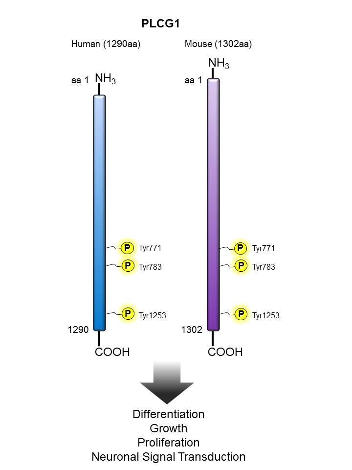
PLCγ


Description:

PLCγ-1 is a ubiquitously expressed isozyme that is activated by phosphorylation of tyrosine 783. It plays important roles in mammalian growth and development, proliferation, differentiation, signal transduction, endocytosis, cytoskeletal reorganization, and activation of ion channels. In the brain, PLCγ-1 is highly expressed and participates in brain development and synaptic transmission. Abnormal expression and activation of PLCγ-1 is associated with various brain disorders such as epilepsy, depression, Huntington's disease and Alzheimer's disease. Research in zebrafish also suggests its important roles in the maturation of granulocytes.

Other Names:

PLCG1, PLC-Gamma-1, NCKAP3

Human to Mouse Sequence Homology:

95.00%

Notable Upstream Kinase(s):

ABL, EGFR, SYK, ITK, RET,

Notable Phosphatase(s):

PTP1C

Product Listing:

See products for PLCγ

Antigen References:

1. Jang HJ, et al. 2013. Adv. Biol. Regul. 53:51.
2. Krawczyk P, et al. 2011. Postepy. Hig. Med. Dosw. (Online). 65:470.
3. Matsumoto K, et al. 2013. Biomaterials. 34: 5988.
4. Lo Vasco VR, et al. 2013. J. Cell Commun. Signal. 7:141.
5. Jing CB, et al. 2013. Dev. Biol. 374:24.
6. Gierschik P. et al. 2012. Structure. 12:1989.

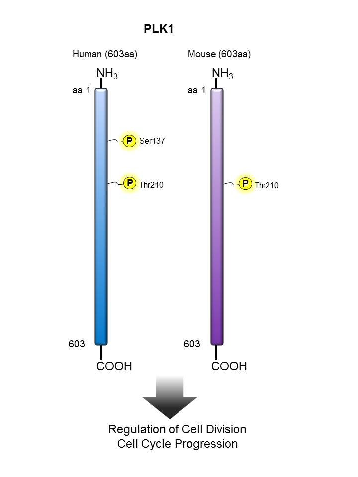
PLK1


Description:

PLK-1 (polo-like kinase 1) is a member of te serine/threonine protein kinase family, cdc5/polo subfamily. Highly homologous to polo-like kinase (Drosophila), PLK-1 contains two polo box domains with a predicted molecular weight of 68 kD. This nuclear protein is highly expressed in placenta and colon and has been shown to regulate cdc2/cyclin B through phosphorylation and activation of cdc25c phosphatase. PLK-1 may also be required for cell division; depletion of PLK-1 results in apoptosis. PLK-1 is upregulated by growth stimulating agents and is regulated by cell cycle position (highest in G2/M phase, declining to nearly undetectable levels after mitosis and throughout G1). PLK-1 is modified by phosphorylation (Thr210 is the major phosphorylation site in activated PLK-1 from mitotic cells) and has been shown to interact with nuclear distribution gene C. The 2A3 antibody recognizes human phosphorylated PLK-1 (Thr210) and has been shown to be useful for Western blotting. To increase specificity, it is recommended that the 2A3 antibody be used for Western blotting after immunoprecipitation with the pan-specific PLK-1 3F8 antibody.

Other Names:

Serine/Threonine protein kinase PLK, Polo-like kinase (PLK), Serine-threonine protein kinase 13

Human to Mouse Sequence Homology:

95%

Notable Upstream Kinase(s):

AuroraA, AuroraB

Notable Phosphatase(s):

PPP1CA

Product Listing:

See products for PLK1

Antigen References:

1. Hamanaka R, et al. 1994. Cell Growth Differ. 5:249.
2. Lake RJ, et al. 1993. Mol. Cell. Biol. 13:7793.
3. Holtrich U, et al. 1994. P. Natl. Acad. Sci. USA 91:1736.

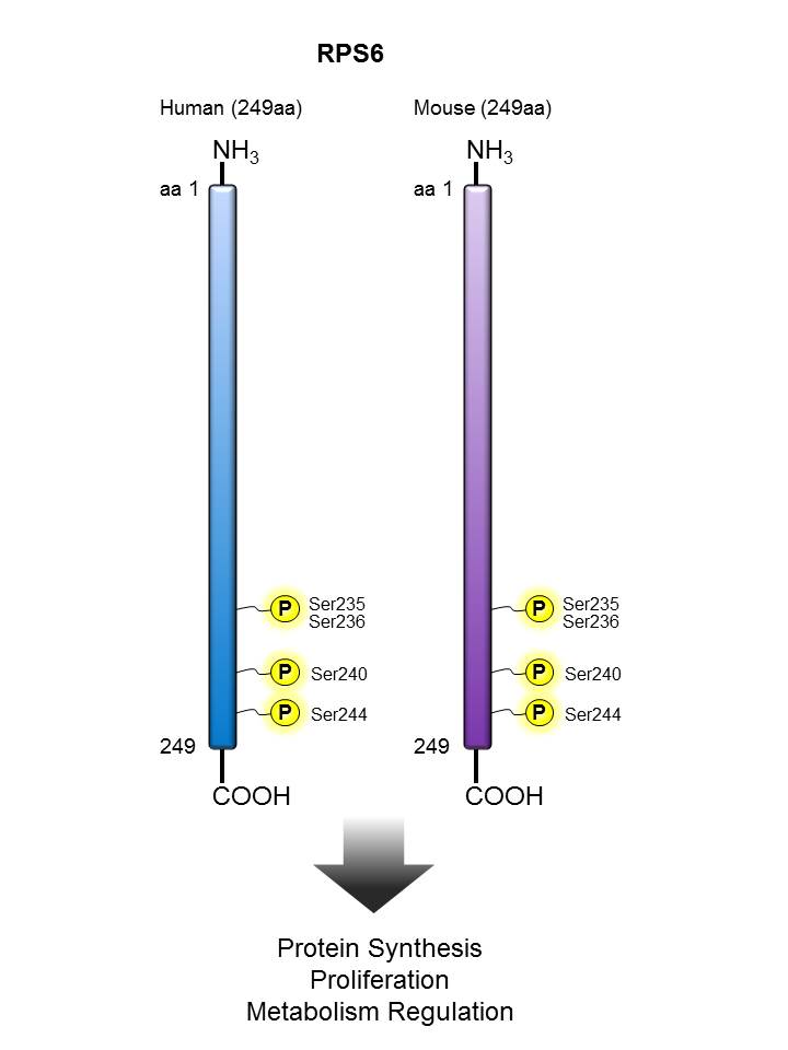
RPS6


Description:

The eukaryotic ribosome is composed of 40S and 60S subunits, which is involved in the physiological process of protein translation. In many organisms, ribosomal proteins undergo various post-translational modifications, including phosphorylation, acetylation, methylation, O-linked b-N-acetylglucosaminylation, and ubiquitylation. Ribosomal proteinS6 (RPS6), the first identified ribosomal protein which can be phosphorylated, is a component of eukaryotic ribosomal 40S subunit. Functional studies of RPS6 phosphorylation suggested its role in the regulation of protein synthesis, cell proliferation, and glucose homeostasis. The phosphorylation of RPS6 can be triggered by multiple stimuli including growth factors, tumor promoting agents, and mitogens. The inhibition of RPS6 phosphorylation was found mainly under stress conditions such as hypoxia, heat shock, and hyperosmolarity. RPS6 phosphorylation is also commonly used as a marker for neuronal activity and a read out of mammalian target of rapamycin complex 1 (mTORC1) activity.

Other Names:

40S ribosomal protein S6, Ribosomal Protein S6

Human to Mouse Sequence Homology:

100%

Notable Upstream Kinase(s):

P70S6K, P90RSK, PKCD

Notable Phosphatase(s):

PPP5C

Product Listing:

See products for RPS6

Antigen References:

1. Kressler A, et al. 2010. Biochim. Biophys. Acta. 1803:673.
2. Biever A, et al. 2015. Front. Mol. Neurosci. 8:75.
3. Ventimiglia FA, Wool IG. 1974. Proc. Natl. Acad. Sci. USA 71:350.
4. Lastick SM, et al. 1977. Mol. Gen. Genet. 152:223.
5. Ruvinsky I, et al. 2005. Genes Dev. 19:2199.
6. Meyuhas O. 2008. Int. Rev. Cell. Mol. Biol. 268:1.
7. Meyuhas O. 2015. Int. Rev. Cell. Mol. Biol. 320:41.

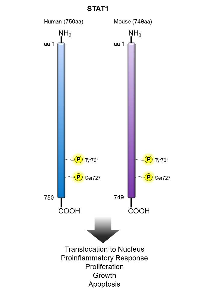
STAT1


Description:

STAT1, also known as signal transduction and activator of transcription 1, is a ubiquitously expressed cytoplasmic protein and is activated in response to cytokine signaling, including IFN-α, IFN-γ, EGF, PDGF, and IL-6. Upon activation, STAT1 is phosphorylated by receptor-associated kinases, translocates to the nucleus, and functions as a transcription factor. Two isoforms of STAT1, with apparent molecular weights of 88 and 91 kD, exist as a result of alternative RNA processing. STAT1 is involved in IFN-mediated immune responses, and STAT1-deficient mice are highly sensitive to bacterial and viral infections.

Other Names:

Signal transducer and activator of transcription 1 (STAT1), Transcription factor ISGF-3 components p91/p84

Human to Mouse Sequence Homology:

92.30%

Notable Upstream Kinase(s):

JAK1, JAK2, SRC, CAMK2G, ERK1, P38A, PKCD,ALK

Notable Phosphatase(s):

PTPN13, PTPN2, SHP2

Product Listing:

See products for STAT1

Antigen References:

1. Durbin JE, et al. 1996. Cell. 84:443.
2. Darnell JE Jr, et al. 1994. Science 264:1415.
3. Chen X, et al. 1998. Cell. 93:827.
4. Ramana CV, et al. 2000. Oncogene. 19:2619.

STAT3


Description:

Tyrosine phosphorylation of STAT3 at Tyr705 occurs in response to LIF, IL-6, leptin, OSM, EGF, PDGF, and HGF. It plays a key role in cell growth and apoptosis through mediating expression of a variety of genes in response to the stimuli.

Other Names:

Signal transducer and activator of transcription 3, Acute-phase response factor, APRF, HIES

Human to Mouse Sequence Homology:

99.90%

Notable Upstream Kinase(s):

JAK2, RET, PKM2, ALK

Notable Phosphatase(s):

PTPRT, PTPN13, DUSP2

Product Listing:

See products for STAT3

Antigen References:

1. Akira S, et al. 1994. Cell 77:63.
2. Zhang X, et al. 1995. Science 267:1990.
3. Sanchez-Margalet V, et al. 2001. Cell. Immunol. 211:30.
4. Simon A, et al. 2000. Science 290:144.
5. Hoey T, et al. 1999. Adv. Immunol. 71:145.

STAT4


Description:

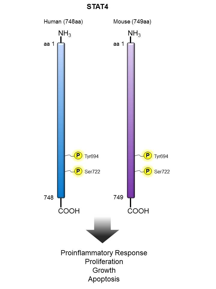
STAT4 is a member of the signal transducer and activator of transcription factors (STAT) family, transducing signals induced by IL-12, IL-23, and IFN-γ stimulation in T cells and monocytes. Upon activation, STAT4 is phosphorylated by receptor-associated JAK kinases, homodimerized through its SH2 domain, translocated to the nucleus and, in turn, transactivates target genes. Activation of STAT4 leads to production of IFN-γ and differentiation of Th1 and Th17 cells. STAT4 deficient mice exhibit impaired Th1 responses and abrogation of cytocytic functions of T and NK cells, suggesting STAT4 is critical in cell-mediated immune responses. Single nucleotide polymorphisms (SNPs) in the STAT4 gene have been shown to correlate with susceptibility to autoimmune diseases, such as rheumatoid arthritis (RA) and systemic lupus erythematosus (SLE).

Other Names:

Signal transducer and activator of transcription 4 (STAT4), SLEB11

Human to Mouse Sequence Homology:

94.50%

Notable Upstream Kinase(s):

JAK2, TYK2

Notable Phosphatase(s):

PTPN13

Antigen References:

1. Svensson A, et al. 2012. J. Virol. 86:9409.
2. Huang Y, et al. 2011. Blood. 118:6793.
3. Good SR, et al. 2009. J. Immunol. 183:3839.
4. O'Malley JT, et al. 2008. J. Immunol. 181:5062.
5. Korman BD, et al. 2008. Curr. Allergy Asthma Rep. 8:398.
6. Wurster AL, et al. 2000. Oncogene. 19:2577.

STAT5


Description:

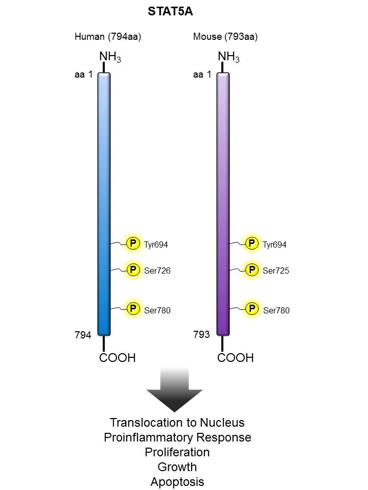
STAT5 has gene transactivation activity and plays a key role in lymphocyte differentiation and proliferation. Tyrosine phosphorylation of STAT5, induced by cytokine stimulation, results in dimerization through its SH2 domains. The dimerized STAT5 proteins translocates from the cytoplasm to the nucleus and subsequently regulates gene transcription. 

Other Names:

Signal transducer and activator of transcription 5 (STAT5), MGF

Human to Mouse Sequence Homology:

96.30%

Notable Upstream Kinase(s):

JAK2, ERK1/2,

Notable Phosphatase(s):

PTPN13

Product Listing:

See products for STAT5

Antigen References:

1. Teglund S, et al. 1998. Cell 93:841.
2. Moriggl R, et al. 1999. Immunity 10:249.
3. Peschon JJ, et al. 1994. J. Exp. Med. 180:1955.
4. Lynn M, et al. 2011. Ann. N.Y. Acad. Sci. 1217:18.
5. Malin S, et al. 2010. Curr. Opin. Immunol. 22:168.

STAT6


Description:

STAT6 is a member of the signal transducer and activator of transcription (STAT) family, activating gene expression in response to IL-4 and IL-13 stimulation. Upon cytokine stimulation, the receptor is phosphorylated by the associated Janus Kinases (Jak), followed by recruiting cytoplasmic STAT6. The Tyr641 residue of STAT6 is, in turn, phosphorylated by Jak. Phosphorylated STAT6 forms homodimers, transclocates to the nucleus, and regulates transcription of target genes. STAT6 plays crucial roles in differentiation of T helper 2 (Th2) cells, class switch of immunoglobulins in B cells, expression of cell surface markers such as MHC class II, and the development of allergic inflammation.

Other Names:

Signal transducer and activator of transcription 6, IL-4 Stat

Human to Mouse Sequence Homology:

85.10%

Notable Upstream Kinase(s):

JAK1, JAK3, ALK

Notable Phosphatase(s):

PTPN13

Product Listing:

See products for STAT6

Antigen References:

1. Goenka S, et al. 2011. Immunol. Res. 50:87.
2. Wurster AL, et al. 2000. Oncogene 19:2577.
3. Akira S. 1999. Stem Cells 17:138.
4. Zamorano J, et al. 2005. J. Immunol. 174:2843.
5. David M, et al. 2001. Oncogene 20:6660.
6. Takeda K, et al. 1996. Nature 380:627.

Stathmin/OP18


Description:

Stathmin/Op18 is a 21 - 23 kD member of the Stathmin family. This protein is localized in the cytoplasm and becomes nuclear during the S/G2 phase of the cell cycle. Stathmin/Op18 acts as a microtubule destabilizer in mitotic spindle regulation and sequesters tubulin dimers into assembly-incompetent complexes. This protein participates as an intracellular relay integrating regulatory signals of the cellular environment and has been shown to be involved in megakaryocyte polyploidization. Stathmin/Op18 can be phosphorylated in response to heat; Ser62 phosphorylation reduces tubulin binding ability. This protein can also be modified by acetylation. Stathmin/Op18 has been shown to interact with tubulin, KIST, CaM kinase II and IV, Cdc2, MAPK, and Cdk1. The Poly6202 antibody recognizes human phosphorylated stathmin/Op18 (Ser16) and has been shown to be useful for Western blotting.

Other Names:

Oncoprotein 18

Human to Mouse Sequence Homology:

98.70%

Notable Upstream Kinase(s):

CDK1, CDK2, ERK1, CAMK2B, CAMK4, PAK1, PKACA, RSK2

Notable Phosphatase(s):

PP2A, PP2B

Antigen References:

1. Sobel A, et al. 1989. J. Biol. Chem. 264:3765.
2. Labdon J, et al. 1992. J. Biol. Chem. 267:3506.
3. Rubin C, et al. 2003. Exp. Hematol. 31:389.
4. Honnappa S, et al. 2003. J. Biol. Chem. 278:38926.

SYK


Description:

Syk is a cytoplasmic 72 kD non-receptor tyrosine kinase that is widely expressed in hematopoietic cells. The Syk kinase associates with a number of proteins including c-Src, Lck, Lyn, Fyn, Vav1, STAT3, SHP1, Grb2, and LAT to couple immunoreceptors to downstream signaling events mediating cellular proliferation, differentiation, and phagocytosis. Syk is modified by phosphorylation on multiple tyrosine sites. The SYK-01 monoclonal antibody (raised against amino acids 5-360 of human Syk) recognizes human, mouse, and rat and Syk and has been shown to be useful for Western blotting, immunoprecipitation, and immunohistochemistry.

Other Names:

Spleen tyrosine kinase, protein tyrosine kinase SYK

Human to Mouse Sequence Homology:

91.70%

Notable Upstream Kinase(s):

SRC, SYK (autophosphorylation),

Notable Phosphatase(s):

PTP1B

Product Listing:

See products for SYK

Antigen References:

1. Taniguchi T, et al. 1991. J. Biol. Chem. 266:15790.
2. Toyabe S-L, et al. 2001.Immunology 103:164.

Tau


Description:

Tau proteins are microtubule-associated proteins (MAPs) that are abundant in neurons of the central nervous system, and are expressed at low levels in CNS astrocytes, oligodendrocytes, and elsewhere. Tau's main function, microtubule assembly and stability, is regulated by phosphorylation. Abnormal hyperphosphorylation of Tau results in a loss of the protein’s function, impairs neuronal homeostasis, and ultimately results in the formation of aberrant aggregates, neurofibrillary tangles, that are a pathological hallmark of a group of neurodegenerative diseases, such as Alzheimer’s disease, which are termed tauopathies.

Other Names:

Microtubule-associated protein tau, PHF-tau, paired helical filament-tau, neurofibrillary tangle protein, microtubule-associated protein tau, isoform 4, G protein beta1/gamma2 subunit-interacting factor 1, DDPAC, FTDP-17, MAPTL, MSTD, MTBT1, MTBT2, PPND

Human to Mouse Sequence Homology:

73.40%

Notable Upstream Kinase(s):

CDK5, GSK3, PKA, JNK, CAMK2, P38

Notable Phosphatase(s):

PP2A, PP2B, PTPA,

Product Listing:

See products for Tau


TDP43


Description:

TDP43 (aka TARDBP) was originally identified as a protein which binds to the transactivation response (TAR) sequence found in the long terminal repeat of the HIV-1 virus genome. TDP43 contains two copies of a ~90 amino acid RNA recognition motif (RRM) found in many proteins which bind single stranded RNA. TDP-43 localizes to the nucleus. In patients with frontotemporal lobar degeneration and amyotrophic lateral sclerosis, it is absent from the nucleus of affected neurons but it is the primary component of cytoplasmic ubiquitin-positive inclusion bodies.  Ubiquitously expressed. In particular, expression is high in pancreas, placenta, lung, genital tract and spleen. Hyperphosphorylated, ubiquitinated, and cleaved in hippocampus, neocortex, and spinal cord from individuals affected with ALS and FTLDU.

Other Names:

TAR DNA-binding protein-43, TDP 43, TARDBP, ALS10

Human to Mouse Sequence Homology:

96.10%

Notable Upstream Kinase(s):

CK1 (δ,ε)

Notable Phosphatase(s):

PPP3C

Product Listing:

See products for TDP43


TIF1β/KAP1


Description:

Transcriptional intermediary factors (TIFs) are a group of transcriptional coactivators and corepressors that regulate gene expression by modulating chromatin structure and assembly of transcription initiation complexes. TIF1β is a member of the TIF1 subfamily of chromatin-associated TIFs that play a key role in many developmental and physiological processes. Studies using knockout mice reveal the important function of TIF1β in regulating genomic imprinting, T cell activation, and T cell tolerance.

Other Names:

TRIM28 pS473, KAP-1 pS473 , KRIP-1 pS473

Human to Mouse Sequence Homology:

93.20%

Notable Upstream Kinase(s):

ATM, DNA-PK, CHK2, PRKD1

Notable Phosphatase(s):

PPP1CA, PPP4C

Product Listing:

See products for TIF1β/KAP1

Antigen References:

1. Messerschmidt DM, et al. 2012. Science 335:1499.
2. Chikuma S, et al. 2012. Nat. Immunol. 13:596.

Zap70


Description:

ZAP70 was identified in TCR-stimulated Jurkat cells. It is an inactive cytosolic tyrosine kinase that is recruited to a transmembrane receptor lacking intrinsic catalytic activity. ZAP70 and the related spleen tyrosine kinase (Syk) play a critical role in T-cell development and activation. This enzyme, which is phosphorylated on tyrosine residues upon T-cell antigen receptor (TCR) stimulation, functions in the initial step of TCR-mediated signal transduction in combination with the Src family kinases, Lck and Fyn. ZAP70 activation can be regulated by binding to phosphorylated ITAMs of the TCR and by phosphorylation of multiple tyrosine residues on ZAP70. Phosphorylation of Tyr315 and Tyr319 are essential for ZAP70 positive regulation of T-lymphocyte activation whereas Tyr292 has a negative regulatory role.Phosphorylated Tyr319 is a positive regulator of ZAP70, which triggers the binding of Lck and activation of NFAT and IL-2 induction. The binding of Lck to phosphorylated Tyr319 promotes Lck mediated phosphorylation of Tyr493, and facilitates activation of downstream signaling. The importance of Tyr319 phosphorylation in positive regulation of ZAP70 was confirmed in a mouse model with Tyr319F ZAP70 mutant expression, which resulted in severe defects in calcium mobilization and thymocyte selection.

Other Names:

Phospho Zeta-Chain (TCR) Associated Protein Kinase 70 kD, Phospho SRK, Phospho 70 KD Zeta-Chain Associated Protein, Phospho Tyrosine-protein kinase ZAP-70

Human to Mouse Sequence Homology:

93.50%

Notable Upstream Kinase(s):

LCK, ZAP70 (autophosphorylation), ABL

Notable Phosphatase(s):

PTPN22

Product Listing:

See products for Zap70

Antigen References:

1. Chan AC, et al. 1991. Proc. Natl. Acad. Sci. 88:9166.
2. Arpaia E, et al. 1994. Cell 76:947.
3. Chan AC, et al. 1994. Science 264:1599.
4. Negishi I, et al. 1995. Nature 376:435.
5. Wang H, et al. 2010. Cold Spring Harb. Perspect. Biol. 2:a002279.
6. Klammt C, et al. 2015. Nat. Immunol. 16:961.
7. Weiss A. 1993. Cell 73:209.
8. Gong Q, et al. 2001. J. Exp. Med. 194:507.

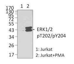
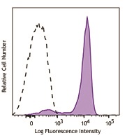
We offer antibody clones raised against phosphorylated targets that have been validated for use in multiple experimental applications. For example, clone 4B11B69 raised against phosphorylated ERK1/2 (Tyr202/Tyr204) has been validated in-house for successful use in western blotting, immunofluorescence, and intracellular flow cytometry.
Alexa Fluor® 647 anti-ERK1/2 Phospho (Thr202/Tyr204) Antibody (clone 4B11B69)

Intracellular Flow Cytometry (ICFC):


Human peripheral blood lymphocytes were stimulated with (filled histogram), or without (open histogram), Cell Activation Cocktail (without Brefeldin A) for 15 minutes, then fixed with Fixation Buffer, permeabilized with True-Phos™ Perm Buffer, and intracellularly stained with ERK1/2 Phospho (Thr202/Tyr204) (Clone 4B11B69) Alexa Fluor® 647.
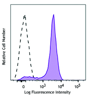
 

C57BL/6 mouse splenocytes were stimulated with (filled histogram) or without (open histogram) Cell Activation Cocktail (without Brefeldin A) for 15 minutes, then fixed with Fixation Buffer, permeabilized with True-Phos™ Perm Buffer, and intracellularly stained with ERK1/2 Phospho (Thr202/Tyr204) (Clone 4B11B69) Alexa Fluor® 647.

Superior Signal-to-Noise Ratio:
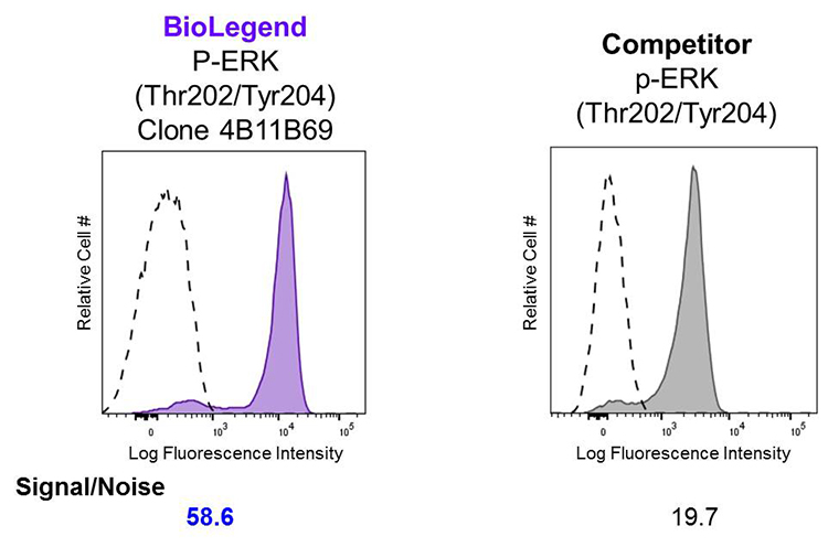
 

Human peripheral blood lymphocytes were stimulated with (filled histogram) or without (open histogram) Cell Activation Cocktail (without Brefeldin A) for 15 minutes, then fixed with Fixation Buffer, permeabilized with True-Phos™ Perm Buffer, and intracellularly stained with Alexa Fluor® 647 ERK1/2 Phospho (Thr202/Tyr204). Clone 4B11B69 was used at 0.125 µg/test and compared side-by-side against the competitor's product, which was used at 0.1 µg/test.


Products Referenced Above:

Product Name Catalog #
Alexa Fluor® 647 anti-ERK1/2 Phospho (Thr202/Tyr204) Antibody 675503 | 675504
Cell Activation Cocktail (without Brefeldin A) 423301 | 423302
Fixation Buffer 420801
True-Phos™ Perm Buffer 425401


Purified anti-ERK1/2 Phospho (Thr202/Tyr204) Antibody (clone 4B11B69)

Western Blot (WB):


Left: Untreated (lane 1) and PMA-treated (lane 2) Jurkat whole cell lysates were resolved by electrophoresis, transferred to nitrocellulose, and probed with purified monoclonal anti-ERK1/2 Phospho (Thr202/Tyr204) (clone 4B11B69) antibody. Proteins were visualized using an anti-mouse-IgG secondary conjugated to HRP (Cat. No. 405306) and chemiluminescence detection.

Immunofluorescence (IF):


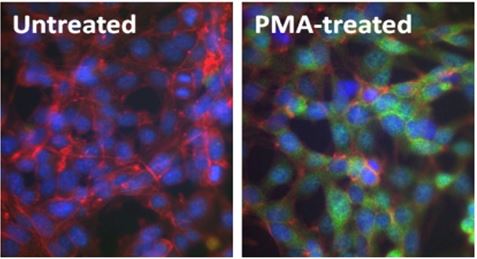
Above: Untreated (left) and PMA-treated (20 nM PMA treatment for 10 minutes, right) mouse NIH-3T3 cells were stained with purified anti-ERK1/2 Phospho (Thr202/Tyr204) (clone 4B11B69) antibody, followed by staining with DyLight™ 488 conjugated goat anti-mouse IgG (green, Cat. No. 405310) antibody and Alexa Fluor® 594 conjugated phalloidin (red). Nuclei were stained with DAPI (blue).

Products Referenced Above:

Product Name Catalog #
Purified anti-ERK1/2 Phospho (Thr202/Tyr204) Antibody 675502
HRP Goat anti-mouse IgG (minimal x-reactivity) Antibody (WB) 405306
DyLight™ 488 Goat anti-mouse IgG (minimal x-reactivity) Antibody (IF) 405310
DAPI (4', 6-Diamidino-2-Phenylindole, Dilactate) (IF) 422801


Phospho-target Antibodies for Neuroscience

With the acquisition of Covance's antibody product portfolio, BioLegend now offers antibodies targeted towards phosphorylated proteins implicated in the biology of the brain, and neurodegenerative diseases.

Purified anti-Tau Phospho (Thr181) Antibody Clone M7004D06

Immunohistochemistry (IHC):


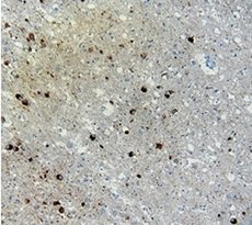
(Left) IHC staining of anti-Tau Phospho (Thr181) antibody (clone M7004D06) on formalin-fixed paraffin-embedded Alzheimer's brain tissue. Following antigen retrieval using Sodium Citrate, the tissue was incubated with the primary antibody at 10 µg/ml for 60 minutes at room temperature. BioLegend's Ultra Streptavidin (USA) HRP Detection Kit was used for detection.

Western Blot (WB):


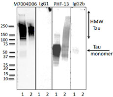
(Left) Western blot of anti-Tau Phospho (Thr181) (clone M7004D06), anti-Tau Phospho (Ser396) (clone PHF-13) antibodies, and isotype-matched control IgG1 and IgG2b. Lane 1: 20 µg of Parkinson’s disease human brain lysate; Lane 2: 2 µg of Tau paired helical filaments. Blots were incubated overnight at 4°C with 10 µg/ml of the primary antibody, followed by horseradish peroxidase labeled goat anti-mouse secondary antibody. Western blot of anti-Tau Phospho (Ser396) (clone PHF-13) serves as a positive control for Tau staining.

Products Referenced Above:

Product Name Catalog #
Purified anti-Tau Phospho (Thr181) Antibody (Clone M7004D06) 846602
Purified anti-Tau Phospho (Ser396) Antibody (Clone PHF-13) 829001
Ultra Streptavidin (USA) HRP Detection Kit (Multi-Species) (IHC) 929701
Purified anti-Tau Phospho (Ser396) Antibody  829001

Kinases

Kinases are enzymes that catalyze the addition of phosphoryl groups to certain amino acid residues on target molecules, which is called phosphorylation. To do this, a kinase will bind to ATP, hydrolyze the gamma (γ) phosphoryl, and bind to its target molecule, generating ADP in the process. Then, it transfers the phosphoryl group to the target molecule, dissociates, binds to a new ATP molecule. This process can then be repeated.


Phosphorylation Targets

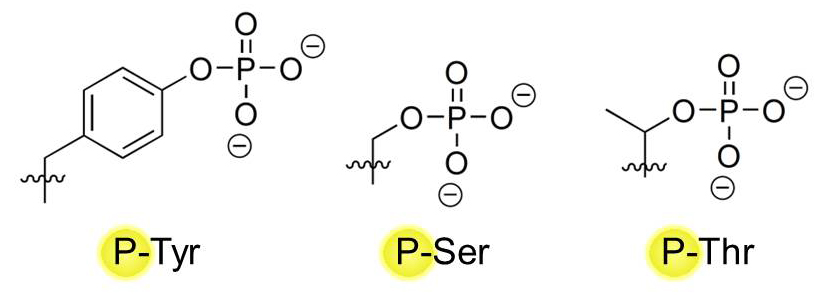
Proteins make up the majority of target molecules that can be phosphorylated by kinases, but lipids (e.g. Phosphatidylinositol-4,5-bisphosphate 3, or PI3) can also be phosphorylated. On proteins, three major amino acid residues serve as major sites of phosphorylation: Tyrosines (Tyr, Y) Serines (Ser, S), and Threonines (Thr, T). They all possess a hydroxyl (-OH) group in their variable chain that can serve as a placement site for a phosphate group by kinases:
 

Phosphatases

Phosphatases work in reverse to kinases, and are responsible for removing phosphate groups from their targets. Dephosphorylation that is catalyzed by phosphatases is important for maintaining homeostatic balance in the biological system – once a protein is phosphorylated for activation, it must be turned off via dephosphorylation by phosphatases or by protein degradation. Constitutively active phosphorylation signal pathways are hallmarks of numerous malignancies, including cancer.


True-Phos™ Perm Buffer


Finding the right conditions to stain for phosphorylated intracellular targets for flow cytometric analysis can be tricky. At BioLegend, we have developed the True-Phos™ Perm Buffer for flow cytometry, which is specifically designed to help detect our highly sensitive phosphorylation-specific antibodies. True-Phos™ Perm Buffer is not designed for use in western blotting or microscopy applications.

Representative Data:
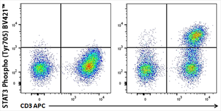


Human whole blood unstimulated (left panel) or stimulated with IL-6 for 15 minutes (right panel) were treated with RBC Lysis/Fixation Solution (10X), permeabilized with True-Phos™ Perm Buffer, and stained with CD3 APC and STAT3 Phospho (Tyr705) Brilliant Violet 421™


HeLa cells were treated with Nocodazole for 24 hours (right panel) or untreated (left panel), fixed with Fixation Buffer, permeabilized with True-Phos™ Perm Buffer, then stained with DAPI and Histone H3 Phospho (Ser10) Alexa Fluor® 647.

Products Referenced Above:

Product Name Catalog #
True-Phos™ Perm Buffer 425401
RBC Lysis/Fixation Solution (10x) 422401
Fixation Buffer 420801
Brilliant Violet™ 421 phospho STAT3 (Tyr705) 651009 | 651010
Alexa Fluor® 647 phospho Histone H3 (Ser10) 650805 | 650806
ProductsHere

Login / Register
Remember me
Forgot your password? Reset password?
Create an Account