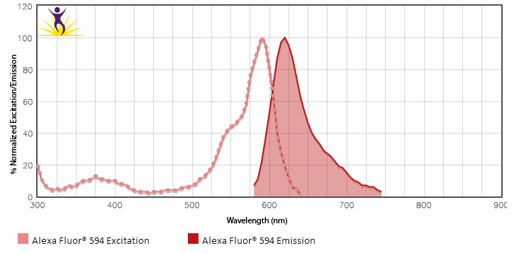
For multicolor fluorescence microscopy, bright fluorescence with little background is critical for specific detection of antigens. Furthermore, directly labeled antibodies are essential, since the usage of secondary antibodies may be limited when using multiple antibodies. BioLegend now introduces our line of directly conjugated Alexa Fluor® 594 antibodies for immunofluorescence microscopy. Alexa Fluor® 594 is a bright, stable fluorophore emitting into the red range of the color spectrum, well-suited for imaging applications.

Alexa Fluor® and Pacific Blue™ are trademarks of Life Technologies Corporation.

Alexa Fluor® 700 is generally not recommended for use in microscopy applications.

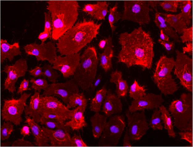
Human/mouse CD44, clone IM7

Human/mouse CD44, clone IM7

HUVEC cells were fixed with 1% paraformaldehyde (PFA) for 10 minutes and blocked with 5% FBS for 30 minutes. Then the cells were stained with 5 µg/ml of anti-human/mouse CD44 (clone IM7) Alexa Fluor® 594 (red) in blocking buffer for 3 hours at room temperature. Nuclei were counterstained with DAPI and are shown in blue. The image was captured with 40x objective.

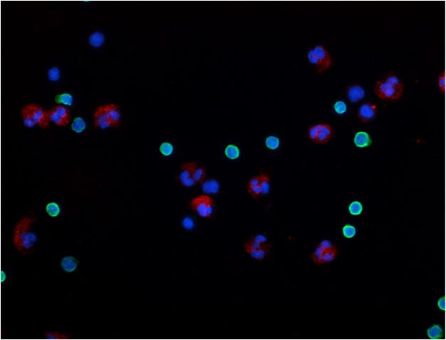
Human CD11b, clone ICRF44

Human CD11b, clone ICRF44

Human peripheral mononuclear cell and neutrophil mixed cells were fixed with 2% paraformaldehyde (PFA) and then stained with 5 µg/ml CD11b (clone ICRF44) Alexa Fluor® 594 (red) and 5 µg/ml CD3 (clone UCHT1) Alexa Fluor® 488 (green) for 30 minutes at room temperature. Nuclei were counterstained with DAPI and are shown in blue. The image was captured by 40X objective.

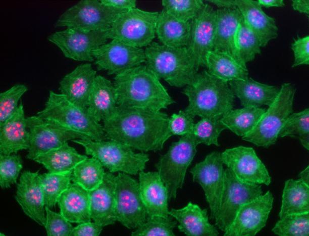
Human Ki-67, clone Ki-67

Human Ki-67, clone Ki-67

HeLa cells were fixed with 1% paraformaldehyde (PFA) for 10 minutes, permeabilized with 0.5% Triton X-100 for 10 minutes and blocked with 5% FBS for 30 minutes. Then the cells were intracellular stained with 2.5 µg/ml of Ki-67 (clone Ki-67) Alexa Fluor® 594 (red) in blocking buffer overnight at 4°C and followed by Alexa Fluor® 488 Phalloidin (green) staining for 20 minutes. Nuclei were counterstained with DAPI and are shown in blue. The image was captured with 40x objective.

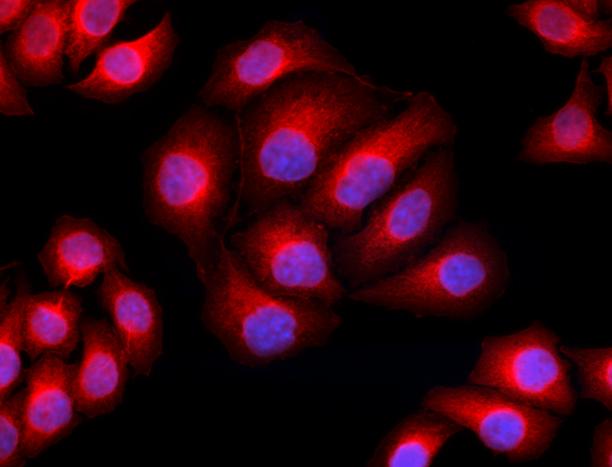
Tubulin-alpha, clone 10D8

Tubulin-alpha, clone 10D8

HeLa cells were fixed with 1% paraformaldehyde (PFA) for 10 minutes, permeabilized with 0.5% Triton X-100 for 10 minutes and blocked with 5% FBS for 30 minutes. Then the cells were intracellular stained with 5 µg/ml of Tubulin-alpha (clone 10D8) Alexa Fluor® 594 (red) in blocking buffer for 3 hours at room temperature. Nuclei were counterstained with DAPI and are shown in blue. The image was captured with 40x objective.

ex em

Alexa Fluor® 594 excites optimally at 590 nm and emits optimally at 617 nm.


Compare with other fluorophores on our Fluorescence Spectra Analyzer.

ProductsHere

Login / Register
Remember me
Forgot your password? Reset password?
Create an Account