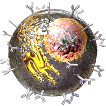
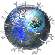
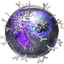
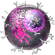
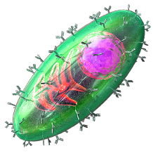
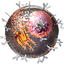
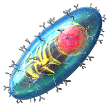
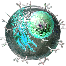
Transcription factors are proteins that regulate the transcription of genes, or the production of mRNA from DNA. Their unique feature is their ability to bind DNA at sequences known as promoter or enhancer regions. Once the transcription factor binds to an enhancer region, this can cause stimulation or repression of gene transcription. Promoter sequences are found at the beginning of the gene and serve as the transcription start site where the transcription initiation complex is formed. Unlike promoter sequences, enhancer or regulatory sequences can be found at sites that are distal to the gene that is being transcribed. It is the action of transcription factors that determines the differentiation fate of cells. For example, the expression of T-bet and FOXP3 transcription factors (along with specific cytokines) will differentiate T cells into T regulatory cells. This page describes the transcription factors expressed by various cell types, including T cells, B cells, monocytes, macrophages, and dendritic cells, as well subtypes of each.


For nuclear protein flow cytometry staining, BioLegend's True-Nuclear™ Transcription Factor Buffer Set has been specially formulated for intracellular staining with minimum effect on the surface fluorochrome staining. Additionally, our True-Phos™ Perm Buffer, is designed for use with antibodies for phospho-proteins (e.g. p-STAT6, p-ERK, etc.) and other intracellular targets (e.g. Cyclin B1, pan-STAT3, etc.). Not all cell surface markers are compatible with BioLegend's True-Phos™ Perm Buffer. View our complete list of Transcriptional Regulator Antibodies.

Please select a cell

B cell

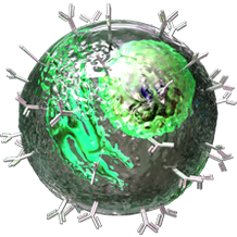
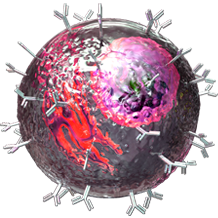
B cells are a lineage of lymphocytes originally discovered in the Bursa of Fabricus in birds (hence the B in B cells). B cell development in mammals was mainly found to occur in the bone marrow, as they do not have a Bursa of Fabricus. B cells are not only responsible for antibody production, but are also involved in several other areas of immunology.

Transcription Factors

BACH2
Bcl-6
Blimp-1
IRF4
IRF8
Pax-5
SPI1
STAT5
XBP-1s
*Note: Not all Transcription Factors listed are constitutively expressed in each cell type shown. Some factors may be specific to various cell stages or subsets within the known cell types.

Related Pages

Maturation Markers
B cells
Essential Markers for Phenotyping
B Cell Products
Bio-Bit - B All You Can B

Related Pathways

Th17 Biology
TH2 Pathway (Humoral Immune Response)
TH1 Pathway (Cellular Immune Response)
T Follicular Helper Cell Pathway
Regulatory T Cells
Neurodegeneration
Immunologic Networks 2011
Cytokine Network
Chemokine Receptor Biology
B cell Development and Activation
B-Cell Receptor Pathway
BAFF in B-Cell Signaling

Mature B cell

Upon exiting the bone marrow, Mature B Cells will express both IgM and IgD on their cell surface. At this point, the cells will have acquired functional competence, hence their title of “mature”. They may be called naïve if they have not yet encountered their specific antigen.

Transcription Factors

BACH2
IRF8
Pax-5
*Note: Not all Transcription Factors listed are constitutively expressed in each cell type shown. Some factors may be specific to various cell stages or subsets within the known cell types.

Related Pages

Maturation Markers
B cells
Essential Markers for Phenotyping
B Cell Products

Related Pathways

TH2 Pathway (Humoral Immune Response)
Immunologic Networks 2011
Cytokine Network
Chemokine Receptor Biology
BAFF in B-Cell Signaling

Follicular B cell

Follicular B Cells can be found within structures called follicles (which contain germinal centers) in secondary and tertiary lymphoid organs like the spleen, Peyer's patches, and lymph nodes. Unlike Marginal Zone B Cells, Follicular B Cells are capable of recirculating throughout the body. They are capable of becoming either Memory B Cells or Long Lived Plasma Cells. In most instances, these cells require T Cells for full activation.

Transcription Factors

BACH2
Bcl-6
Pax-5
*Note: Not all Transcription Factors listed are constitutively expressed in each cell type shown. Some factors may be specific to various cell stages or subsets within the known cell types.

Related Pages

Maturation Markers
B cells
Essential Markers for Phenotyping
B Cell Products

Related Pathways

T Follicular Helper Cell Pathway
Immunologic Networks 2011
Cytokine Network
Chemokine Receptor Biology
BAFF in B-Cell Signaling

Marginal Zone B cell

Marginal Zone (MZ) B Cells are a mature form of B Cells that home specifically to the MZ of the spleen. As such, they specifically guard against blood-borne pathogens they would be likely to encounter in the spleen. They typically have a lower threshold of activation compared to Follicular B Cells and can develop into Short Lived Plasma Cells, generating “natural antibodies” in a T Cell independent manner.

Transcription Factors

BACH2
IRF8
Pax-5
SPI1
*Note: Not all Transcription Factors listed are constitutively expressed in each cell type shown. Some factors may be specific to various cell stages or subsets within the known cell types.

Related Pages

Maturation Markers
B cells
Essential Markers for Phenotyping
B Cell Products

Related Pathways

Immunologic Networks 2011
Cytokine Network
Chemokine Receptor Biology
BAFF in B-Cell Signaling

Short lived plasma cell

Upon encountering antigen, Mature B Cells can develop into plasmablasts, which further differentiate into Plasma Cells. As indicated by their names, Short Lived Plasma Cells (SLPC) only persist for a few days, rapidly churning out antibody before undergoing apoptosis. SLPCs can typically be found in the red pulp of the spleen or in medullary chords of lymph nodes.

Transcription Factors

Blimp-1
IRF4
XBP-1s
*Note: Not all Transcription Factors listed are constitutively expressed in each cell type shown. Some factors may be specific to various cell stages or subsets within the known cell types.

Related Pages

How to Make an Antibody
Maturation Markers
B cells
Essential Markers for Phenotyping
B Cell Products

Related Pathways

Th17 Biology
TH2 Pathway (Humoral Immune Response)
T Follicular Helper Cell Pathway
Immunologic Networks 2011
Chemokine Receptor Biology
Cytokine Network
BAFF in B-Cell Signaling

Memory B cell

Memory B Cells develop after a primary infection within the host. After encountering an antigen, a subset of cells that developed from an individual cell can retain the experience and memory of its target. This leads to a quicker immune response should that antigen should ever reintroduce itself to the body.

Transcription Factors

BACH2
IRF8
Pax-5
SPI1
*Note: Not all Transcription Factors listed are constitutively expressed in each cell type shown. Some factors may be specific to various cell stages or subsets within the known cell types.

Related Pages

Maturation Markers
B cells
Essential Markers for Phenotyping
B Cell Products

Related Pathways

Immunologic Networks 2011
Cytokine Network
Chemokine Receptor Biology
BAFF in B-Cell Signaling

Long lived plasma cell

Upon encountering antigen, Mature B Cells can develop into plasmablasts, which further differentiate into Plasma Cells. Plasma Cells are, quite frankly, powerhouses of antibody production. They generate a single class of immunoglobulin designated for a specific antigen. Antibody production can continue for days or months until the target is neutralized.

Transcription Factors

Blimp-1
IRF4
XBP-1s
*Note: Not all Transcription Factors listed are constitutively expressed in each cell type shown. Some factors may be specific to various cell stages or subsets within the known cell types.

Related Pages

How to Make an Antibody
Maturation Markers
B cells
Essential Markers for Phenotyping
B Cell Products

Related Pathways

Th17 Biology
TH2 Pathway (Humoral Immune Response)
T Follicular Helper Cell Pathway
Immunologic Networks 2011
Cytokine Network
Chemokine Receptor Biology
BAFF in B-Cell Signaling

B1 cell

B-1 Cells are considered an innate lymphocyte that appears early on in the development of the host. This has led to their being named “B-1”, while conventional B Cells that appear later on are termed “B-2”. B-1 Cells make up roughly 5% of mouse and human total B cell population. B-1 Cells arise from stem cell populations and exhibit the ability to self-renew. Typically, they only express IgM on their surface, with no IgG or IgD. B-1 cells are associated with a quick, innate immune response as they express a limited range of “natural”, lower-affinity antibodies, do not undergo affinity maturation, do not require T cell help, and do not have a proclivity to develop into Memory B cells.

Transcription Factors

BACH2
IRF8
Pax-5
SPI1
*Note: Not all Transcription Factors listed are constitutively expressed in each cell type shown. Some factors may be specific to various cell stages or subsets within the known cell types.

Related Pages

Maturation Markers
B cells
Essential Markers for Phenotyping
B Cell Products

Related Pathways

Immunologic Networks 2011
Chemokine Receptor Biology
Cytokine Network
BAFF in B-Cell Signaling

Breg

Regulatory B Cells, or Bregs, are a subtype of B Cells capable of downregulating inflammation and inducing tolerance. However, these cells can be phenotypically difficult to identify, as they bear many common markers found on other B Cell types. The most well studied Bregs may be the B-10 and T2-MZP (Transitional 2 Marginal Zone Precursor) population. Many Breg subclasses exact their immunosuppression through IL-10. No specific transcription factors have been identified that define either mouse or human Bregs, to date. Since Bregs can be induced in vitro and in vivo, it's possible that certain factors that are present in their microenvironment play a role in Breg induction.

Transcription Factors

Blimp-1
IRF4
*Note: Not all Transcription Factors listed are constitutively expressed in each cell type shown. Some factors may be specific to various cell stages or subsets within the known cell types.

Related Pages

Maturation Markers
B cells
Essential Markers for Phenotyping
B Cell Products
Bio-Bit - B All You Can B

Related Pathways

Immunologic Networks 2011
Cytokine Network
Chemokine Receptor Biology
BAFF in B-Cell Signaling

Germinal Center B cell

Germinal centers (GCs) form inside peripheral lymphoid organs in response to T cell-dependent antigens. B cells present inside the GCs differentiate into plasma cells and memory B cells, where they undergo affinity maturation and are selected for their antibody repertoires. Germinal center B cells are the fastest proliferating cells in the human body.

Transcription Factors

BACH2
Bcl-6
Pax-5
*Note: Not all Transcription Factors listed are constitutively expressed in each cell type shown. Some factors may be specific to various cell stages or subsets within the known cell types.

Related Pages

Maturation Markers
B cells
Essential Markers for Phenotyping
B Cell Products

Related Pathways

Immunologic Networks 2011
Cytokine Network
Chemokine Receptor Biology
BAFF in B-Cell Signaling

Macrophage/Monocyte

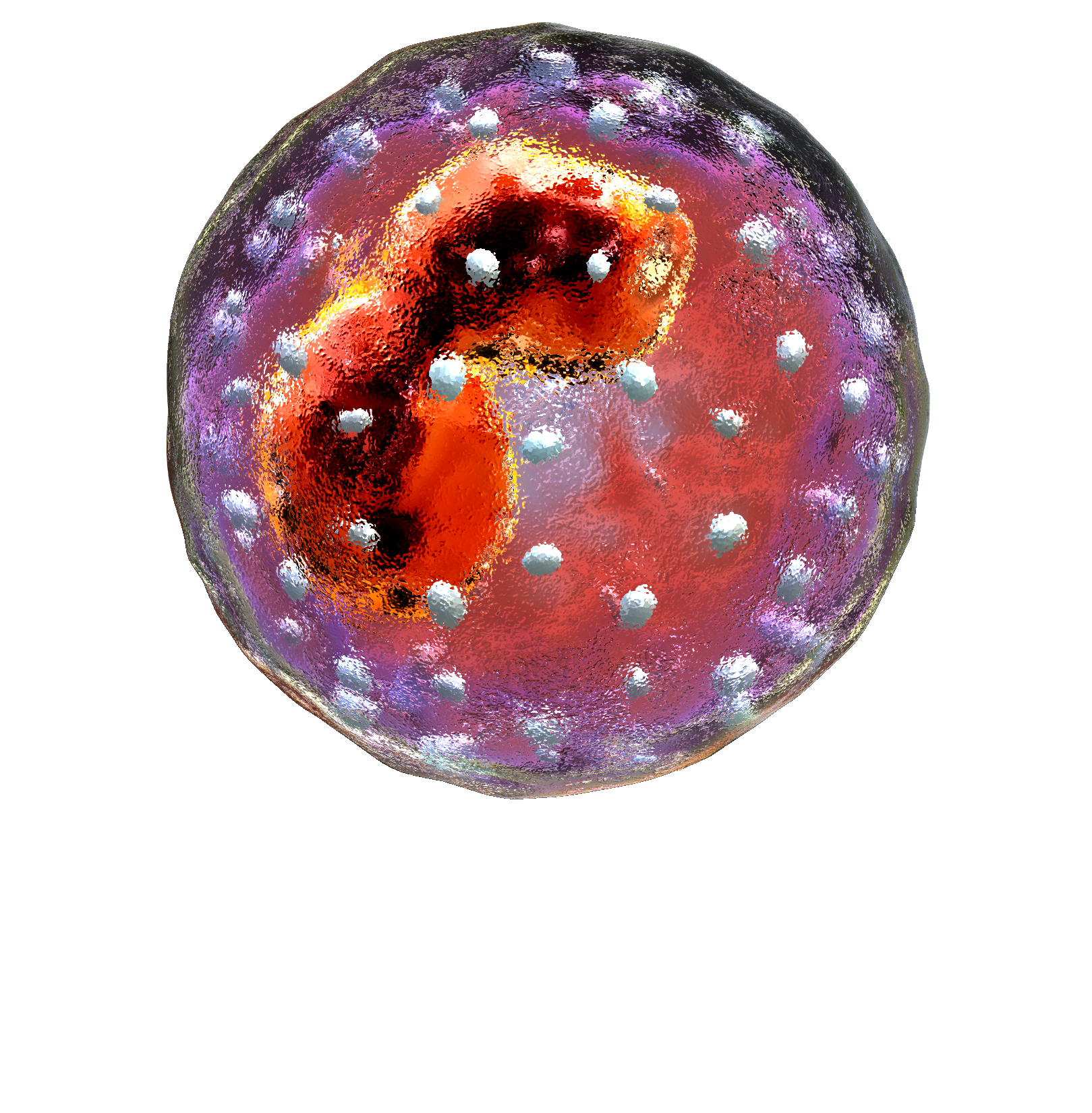
Monocytes are a population of circulating white blood cells with the potential to differentiate into macrophages and dendritic cells in tissue. Monocytes develop in the bone marrow and can be subdivided based on size, trafficking and innate immune receptor expression. Monocytes are key in anti-microbial responses, but are also known to be involved with some inflammatory diseases.

Monocytes can be drawn into tissues by inflammatory signals or chemokines, where they develop into macrophages. Macrophages specialize in phagocytosis and can also help with pathogen killing and wound repair. Their function and phenotype can depend on their tissue environment. M1, or classical, macrophages promote Th1 responses and inflammation, while M2, or, alternative macrophages, support immunosuppression and wound healing/tissue repair.

Transcription Factors

ATM
c-Jun
C/EBP β
CREB
IRF1
IRF4
IRF5
IRF7
IRF8
Ki-67
myeloid antigen
NF-κB p65
p53
PCNA
PPARγ
SPI1
SSRP1
STAT1
STAT3
STAT6
XBP-1s
*Note: Not all Transcription Factors listed are constitutively expressed in each cell type shown. Some factors may be specific to various cell stages or subsets within the known cell types.

Maturation Markers

View Macrophage/Monocytes maturation markers

Related Pages

Cell Markers
Macrophages
Monocytes
Essential Markers for Phenotyping
Monocyte/Macrophage Products

Related Pathways

Tregs in Tumor Escape
TH2 Pathway (Humoral Immune Response)
TH1 Pathway (Cellular Immune Response)
Neurodegeneration
Innate Immune Signaling
Immunologic Networks 2011
Dendritic Cell, Monocyte, and Macrophage Biology
Chemokine Receptor Biology
Cytokine Network
Cancer Immunoediting

Monocyte

Monocytes are a population of circulating white blood cells with the potential to differentiate into tissue macrophages and dendritic cells (DCs). Monocytes are derived from precursors in the bone marrow and can be subdivided into subsets that differ in size, trafficking and innate immune receptor expression. Monocytes mediate host antimicrobial defense and are also implicated in many inflammatory diseases, including atherosclerosis.

Adapted from Lawrence and Natoli. 2011. Nat Rev Immunol 11:750.

Transcription Factors

c-Jun
C/EBP β
IRF1
IRF4
IRF5
IRF8
NF-κB p65
SPI1
STAT1
STAT3
*Note: Not all Transcription Factors listed are constitutively expressed in each cell type shown. Some factors may be specific to various cell stages or subsets within the known cell types.

Related Pages

Dendritic Cells
Macrophages
Monocytes
Essential Markers for Phenotyping
Monocyte/Macrophage Products

Related Pathways

TH2 Pathway (Humoral Immune Response)
Neurodegeneration
Innate Immune Signaling
Immunologic Networks 2011
Dendritic Cell, Monocyte, and Macrophage Biology
Cytokine Network
Chemokine Receptor Biology
Cancer Immunoediting

M1

M1 macrophages have an activation phenotype that is associated with increased microbicidal activity and antigen-presenting function. This activation phenotype is modelled in vitro by IFNγ and/or LPS stimulation. M1 macrophages have different phenotypes in mice and humans, as the human M1 macrophage does not induce NOS2 in response to IFNγ/LPS treatment. In both mouse and human models, M1 macrophages are associated with increased IL-12 and MHC class II expression.

Transcription Factors

CREB
IRF5
NF-κB p65
SPI1
STAT1
*Note: Not all Transcription Factors listed are constitutively expressed in each cell type shown. Some factors may be specific to various cell stages or subsets within the known cell types.

Maturation Markers

View Macrophage/Monocytes maturation markers

Related Pages

Cell Markers
Macrophages
Essential Markers for Phenotyping
Monocytes/Macrophage Products

Related Pathways

Neurodegeneration
Innate Immune Signaling
Immunologic Networks 2011
Dendritic Cell, Monocyte, and Macrophage Biology
Cytokine Network
Chemokine Receptor Biology
Cancer Immunoediting

M2

M2 macrophages are associated with parasitic infections and Th2 immune responses, as well as anti-inflammatory and homeostatic functions linked to wound healing, fibrosis and tissue repair. This phenotype is usually induced with IL-4 and/or IL-13 stimulation. Like M1 macrophages, mice and human M2 macrophages have differing activation markers. In mice, this phenotype is associated with resistin-like-α, arginase 1, chitinase 3-like 3, IL-10, and CD206 expression. In humans, there is no induction of resistin-like-α, arginase 1 and chitinase 3-like 3. Instead, there is an increase in indoleamine 2,3-dioxygenase expression.

Transcription Factors

C/EBP beta
CREB
IRF4
PPARgamma
SPI1
STAT6
*Note: Not all Transcription Factors listed are constitutively expressed in each cell type shown. Some factors may be specific to various cell stages or subsets within the known cell types.

Maturation Markers

View Macrophage/Monocytes maturation markers

Related Pages

Cell Markers
Macrophages
Essential Markers for Phenotyping
Monocytes/Macrophage Products

Related Pathways

Neurodegeneration
Innate Immune Signaling
Immunologic Networks 2011
Dendritic Cell, Monocyte, and Macrophage Biology
Chemokine Receptor Biology
Cytokine Network
Cancer Immunoediting

Dendritic Cell

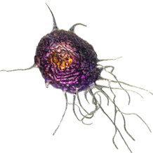
Dendritic cells (DCs) are immune cells that function as antigen-presenting cells of the immune system, interacting with T cells and B cells to trigger and modulate the adaptive immune response. They act as messengers between the innate and adaptive immunity. Dendritic cells are present in tissues in contact with the external environment, such as the skin (where there is a specialized dendritic cell type called Langerhans cells) and the inner lining of the nose, lungs, stomach and intestines. They can also be found in an immature state in the blood, but migrate to lymph nodes where they stimulate T and B cells.

Transcription Factors

ATM
BATF
Bcl-6
GFI1
ID2
Ikaros
IRF2
IRF4
IRF7
IRF8
Ki-67
NF-κB p65
NFIL3
p53
PCNA
RelB
SPI1
SSRP1
STAT3
STAT5
TCF4 (E2-2)
*Note: Not all Transcription Factors listed are constitutively expressed in each cell type shown. Some factors may be specific to various cell stages or subsets within the known cell types.

Maturation Markers

View Dendritic Cell maturation markers

Related Pages

Cell Markers
Dendritic Cells
Essential Markers for Phenotyping
Dendritic Cell Products
Bio-Bit - Dendritic Cells at the Center of the Immune System

Related Pathways

Tregs in Tumor Escape
Th17 Biology
TH2 Pathway (Humoral Immune Response)
TH1 Pathway (Cellular Immune Response)
T Follicular Helper Cell Pathway
Regulatory T Cells
Innate Immune Signaling
Immunologic Networks 2011
Dendritic Cell, Monocyte, and Macrophage Biology
Dendritic Cells
Cytokine Network
Chemokine Receptor Biology
Cancer Immunoediting

Plasmacytoid Dendritic Cell

Plasmacytoid dendritic cells (pDCs) are innate immune cells that circulate in the blood and are found in peripheral lymphoid organs. In humans, pDCs express the surface markers CD123, BDCA-2(CD303) and BDCA-4(CD304), but do not express CD11c or CD14, which distinguishes them from conventional dendritic cells or monocytes, respectively. Mouse pDC express CD11c, B220, BST-2 (mPDCA) and Siglec-H and are negative for CD11b. Upon stimulation and subsequent activation, these cells produce large amounts of type I interferon, which are critical anti-viral compounds mediating a wide range of effects.

Transcription Factors

BATF
GFI1
Ikaros
IRF2
IRF4
IRF7
IRF8
NFIL3
SPI1
TCF4 (E2-2)
*Note: Not all Transcription Factors listed are constitutively expressed in each cell type shown. Some factors may be specific to various cell stages or subsets within the known cell types.

Maturation Markers

View Dendritic Cell maturation markers

Related Pages

Cell Markers
Dendritic Cells
Essential Markers for Phenotyping
Dendritic Cell Products
Bio-Bit - Dendritic Cells at the Center of the Immune System

Related Pathways

Tregs in Tumor Escape
Innate Immune Signaling
Immunologic Networks 2011
Dendritic Cell, Monocyte, and Macrophage Biology
Dendritic Cells
Chemokine Receptor Biology
Cytokine Network
Cancer Immunoediting

Conventional DC

Conventional DCs specialize in antigen presentation and processing. They can be further divided into two subgroups, based on their tissue localization and migratory pathways. There are migratory DCs, which develop from precursors that reside in peripheral tissues and then migrate to regional lymph nodes. These migratory DCs are not found in the spleen.

Transcription Factors

BATF
Bcl-6
GFI1
ID2
Ikaros
IRF4
IRF8
NFIL3
RelB
SPI1
STAT3
STAT5
*Note: Not all Transcription Factors listed are constitutively expressed in each cell type shown. Some factors may be specific to various cell stages or subsets within the known cell types.

Maturation Markers

View Dendritic Cell maturation markers

Related Pages

Cell Markers
Dendritic Cells
Essential Markers for Phenotyping
Dendritic Cell Products
Bio-Bit - Dendritic Cells at the Center of the Immune System

Related Pathways

Tregs in Tumor Escape
T Follicular Helper Cell Pathway
Innate Immune Signaling
Immunologic Networks 2011
Dendritic Cell, Monocyte, and Macrophage Biology
Dendritic Cells
Chemokine Receptor Biology
Cytokine Network
Cancer Immunoediting

Monocyte-derived DC

Monocyte-derived DCs are professional antigen presenting cells recruited during infections with certain microorganisms, and serve as emergency back-up in acute inflammation states. These DCs can be induced by stimulation with GM-CSF and IL-4, and express CD11c, MHC II, CD24 and CD209a (DC-SIGN) while losing expression of M-CSFR and Ly-6C.

Transcription Factors

BATF
IRF4
NFIL3
SPI1
*Note: Not all Transcription Factors listed are constitutively expressed in each cell type shown. Some factors may be specific to various cell stages or subsets within the known cell types.

Maturation Markers

View Dendritic Cell maturation markers

Related Pages

Cell Markers
Dendritic Cells
Essential Markers for Phenotyping
Dendritic Cell Products
Bio-Bit - Dendritic Cells at the Center of the Immune System

Related Pathways

Innate Immune Signaling
Immunologic Networks 2011
Dendritic Cell, Monocyte, and Macrophage Biology
Dendritic Cells
Cytokine Network
Chemokine Receptor Biology

T cell

CD4 T helper cells are the primary orchestrators of the adaptive immune response, mediating a variety of cellular and humoral responses against pathogens and cancer. Although they lack any capacity to directly kill or engulf pathogens, they are powerful activators of effector cells such as macrophages, cytotoxic T cells, and B cells. On the other hand, regulatory T cells (Tregs) are potent suppressors of the immune response important in limiting the immune reaction. Recent advances have led to the discovery of a diverse set of T helper subsets, each with unique functions.

Transcription Factors

Aiolos
ATM
BATF
Bcl-6
Blimp-1
FOXP3
GATA3
Helios
Ikaros
IRF4
IRF7
Ki-67
NF-κB p65
p53
PCNA
SSRP1
STAT1
STAT4
T-bet
TCL1
Th-POK
ZAP-70
*Note: Not all Transcription Factors listed are constitutively expressed in each cell type shown. Some factors may be specific to various cell stages or subsets within the known cell types.

Maturation Markers

View T cell maturation markers

Related Pages

Cell Markers
γδ T Cells
T Helper Types
Essential Markers for Phenotyping
Costimulatory Molecules Category
T cell Receptor Products
Th1 Cell Products
Th2 Cell Products
Th17 Cell Products
Treg Cell Products

Related Pathways

Tregs in Tumor Escape
Th17 Biology
TH2 Pathway (Humoral Immune Response)
TH1 Pathway (Cellular Immune Response)
T Follicular Helper Cell Pathway
T-Cell Receptor Signaling
Regulatory T Cells
Neurodegeneration
Innate Immune Signaling
Immunologic Networks 2011
Gamma-Delta T cells
Chemokine Receptor Biology
Cytokine Network
CD4 and CD8 T-Cell Lineage
CD28 Signaling in T-Helper Cell
Cancer Immunoediting

T Follicular Helper

Tfh cells provide essential help to the production of affinity matured B cells in germinal centers. They are distinguishable from other T helper types by a number of criteria, including expression of the transcription factor BCL-6 and CXCR5, the receptor for CXCL13 that recruits Tfh cells to follicles. Unlike Th2 cells, Tfh cells produce IL-21 which potently stimulates the differentiation of B cells into antibody-forming cells.

Transcription Factors

BATF
Bcl-6
c-MAF
IRF4
STAT3
STAT5
*Note: Not all Transcription Factors listed are constitutively expressed in each cell type shown. Some factors may be specific to various cell stages or subsets within the known cell types.

Related Pages

Cell Markers
T Helper Types
Essential Markers for Phenotyping
Bio-Bit - PD-1 and T Follicular Regulatory Cells
Bio-Bit - The Role of T Follicular Helper Cells in HIV

Related Pathways

Th17 Biology
T Follicular Helper Cell Pathway
T-Cell Receptor Signaling
Regulatory T Cells
Immunologic Networks 2011
Chemokine Receptor Biology
CD4 and CD8 T-Cell Lineage
Cytokine Network
CD28 Signaling in T-Helper Cell

Th1

Th1 cells are IFN-γ + IL-12 polarized T helper cells that drive the killing efficacy of macrophages and proliferation of cytotoxic CD8 T cells in response to pathogens. They are characterized by the expression of the transciption factor T-bet and produce cytokines: IL-2, IFN-γ, and TNF.

Transcription Factors

IRF8
STAT1
STAT4
T-bet
*Note: Not all Transcription Factors listed are constitutively expressed in each cell type shown. Some factors may be specific to various cell stages or subsets within the known cell types.

Maturation Markers

View T cell maturation markers

Related Pages

Cell Markers
T Helper Types
Essential Markers for Phenotyping
Th1 Cell Products

Related Pathways

Tregs in Tumor Escape
Th17 Biology
TH2 Pathway (Humoral Immune Response)
TH1 Pathway (Cellular Immune Response)
T Follicular Helper Cell Pathway
T-Cell Receptor Signaling
Regulatory T Cells
Neurodegeneration
Innate Immune Signaling
Immunologic Networks 2011
Cytokine Network
Chemokine Receptor Biology
CD4 and CD8 T-Cell Lineage
CD28 Signaling in T-Helper Cell
Cancer Immunoediting

Th2

Th2 cells are IL-4 polarized T helper cells that stimulate the humoral response to infections, including B cell proliferation, class-switching, and increased antibody production. They are characterized by the expression of the transciption factor GATA3 and produce cytokines: IL-4, IL-5, IL-6, IL-10, and IL-13.

Transcription Factors

Blimp-1
c-MAF
GATA3
Ikaros
IRF4
NF-κB p65
STAT5
*Note: Not all Transcription Factors listed are constitutively expressed in each cell type shown. Some factors may be specific to various cell stages or subsets within the known cell types.

Maturation Markers

View T cell maturation markers

Related Pages

Cell Markers
T Helper Types
Essential Markers for Phenotyping
Th2 Cell Products

Related Pathways

Th17 Biology
TH2 Pathway (Humoral Immune Response)
TH1 Pathway (Cellular Immune Response)
T Follicular Helper Cell Pathway
T-Cell Receptor Signaling
Regulatory T Cells
Innate Immune Signaling
Immunologic Networks 2011
Cytokine Network
Chemokine Receptor Biology
CD4 and CD8 T-Cell Lineage
CD28 Signaling in T-Helper Cell
Cancer Immunoediting

Th9

Th9 cells are a recently characterized subset of T helper cells that play a role in protection against extracellular parasites, particularly nematodes. Th9 cells are polarized from naive cells via IL-4 and TGF-β. While they are distinct from Th2, Th9 cells can be derived from Th2 cells through the action of TGF-β. Despite producing anti-inflammatory IL-10, they are associated with inflammatory diseases such as allergic inflammation.

Transcription Factors

BATF
GATA3
IRF4
NF-κB p65
SPI1
STAT6
*Note: Not all Transcription Factors listed are constitutively expressed in each cell type shown. Some factors may be specific to various cell stages or subsets within the known cell types.

Related Pages

Cell Markers
T Helper Types
Activation Bundles
Essential Markers for Phenotyping

Related Pathways

T-Cell Receptor Signaling
Immunologic Networks 2011
Cytokine Network
Chemokine Receptor Biology
CD4 and CD8 T-Cell Lineage
CD28 Signaling in T-Helper Cell

Th17

Th17 cells are a unique subset of activated T helper cells that normally provide anti-microbial immunity at epithelial and mucosal barriers. They are also implicated in a variety of autoimmune diseases. Th17 cells are characterized by their expression of intracellular RORγt and STAT3 and production of cytokines: IL-17A, IL-17F, IL-21 and IL-22. In humans, they can be identified using cell surface markers for CD4, CD161 and CCR6.

Transcription Factors

BATF
c-MAF
HIF1a
IRF4
RORα
ROR γ
STAT3
T-bet
*Note: Not all Transcription Factors listed are constitutively expressed in each cell type shown. Some factors may be specific to various cell stages or subsets within the known cell types.

Maturation Markers

View T cell maturation markers

Related Pages

Cell Markers
T Helper Types
Th17
Activation Bundles
Essential Markers for Phenotyping
Th17 Cells Products

Related Pathways

Th17 Biology
T Follicular Helper Cell Pathway
T-Cell Receptor Signaling
Regulatory T Cells
Neurodegeneration
Innate Immune Signaling
Immunologic Networks 2011
Cytokine Network
Chemokine Receptor Biology
CD4 and CD8 T-Cell Lineage
CD28 Signaling in T-Helper Cell

Th22

Th22 cells are a new subset of pro-inflammatory T helper cells whose normal function is yet to be fully defined, although they are suggested to be involved in skin immunity. They are implicated in a variety of autoimmune diseases, such as rheumatoid arthritis, Crohn's disease, psoriasis, and atopic dermatitis. Although displaying some similarities to Th17 cells with cell surface receptors and the production of IL-22, Th22 cells are distinguished by their polarization by TNF-α, expression of the aryl hydrocarbon receptor and CCR10, and their lack of IL-17 production.

Transcription Factors

AHR
STAT3
*Note: Not all Transcription Factors listed are constitutively expressed in each cell type shown. Some factors may be specific to various cell stages or subsets within the known cell types.

Related Pages

Cell Markers
T Helper Types
Essential Markers for Phenotyping

Related Pathways

T-Cell Receptor Signaling
Innate Immune Signaling
Immunologic Networks 2011
Cytokine Network
Chemokine Receptor Biology
CD4 and CD8 T-Cell Lineage
CD28 Signaling in T-Helper Cell

Treg

T regulatory cells (also known as Tregs or Regulatory T cells) are essential cells in the immune system that suppress immune responses of other cells, designed to limit excessive reactions and prevent autoimmunity. Tregs are characterized by the expression of CD4, CD25, and Foxp3, while lacking CD127. CD4+Foxp3+ regulatory T cells have been referred to as "naturally-occurring" regulatory T cells to distinguish them from "suppressor" T cell populations that are generated in vitro. While other variants of suppressive T cells do exist, such as CD8 suppressor cells, Th3 and Tr-1 cells, Tregs are classically defined as CD4+CD25+FOXP3+ cells.

Transcription Factors

FOXP3
Helios
Smad3
STAT5
T-bet
*Note: Not all Transcription Factors listed are constitutively expressed in each cell type shown. Some factors may be specific to various cell stages or subsets within the known cell types.

Maturation Markers

View T cell maturation markers

Related Pages

Cell Markers
T Helper Types
Treg Resources
Activation Bundles
Essential Markers for Phenotyping
Treg Cell Products
Bio-Bit - The Treg-Protector™
Bio-Bit - True-Nuclear™ Treg Flow Kits

Related Pathways

Tregs in Tumor Escape
Th17 Biology
TH2 Pathway (Humoral Immune Response)
TH1 Pathway (Cellular Immune Response)
T Follicular Helper Cell Pathway
T-Cell Receptor Signaling
Regulatory T Cells
Neurodegeneration
Immunologic Networks 2011
Cytokine Network
Chemokine Receptor Biology
CD4 and CD8 T-Cell Lineage
CD28 Signaling in T-Helper Cell
Cancer Immunoediting
ProductsHere

Login / Register
Remember me
Forgot your password? Reset password?
Create an Account