 |
| We now provide antibody sampler kits to analyze multiple parts of a pathway of the Central Nervous System. The Glial Cell Antibody Sampler Kit contains reagents for the detection of common glial cell markers like CX3CR1, GFAP, Myelin, and P2RY12. Additional kits aid in the detection of phospho-specific species or total α-Synuclein/tau, whose aggregates have been heavily implicated in Alzheimer's and Parkinson's disease. For Tau and other targets, we also provide several HRP directly conjugated antibodies, simplifying the western blotting process by shortening the overall protocol. |
| Glial Cell Marker Antibody Sampler Kit |
 |
 |
| IHC staining of purified anti-P2RY12 antibody (clone S16007D) on FFPE mouse brain tissue. Nuclei were counterstained with DAPI. Scale bar: 50 µm |
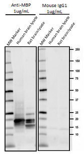
Western blot of anti-Myelin Basic Protein antibody (clone P82H9) and isotype-matched IgG1 control. MW: molecular weight marker; human brain lysates: 20 µg; rat brain lysates: 20 µg. |
α-Synuclein Antibody Sampler Kit |
Tau Antibody Sampler Kit |
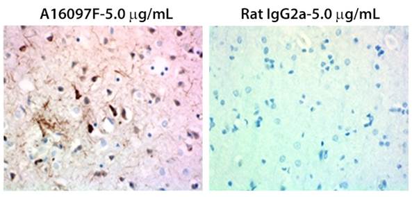
| IHC staining of α-synuclein deposits with purified anti-α-Synuclein, C-Terminal Truncated antibody (clone A15127A) on FFPE Parkinson's disease brain tissue. | IHC staining of anti-Tau, 368-441 antibody (clone A161097F) and rat IgG2a isotype control on FFPE Alzheimer's disease brain tissue. |
| Description | Cat No. | Specificities | Clones |
|---|---|---|---|
| Glial Cell Marker Antibody Sampler Kit | 899904 | P2RY12, CX3CR1, GFAP, Myelin CNPase, Myelin Basic Protein | S16007D, 8E12.D9, SMI 24, SMI 91, P82H9 |
| α-Synuclein Antibody Sampler Kit | 899903 | α-Synuclein Phospho (Tyr39), α-Synuclein Phospho (Ser129), α-Synuclein (80-96), α-Synuclein (C-Terminal Truncated x-122), α-Synuclein (117-122) | A15119B, P-syn/81A, A15115A, A15127A, A15126D |
| Tau Antibody Sampler Kit | 899902 | Tau Phospho (Ser262), Tau Phospho (Thr181), Tau (1-223), Tau (368-441) | A15091A, M7004D06, A16103A, A16097F |
 |
|
189-195 Antibody  Western blot of HRP anti-Tau, 189-195 antibody (clone 39E10). Lane 1: Molecular weight marker; Lane 2: 20 µg of normal human brain lysate; Lane 3: 20 µg of mouse brain lysate; Lane 4: 20 µg of rat brain lysate. The blot was incubated with 1.0 µg/mL of the primary antibody overnight at 4°C. Enhanced chemiluminescence (Cat. No. 426302) was used as the detection system. |
 |
| Peer reviews are important, even for products! You can write a review about a BioLegend neuroscience product in exchange for a $20 Amazon gift card. Write a review... |
*Any references to promotions on this page may not be valid at this time. View our promotions page for the most up-to-date promotions.
 Login / Register


Follow Us