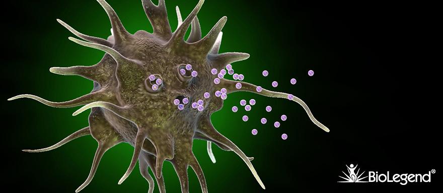
Exosomes are small cell-derived vesicles of 40-100 nm in size and are of endocytic origin. Initially, exosomes were regarded as the vehicles for the removal of unnecessary cellular proteins. Lately, exosomes are considered as important drivers of intercellular communication. Practically every cell secretes exosomes and these nanovesicles are representatives of the cell of origin. Exosomes are found in all biofluids including blood, milk, urine, sweat, tears, and culture supernatant. Explore the biology of exosomes below.


Featured Products

Isolate human exosomes via CD9, CD63, or CD81 with our MojoSort™ Microbead kits. These kits are compatible with a variety of samples and feature short incubation times and no pre-enrichment.


The NEXTFLEX™ Small RNA-Seq Kit v4 offers a simplified exosomal miRNA-Seq library prep protocol with a gel-free workflow. Using this workflow, researchers can achieve a high number of reads aligning to mature miRNA and low adapter dimers, even with the very low miRNA inputs characteristic of exosome samples.


Biogenesis

Early endosomes loaded with ubiquitinated proteins, upon recognition by ESCRT (Endosomal Sorting Complex Required for Transport), allows the formation of intraluminal vesicles (ILVs), which in turn become multivesicular bodies (MVBs), some of which are degraded in lysosomes. The fusion of MVBs with the plasma membrane causes the release of exosomes into the extracellular space.


Composition

Exosomes contain a complex composition of molecules, including proteins, lipids, microRNA, and mRNA, which are cataloged in the EXoCarta database (https://www.exocarta.org/). The most common exosomal proteins are membrane transporters and fusion proteins (Annexins, GTPases and flotillin), heat shock proteins, tetraspanins (CD9, CD63 and CD81), MVB synthesis proteins (Alix and TSG101), lipid-related proteins and phospholipases. Proteins such as CD9, CD63, CD81, TSG101, Alix and HSP70 are common to most exosomes. Exosomes are enriched with lipids like cholesterol, sphingolipids, ceramide, glycolipid GM3, and glycerophospholipids containing long, saturated fatty-acyl chains.


Mechanism of Action

Exosomes play a key role in cell-to-cell communication by merging with a recipient cell. Exosomes may remain stably associated with the plasma membrane or are internalized via an endocytic pathway, releasing their contents. The biological property of the target cell can then be altered at the genetic level (exosomal RNA), epigenetic level (exosomal miRNA) or at the protein level. Beneficial (e.g. enhancing the immune status) or detrimental (e.g. disseminating pathogenesis) outcomes are possible with these interactions. An example of a beneficial outcome is the successful suppression of the maternal immune system during pregnancy that is facilitated by exosomes. Placenta-derived exosomes exhibit immunosuppressive properties by the expression of markers such as Fas ligand (FasL), TRAIL, and PD-L1 inducing T cell death and downregulating NK activity via the expression of NKG2D ligands such as MICA/B and RAEγ1/ULBPs. Other placental exosome-associated proteins include CD247, membranous TGFβ1, placental alkaline phosphatase (PLAP), pregnancy specific glycoprotein 3, and trophoblast glycoprotein 5T4.


Characterization of Exosomes

Analysis of exosomes and their contents can reveal a lot about the host cell and also underlying pathophysiological conditions such as cancer, neurological disorders, heart disease, immunological dysregulation, and infectious disease, to name a few. Therefore, exosomes are regarded as potential biomarkers as well as therapeutic agents. Techniques such as flow cytometry, Western blotting, immunofluorescence, electron microscopy and PCR are extensively used to study both the surface proteins as well as internal exosomal contents. BioLegend provides antibodies to many surface and internal protein targets, which can be used to immunophenotype and molecularly characterize exosomes.

Markers for immune cell derived exosomes


Since exosomal contents can indicate the cell of origin, it is reasonable to predict the immune signature of a particular type of exosome. For example, exosomes derived from professional antigen-presenting cells express MHC I, MHC II, adhesion and costimulatory molecules, such as CD80 and CD86. Similarly, NK cell-derived exosomes enclose perforin and granzyme B.


BioLegend offers a comprehensive list of anti-human, mouse and rat antibodies directed towards a wide variety of protein targets (both surface and intracellular) which can help characterize exosomes via applications such as flow cytometry, Western blot, fluorescent microscopy and IHC. Refer to our cell markers page in order to find immune cell specific markers which in turn can help determine markers for exosomes originating from a particular cell type. The last tab above cites specific BioLegend products that have been used in the characterization of exosomes.

image

Tumor exosomal markers

The direct interaction between tumor cells and their environment plays a key role in tumor progression, for which exosomes play an important role in establishing cell-cell contact. Exosomes released into the extracellular milieu can promote angiogenesis, tumor proliferation, and thrombosis, in addition to the remodeling of the extracellular matrix. Exosomal markers for some of the common cancers are described below.

Disease Exosomal markers
Melanoma CD63, CD146, Caveolin-1, TYRP1, TYRP2, VLA-4, HSP70, HSP90
Glioblastoma CD63, EGFR, Podoplanin, IDH1 (R132H mutant), EGFRVIII
Prostate cancer Survivin, PSA, PCA3
Bladder cancer EGF, CD227 (Mucin-1), Gs alpha subunit, retinoic acid induced protein 3
Breast cancer CD24, CD340(erbB2/HER-2) , CD326 (EpCAM)
Ovarian cancer CD44, CD171, CD24, ADAM10, CD147, claudin, CD326, MMP2 , MMP9, uPA
Gastric cancer CD196 (CCR6), CD340(erbB2/HER-2), CD147 (EMMPRIN), C-MET
Renel cell carcinoma CD49e, CD49f, CD105/Endoglin
Tumor cell proliferation TGF-b1, FGF basic, MCP-1/CCL2, XIAP, SCF
Epithelial to Mesenchymal Transition (EMT) ILK1, Caveolin-1, MMPs, Wnt-11, CD49c

Exosomes in Neurological Disorders


Exosomes are proposed to be novel mediators in the normal development and physiology of the nervous system, playing functional roles during development and regeneration of neurons, and as mediators of cell-cell communication between neurons, oligodendrocytes, and other cells within the CNS.


On the other hand, recent research indicates that exosomes can spread “toxic” forms of mutated or misfolded proteins that are associated with neurodegenerative diseases. These include Amyloid beta, α-Synuclein, and Prion protein (PrP), which are associated with Alzheimer's disease, Parkinson's disease, and Prion disease, respectively.


BioLegend offers antibodies against a wide variety of proteins that can help characterize exosomes originating from various cell types of the CNS.

Publications citing the use of BioLegend products for the characterization of Exosomes

Citation Pubmed Products
Exosomes released from macrophages infected with intracellular pathogens stimulate a proinflammatory response in vitro and in vivo. PubMed Human CD63, CD81, CD86
Cancer exosomes express CD39 and CD73, which suppress T cells through adenosine production. PubMed Human CD39, CD73
Plasma-derived MHC class II+ exosomes from tumor-bearing mice suppress tumor antigen-specific immune responses. PubMed Mouse Alix
CD169 mediates the capture of exosomes in spleen and lymph node. PubMed,
Suppl. Data
Mouse CD24
Exosomes from red blood cell units bind to monocytes and induce proinflammatory cytokines, boosting T-cell responses in vitro. PubMed Human CD142
Human B Cell-Derived Lymphoblastoid Cell Lines Constitutively Produce Fas Ligand and Secrete MHCII+FasL+ Killer Exosomes. PubMed Human MHC II
Exosomes released by islet-derived mesenchymal stem cells trigger autoimmune responses in NOD mice. PubMed Mouse CD63, CD81
Development of glomerulus-, tubule-, and collecting duct-specific mRNA assay in human urinary exosomes and microvesicles. PubMed Human CD63
Antigenic composition of single nano-sized extracellular blood vesicles. PubMed Human CD31, CD63, CD81
Urinary podocyte microparticles identify prealbuminuric diabetic glomerular injury. PubMed Annexin V, mouse Podoplanin
Extracellular vesicles in urine of women with but not without kidney stones manifest patterns similar to men: a case control study. PubMed Human EGFR and MCP-1
Lipoprotein-apheresis reduces circulating microparticles in individuals with familial hypercholesterolemia. PubMed Human CD11b, CD144, CD235a
Microvesicles released from human renal cancer stem cells stimulate angiogenesis and formation of lung premetastatic niche. PubMed Human CD49e, CD49f, CD105, MHC I
Exosomes neutralize synaptic-plasticity-disrupting activity of Aβ assemblies in vivo. PubMed Anti-β-Amyloid
A role for the eIF4E-binding protein 4E-T in P-body formation and mRNA decay. PubMed Anti-HA
Exosomes secreted by cortical neurons upon glutamatergic synapse activation specifically interact with neurons. PubMed Anti-GFP
ProductsHere

Login / Register
Remember me
Forgot your password? Reset password?
Create an Account