MitoSpy™ mitochrondrial localization probes are cell-permeant, fluorogenic chemical reagents used for labeling mitochondria of living cells. They can be used for live-cell imaging applications or they can be fixed to facilitate mounting the specimen with antifade for inverted microscopy or confocal imaging applications. MitoSpy™ Orange CMTMRos and MitoSpy™ Red CMXRos can be fixed and permeabilized to allow for co-staining with additional antibodies. Learn more about each of the MitoSpy™ reagents and the applications each are best-suited for.


MitoSpy™ NIR


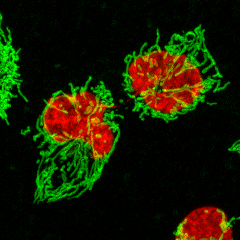
HeLa cells were stained with 0.25µM CytoPhase™ Violet dye (red) for 60 minutes at 37°C. Then 20 nM of MitoSpy™ NIR DiIC1(5) (green) was added for an additional 30 minutes at 37°C. The Z-stack images were captured on an LSM 880 with Airyscan using a 63x Oil objective. The files were Airyscan processed and a Maximum Intensity projection was created using Zen software; image courtesy of the Biophotonics Core Facility at the Salk Institute.


MitoSpy™ Red


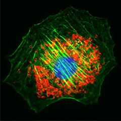
NIH3T3 cells were stained with 100 nM of MitoSpy™ Red CMXRos (red) for 20 minutes at 37°C, fixed with 1% PFA for ten minutes at room temperature, and permeabilized with 1X True Nuclear™ Perm Buffer for ten minutes at room temperature. Then the cells were stained with Flash Phalloidin™ NIR 647 (green) for 20 minutes at room temperature and counterstained with DAPI (blue). The image was captured with a 60x objective.


graph
  • MitoSpy™ Green FM has excitation/emission (ex/em) peaks at 488 nm/520 nm, common for the FITC or Alexa Fluor® 488 filter set. 
  • MitoSpy™ Orange CMTMRos has ex/em peaks of 551 nm/576 nm, common for the PE, TRITC, or Alexa Fluor® 555 filter sets. 
  • MitoSpy™ Red CMXRos has ex/em peaks at 577 nm/598 nm, common for the Texas Red® or Alexa Fluor® 594 filter set. 
  • MitoSpy™ NIR DiIC1(5) has ex/em peaks of 638 nm/658 nm, common for the Alexa Fluor® 647 or APC filter set.

MitoSpy™ Orange, MitoSpy™ Red, and MitoSpy™ NIR localize to the mitochondria based on its membrane potential. This potential is created by the transport of ions across the membrane. Thus, MitoSpy™ Orange, MitoSpy™ Red, and MitoSpy™ NIR work well to indicate cell health as well as localization. On the other hand, MitoSpy™ Green FM’s attraction to the mitochondria is not based on membrane potential. While the full mechanism of MitoSpy™ Green FM is not yet understood, it may have to do with homology between the reagent’s structure and proteins at the mitochondrial surface. Because of this reagent’s membrane potential independence, it can be used to measure mitochondrial mass of individual cells in flow cytometry.

image 1


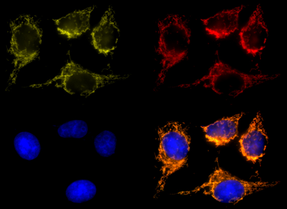
HeLa cells stained with MitoSpy™ Orange (yellow), fixed and permeabilized with 4% PFA and 0.1% Triton X-100 and stained with Cytochrome C Alexa Fluor® 647 (red) and DAPI (blue).


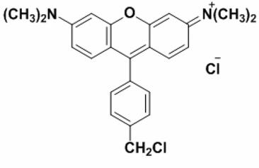
MitoSpy™ Orange structure.

MitoSpy™ Orange structure.

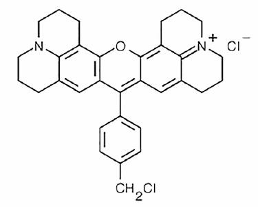
MitoSpy™ Red structure.

MitoSpy™ Red structure.

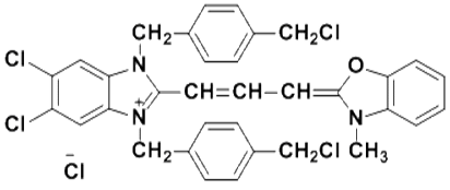
MitoSpy™ Green structure.

MitoSpy™ Green structure.

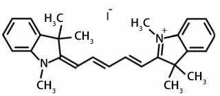
MitoSpy™ NIR structure.

MitoSpy™ NIR structure.

MitoSpy™ Orange and MitoSpy™ Red contain a chloromethyl group that covalently attaches to cysteines in the cell. This gives them a higher probability of remaining in a cell after the sample is fixed and permeabilized. However, as the fix/perm process washes out a large amount of the reagent, a much higher concentration of MitoSpy™ Orange and MitoSpy™ Red is required to get sufficient staining.

After the fix/perm process, most MitoSpy™ Green and MitoSpy™ NIR will be washed away. As such, they are not recommended to be used with samples that require fixation and permeabilization.

*Note that all MitoSpy™ products must be used on live samples. The recommendations below reflect whether MitoSpy™ is successfully retained following the "Retention Condition" treatment.

Retention Condition* Recommended MitoSpy™ Orange Concentration Recommended MitoSpy™ Red Concentration Recommended MitoSpy™ Green Concentration Recommended MitoSpy™ NIR Concentration
Live Cells 50-250 nM 50-250 nM 50-250 nM 1-50 nM
Fixed Cells 50-250 nM 50-250 nM 50-250 nM Not recommended.
Fixed and Permeabilized Cells 250-500 nM 250-500 nM Not recommended. Not recommended.
MitoSpy™ Orange Live staining (50 nM)
PFA-fixed and permeabilized staining (500 nM)
MitoSpy™ Green

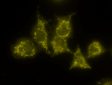
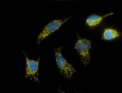
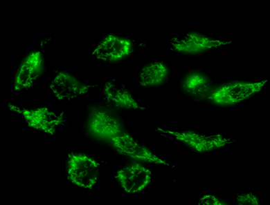
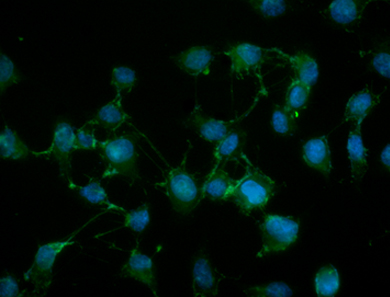
HeLa cells that were stained live with either MitoSpy™ Orange (yellow) or Green (green). Cells that were fixed and permeabilized with 4% PFA and 0.1% Triton X-100 were also stained with DAPI (blue). Photos were taken with a 60x magnification.

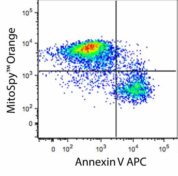
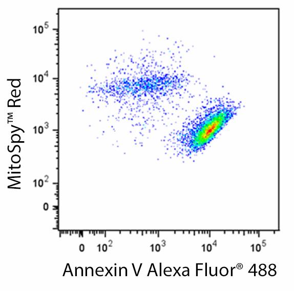
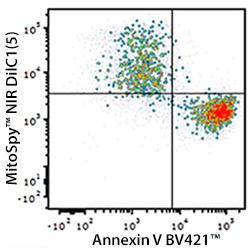
MitoSpy™ OrangeMitoSpy™ Red, and MitoSpy™ NIR can also be used as an indicator of cellular health. When the mitochondria are actively respiring, there is a potential difference across the mitochondrial membrane that is called membrane polarization. If a cell is unhappy due to apoptosis or cell death, this probe will not be strongly attracted to the mitochondria of that cell. In the image below, cells positive for MitoSpy™ Orange, MitoSpy™ Red, MitoSpy™ NIR are alive and healthy, while Annexin V positive events are at the beginning stages of apoptosis. In this flow cytometry application, the MitoSpy™ Orange, MitoSpy™ Red, and MitoSpy™ NIR should not be fixed prior to analysis since there is a substantial loss of reagent with fixation.  An apoptotic phenotype is indicated by a low or negative MitoSpy™ Orange, MitoSpy™ Red, or MitoSpy™ NIR signal.  If reagent is lost with fixation, the resulting loss of signal intensity will confound the ability to accurately phenotype apoptosis.  Fixation of MitoSpy™ Orange and MitoSpy™ Red is only applicable for subcellular localization in imaging applications. Fixation is not recommended at all with MitoSpy™ NIR.

annexin image
annexin v alexa fluor
annexin v bv421

Human T-cell leukemia cell line, Jurkat, was treated for 5 hours with LEAF™ purified anti-CD95 (clone EOS9.1), then stained with an impermeant nucleic acid stain, APC, Alexa Fluor® 488, or Brilliant Violet 421™ Annexin V, and either MitoSpy™ Orange, Red, or NIR as indicated. Nucleic acid stain positive events were excluded from analysis.

ProductsHere

Login / Register
Remember me
Forgot your password? Reset password?
Create an Account