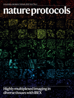
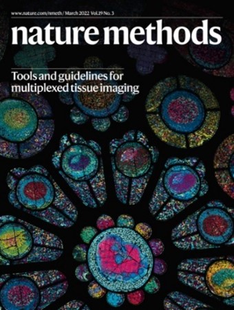
Traditional multiplexed immunofluorescence microscopy is limited by the number of spectrally unique fluorophores that can be imaged simultaneously; 2-7 parameters with common imaging systems. IBEX (Iterative Bleaching Extends multi-pleXity) increases the total number of parameters that can be imaged on a single sample using iterative cycles of antibody labeling and fluorophore inactivation. Importantly, the described method—developed by Dr. Andrea Radtke and colleagues in the laboratory of Dr. Ronald Germain (Center for Advanced Tissue Imaging, Lymphocyte Biology Section, NIAID, NIH)—requires no specialized equipment or proprietary software, making highly-multiplexed microscopy (60+ parameters) more accessible.
The use of information and images from Dr. Radtke and Germain lab colleagues on these informational pages does not imply any endorsement of BioLegend or its products by the US Government.
Image banners and collages generously provided by Dr. Andrea Radtke and Dr. Hiroshi Ichise of the Lymphocyte Biology Section in the National Institute of Allergy and Infectious Diseases (NIAID, NIH).
 Login / Register


Follow Us