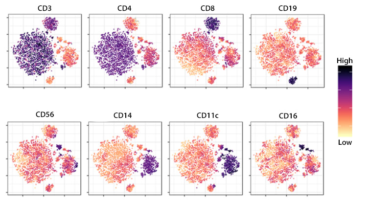
Understanding the role of different immune cells, including how they function to protect against infection and disease, is critical to develop effective treatments for conditions such as autoimmunity, allergy, and cancer. Methods used for cell analysis typically involve identifying individual cells within a heterogeneous population via the detection of cell surface markers or other cell type-specific biomolecules. While cell analysis techniques have traditionally provided either a proteomic or genomic readout, it is increasingly common for these to be combined for multiomic analysis. Where samples such as blood, serum, or tissue biopsy material are derived from both normal and diseased hosts, comparative studies can help reveal mechanisms of disease pathogenesis.


Flow Cytometry and Fluorescence-Activated Cell Sorting (FACS)


Flow cytometry is a technique for characterizing individual cells in suspension based on light scatter and the binding of fluorophore-labeled antibodies to cellular markers. Using a stream of fluid to direct the cells in single file past an interrogation point, it measures key cellular properties that enable identification. Cluster of Differentiation (CD) markers are among the most widely used marker types for flow cytometry and it is common practice for multiple CD markers to be measured in combination. Fluorescence-activated cell sorting (FACS) also involves detecting individual cells in suspension. However, unlike flow cytometry, FACS allows researchers to isolate specific target cells for further downstream studies such as functional assays or clonal expansion.

Multiomics


TotalSeq™ reagents are our antibody-oligonucleotide conjugates that enable protein detection by sequencing at a single-cell level. They can be integrated into existing single-cell transcriptomics, bulk sequencing, or single-cell genomics workflows to reveal distinct immune cell populations that might otherwise be overlooked using techniques such as flow cytometry or single-cell RNA sequencing (scRNA-seq) alone. TotalSeq™ reagents are available as a broad range of human and mouse targets and also include TotalSeq™ Human Universal Cocktails (available in TotalSeq™-A, -B, and -C formats) that each contain over 125 antibodies and their associated isotype controls for a deeper look at immune cells.


Multiomics allows for a greater level of cell characterization.

Magnetic Bead-Based Separation


Magnetic bead-based separation uses antibodies conjugated to magnetic particles for capturing cells from suspension. Following incubation of the sample with the beads, application of a magnetic field separates the cell type of interest from the rest of the population, enriching it for use in downstream applications. An alternative approach to using antibody-conjugated beads is to use biotin-antibody conjugates followed by streptavidin-conjugated magnetic particles. MojoSort™ is our magnetic bead-based separation system comprising magnetic beads conjugated to antibodies or streptavidin. Additionally, the MojoSort™ product range includes magnets, nanobeads for positive selection, and isolation kits (comprised of a biotin-antibody cocktail and streptavidin-conjugated beads) for negative selection by isolating an untouched cell population.

Microscopy


Microscopy-based imaging of cells (immunocytochemistry) and tissues (immunohistochemistry) allows researchers to visualize detail in objects too small to see with the naked eye. It relies on antibodies conjugated to enzymes or fluorophores for detection of specific cellular markers, with many microscopy platforms supporting digital image capture and analysis. While chromogenic detection methods offer the advantage of signal amplification, they are typically used to image only a single marker at a time. Fluorescence-based detection instead allows visualization of multiple markers simultaneously to reveal insights into cellular abundance and localization relative to other cell types present in the sample.

Flex-T™ MHC Tetramers


Being able to identify antigen-specific T cells is critically important to many different fields of research. For example, detecting melanoma-specific CD8+ T cells from a solid tumor in peripheral blood can inform disease progression. To streamline the process of identifying antigen-specific T cells, we developed Flex-T™. Based on the interaction between antigen-presenting cells (APCs) and T cells, through the major histocompatibility complex (MHC) and the T cell receptor (TCR), Flex-T™ comprises MHC monomers loaded with a peptide that can be degraded with a UV light source. To use Flex-T™, researchers simply combine the sample with the loaded monomers then degrade the labile peptide to enable its substitution with the target of interest; the peptide-exchanged monomers are then mixed with fluorophore-labeled streptavidin to form tetramers that can be detected via flow cytometry.


ProductsHere

Login / Register
Remember me
Forgot your password? Reset password?
Create an Account