- Clone
- M5203F01 (See other available formats)
- Regulatory Status
- RUO
- Other Names
- ANG-2, ANGPT2, AGPT2
- Isotype
- Mouse IgG2b, κ
- Ave. Rating
- Submit a Review
- Product Citations
- publications

-
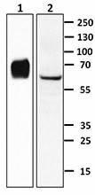
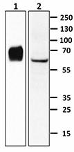
Recombinant human angiopoietin-2 (50 ng, lane 1) and HUVEC cell lysate (30 µg, lane 2) were resolved by electrophoresis, transferred to nitrocellulose, and probed with purified monoclonal anti-Angiopoietin-2 (clone M5203F01) antibody. Proteins were visualized using a goat anti-mouse-IgG secondary antibody conjugated to HRP and chemiluminescence detection. Exposure time was ten seconds for lane 1 and five minutes for lane 2.
| Cat # | Size | Price | Quantity Check Availability | Save | ||
|---|---|---|---|---|---|---|
| 682702 | 100 µg | 212€ | ||||
Angiopoietin-2, also known as ANGPT2, belongs to a family of vascular growth factors. It plays essential roles in embryonic and postnatal angiogenesis. The structure of the mature form of Angiopoietin-2 contains an N-terminal coiled-coil domain, which mediates multimerization of the protein, along with a C-terminal fibrinogen-like domain, which mediates the receptor binding. Both Angiopoietin-1 and Angiopoietin-2 are ligands for the receptor tyrosine kinase, Tie-2. Angiopoietin-2 appears to exist predominantly as a homodimer, but it is also capable of forming multimers while Angiopoietin-1 predominantly exists as multimers. It has been shown that Angiopoietin-2 is able to promote vascular leakage and neutrophil migration. It can also sensitize TNF-α-induced monocyte adhesion. Thus, it is considered a proinflammatory factor. Although Angiopoietin-2 is not required for arterial vascular development, its expression in the tissue suggests that it may play a role in the maintenance of these vessels. This conclusion is also supported by the observation that high concentrations of Angiopoietin-2 activate the same survival signaling pathway as Angiopoietin-1 to prevent endothelial cell apoptosis via Tie-2/Akt. Additionally, Angiopoietin-2 functions as an agonist in the absence of Angiopoietin-1, but it acts as an antagonist in its presence. Human Angiopoietin-2 shares an 86% amino acid sequence homology with its murine counterpart.
Product DetailsProduct Details
- Verified Reactivity
- Human
- Antibody Type
- Monoclonal
- Host Species
- Mouse
- Immunogen
- Human Angiopoietin-2, amino acids (Tyr19-Phe496) (Accession# NP_001138), was expressed with a C-terminal His tag in CHO cells.
- Formulation
- Phosphate-buffered solution, pH 7.2, containing 0.09% sodium azide.
- Preparation
- The antibody was purified by affinity chromatography.
- Concentration
- 0.5 mg/ml
- Storage & Handling
- The antibody solution should be stored undiluted between 2°C and 8°C.
- Application
-
WB - Quality tested
- Recommended Usage
-
Each lot of this antibody is quality control tested by Western blotting. For Western blotting, the suggested use of this reagent is 1.0 - 2.5 µg per ml. It is recommended that the reagent be titrated for optimal performance for each application.
- RRID
-
AB_2566672 (BioLegend Cat. No. 682702)
Antigen Details
- Structure
- Oligomer.
- Distribution
-
Secreted protein, low abundance in nucleus. Placenta, uterus, ovaries, and vascular endothelial cells.
- Function
- Angiopoietin-2 plays a role in angiogenesis and monocyte adhesion. Hyperoxia induces ANGPT2 expression in the lung epithelial cells.
- Interaction
- Monocytes and endothelial cells.
- Ligand/Receptor
- Tie-2.
- Biology Area
- Angiogenesis, Cell Adhesion, Cell Biology, Immunology, Innate Immunity, Signal Transduction
- Molecular Family
- Cytokines/Chemokines, Growth Factors
- Antigen References
-
1. Augustin HG, et al. 2009. Nat. Rev. Mol. Biol. 10:165.
2. Ward NL, Dumont DJ. 2002. Semin. Cell Dev. Biol. 1:19-27.
3. Roviezzo F, et al. 2005. J. Pharmacol. Exp. Ther. 314:738.
4. Sturn DH, et al. 2005. Microcirculation 12:393.
5. Murdoch C, et al. 2007. J. Immunol. 178:7405.
6. Yuan HT, et al. 2009. Mol. Cell Biol. 29:2011.
7. Procopio WN, et al. 1999. J. Biol. Chem. 274:30196.
8. Bhandari V, et al. 2006. Nature Med. 12:1286. - Gene ID
- 285 View all products for this Gene ID
- UniProt
- View information about Angiopoietin-2 on UniProt.org
Related Pages & Pathways
Pages
Related FAQs
Other Formats
View All Angiopoietin-2 Reagents Request Custom Conjugation| Description | Clone | Applications |
|---|---|---|
| Purified anti-Angiopoietin-2 | M5203F01 | WB |
Compare Data Across All Formats
This data display is provided for general comparisons between formats.
Your actual data may vary due to variations in samples, target cells, instruments and their settings, staining conditions, and other factors.
If you need assistance with selecting the best format contact our expert technical support team.
-
Purified anti-Angiopoietin-2
Recombinant human angiopoietin-2 (50 ng, lane 1) and HUVEC c...
 Login / Register


Follow Us