 Login / Register


- Clone
- W16101A (See other available formats)
- Regulatory Status
- RUO
- Other Names
- Phosphatidylinositol 3-kinase regulatory subunit alpha, PI3-kinase subunit p85-alpha, Phosphatidylinositol 3-kinase 85 kDa regulatory subunit alpha, GRB1,
- Isotype
- Rat IgG2a, κ
- Ave. Rating
- Submit a Review
- Product Citations
- publications

-

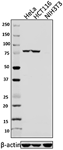
Total cell lysates (15µg protein) from Jurkat, HeLa (negative control) and NIH3T3 cells were resolved by 4-12% Bis-Tris gel electrophoresis, transferred to nitrocellulose, and probed with 17µg/mL purified anti-PIK3R1 (clone W16101A) antibody and competitor’s antibody used at manufactures recommended concentration (upper). Proteins were visualized by chemiluminescence detection using a goat anti-rat-IgG secondary antibody conjugated to HRP for clone W16101A and a donkey anti-rabbit IgG Antibody conjugated to HRP for competitor’s antibody. Direct-Blot™ HRP anti-β-actin Antibody was used as a loading control (lower). Lane M: Molecular Weight ladder. -

NIH3T3 cells were fixed with 4% paraformaldehyde (PFA) for 15 minutes, permeabilized with 0.5% Triton X-100 for 3 minutes, and blocked with 5% FBS for 60 minutes. Then the cells were intracellularly stained with 2 µg/ml anti-PIK3R1 antibody (clone W16101A) overnight at 4°C followed by Alexa Fluor® 594 (red) conjugated goat anti-rat IgG for one hour at room temperature. Nuclei were counterstained with DAPI (blue). The image was captured with a 60X objective.
| Cat # | Size | Price | Quantity Check Availability | Save | ||
|---|---|---|---|---|---|---|
| 697102 | 100 µg | 226€ | ||||
Phosphatidylinositol 3-kinase (PI3K) is a heterodimer comprised of a catalytic subunit encoded by PIK3CA (p110α) and one of a number of regulatory subunits. PIK3R1, also known as PI3K p85-alpha, is an 85kD regulatory subunit of PI3K. Upon activation of the receptor tyrosine kinases, PI3K phosphorylates inositol lipids to phosphatidylinositol-3,4,5-trisphosphate (PIP3), subsequently leads to activation of AKT and downstream effectors. The PI3K signaling regulates various cellular processes such as protein synthesis, cell growth, cell cycle progression, cell proliferation, angiogenesis, and survival. In addition, PI3K pathway has been found to play important roles in tumorigenesis and cancer progression. PIK3R1 appears to be a tumor suppressor due to its ability to stabilize the catalytic subunit of PI3K. Mutations in PIK3R1 have been associated with the autosomal dominant disorder SHORT syndrome.
Product DetailsProduct Details
- Verified Reactivity
- Human, Mouse
- Antibody Type
- Monoclonal
- Host Species
- Rat
- Immunogen
- Human PIK3R1 recombinant protein (1-250 a.a.) expressed in E. coli.
- Formulation
- Phosphate-buffered solution, pH 7.2, containing 0.09% sodium azide.
- Preparation
- The antibody was purified by affinity chromatography.
- Concentration
- 0.5 mg/ml
- Storage & Handling
- The antibody solution should be stored undiluted between 2°C and 8°C.
- Application
-
WB - Quality tested
ICC - Verified - Recommended Usage
-
Each lot of this antibody is quality control tested by Western blotting. For Western blotting, the suggested use of this reagent is 0.2 - 2.0 µg per ml. For immunocytochemistry, a concentration range of 0.5 - 5.0 µg per ml is recommended. It is recommended that the reagent be titrated for optimal performance for each application.
- Application Notes
-
This antibody may not react with the p85 beta based on sequence homology.
- RRID
-
AB_2687110 (BioLegend Cat. No. 697102)
Antigen Details
- Structure
- 724 amino acids with a predicted molecular weight of 83.6 kD. Contains an N-terminal SH3 domain, a Rho-GAP domain, and two C-terminal SH2 domains.
- Distribution
-
Nucleus, cytoplasm, plasma membrane, ER membrane.
- Function
- PIK3R1 is a regulatory subunit of phosphatidylinositol 3-kinase and is involved in the signaling regulating protein synthesis, cell growth, cell cycle progression, cell proliferation, angiogenesis, and survival.
- Interaction
- Forms a heterodimer with the catalytic subunit of PIK3. Interacts with CSF1R, PIK3R2, XBP1, FER, TEK, PTK2, TOM1L1, LIME1, SOCS7, RUFY3, LYN, CBLB, IRS1, IRS4, FGR, HCK, ERBB4, and NTRK1.
- Biology Area
- Cell Biology, Signal Transduction
- Molecular Family
- Protein Kinases/Phosphatase
- Antigen References
-
1. Munkley J, et al. 2015. Oncoscience. 2:755.
2. Huang XP, et al. 2015. FEBS J. 282:579.
3. Wei Y, et al. 2013. Proc. Natl. Acad. Sci. USA. 110:6829.
4. Najib S, et al. 2012. Mol. Cell. Biol. 32:1004.
5. Anderson DH. 2010. Cell Cycle. 9:2055.
6. De Gregorio G, et al. 2007. Oncogene. 26:2039. - Gene ID
- 5295 View all products for this Gene ID
- UniProt
- View information about PIK3R1 on UniProt.org

Related FAQs
Other Formats
View All PIK3R1 Reagents Request Custom Conjugation| Description | Clone | Applications |
|---|---|---|
| Purified anti-PIK3R1 | W16101A | WB,ICC |
Customers Also Purchased
_Antibody_1_WB_062116.jpg)
Compare Data Across All Formats
This data display is provided for general comparisons between formats.
Your actual data may vary due to variations in samples, target cells, instruments and their settings, staining conditions, and other factors.
If you need assistance with selecting the best format contact our expert technical support team.


Follow Us