- Clone
- W18344B (See other available formats)
- Regulatory Status
- RUO
- Other Names
- VASP, Vasodilator Stimulated Phosphoprotein, Vasodilator-Stimulated Phosphoprotein.
- Isotype
- Mouse IgG2b, κ
- Ave. Rating
- Submit a Review
- Product Citations
- publications

-

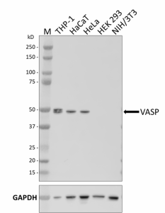
Whole cell extracts (15 µg total protein) from the indicated cell lines were resolved by 4-12% Bis-Tris gel electrophoresis, transferred to a PVDF membrane, and probed with 1 µg/mL of purified anti-VASP antibody (clone W18344B) overnight at 4°C. Proteins were visualized by chemiluminescence detection using HRP goat anti-mouse IgG antibody (Cat. No. 405306) at a 1:3000 dilution. Direct-Blot™ HRP anti-GAPDH antibody (Cat. No. 607904) was used as a loading control at a 1:50000 dilution (lower). Lane M: Molecular weight marker. -

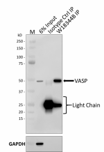
Whole cell extracts (250 µg total protein) prepared from HeLa cells were immunoprecipitated overnight with 2.5 µg of purified mouse IgG2b, κ isotype ctrl antibody (Cat. No. 400302) or purified anti-VASP antibody (clone W18344B). The resulting IP fractions and whole cell extract input (6%) were resolved by 4-12% Bis-Tris gel electrophoresis, transferred to a PVDF membrane and probed with the same purified anti-VASP antibody (overnight at 4°C). Proteins were visualized by chemiluminescence detection using an anti-mouse HRP light-chain specific antibody. Direct-Blot™ HRP anti-GAPDH antibody (Cat. No. 607904) was used as a loading control at a 1:50000 dilution. Lane M: Molecular weight marker. -

HeLa cells were fixed with 4% paraformaldehyde for 10 minutes, permeabilized with methanol for 10 minutes at -20°C, and blocked with 5% FBS for 60 minutes. Cells were then intracellularly stained with 2 µg/mL of either purified mouse IgG2b, κ isotype ctrl antibody (Cat. No. 400302) (panel A) or purified anti-VASP antibody (clone W18344B) (panel B) overnight at 4°C, followed by incubation with Alexa Fluor® 594 goat anti-mouse IgG antibody (Cat. No. 405326) at 2.5 µg/mL. Nuclei were counterstained with DAPI and the image was captured with a 60X objective.
| Cat # | Size | Price | Quantity Check Availability | Save | ||
|---|---|---|---|---|---|---|
| 937501 | 25 µg | 81€ | ||||
| 937502 | 100 µg | 203€ | ||||
Vasodilator-stimulated phosphoprotein (VASP) is part of the Ena-VASP protein family of adaptor proteins which link the cytoskeletal system to signal transduction pathways. The Ena-VASP proteins are involved in cytoskeletal organization, fibroblast migration, platelet activation and axon guidance. VASP interacts with filametous actin formation, so it plays a role in cell adhesion and motility. VASP has three phosphorylation sites, and VASP phosphorylation has been shown to affect actin binding and its effects on actin polymerization. Diseases associated with VASP include thrombosis and clopidogrel resistance.
Product DetailsProduct Details
- Verified Reactivity
- Human
- Antibody Type
- Monoclonal
- Host Species
- Mouse
- Immunogen
- Partial recombinant human VASP
- Formulation
- Phosphate-buffered solution, pH 7.2, containing 0.09% sodium azide
- Preparation
- The antibody was purified by affinity chromatography.
- Concentration
- 0.5 mg/mL
- Storage & Handling
- The antibody solution should be stored undiluted between 2°C and 8°C.
- Application
-
WB - Quality tested
ICC, IP - Verified - Recommended Usage
-
Each lot of this antibody is quality control tested by western blotting. For western blotting, the suggested use of this reagent is 0.25 - 1.0 µg/mL. For immunocytochemistry, a concentration range of 1 - 2 μg/mL is recommended. For immunoprecipitation, the suggested use of this reagent is 2.5 µg/test. It is recommended that the reagent be titrated for optimal performance for each application.
- Application Notes
-
Clone recognizes human VASP.
Clone works well in ICC when cells are permeabilized with either Triton X-100 or methanol.
VASP antibody detects endogenous levels of total VASP protein. In certain cell lines 2 bands running at 46 kD and 50 kD can be detected, which correspond to the non-phosphorylated and phosphorylated forms of VASP, respectively. -
Application References
(PubMed link indicates BioLegend citation) -
- Reinhard M, et al. 1992. EMBO J. 11:2063-2070.
- RRID
-
AB_2876753 (BioLegend Cat. No. 937501)
AB_2876753 (BioLegend Cat. No. 937502)
Antigen Details
- Structure
- VASP is a 380 amino acid protein with a predicted molecular weight of 39 kD. Western blot shows two bands at 46 and 50 kD corresponding to non-phosphorylated and phosphorylated VAST respectively.
- Distribution
-
VASP is mainly localized to the plasma membrane with additional localization to the focal adhesion sites and cell junctions.
- Function
- Cell adhesion, cell motility
- Interaction
- Actin
- Ligand/Receptor
- Robo receptor and Sertoli-Sertoli Cell Junction Dynamics
- Antigen References
-
Wilton KM and Billadeau. 2018. J Immunol. 10:2899-2909
Harker AJ, et al. 2019. Mol Biol Cell. 7:851-62
Bear JE, et al. 2002. Cell. 109:509.
Reinhard M, et al. 1992. EMBO J. 11:2063. - Gene ID
- 7408 View all products for this Gene ID
- UniProt
- View information about VASP on UniProt.org
Related Pages & Pathways
Pages
Other Formats
View All VASP Reagents Request Custom Conjugation| Description | Clone | Applications |
|---|---|---|
| Purified anti-VASP | W18344B | WB,IP,ICC |
Compare Data Across All Formats
This data display is provided for general comparisons between formats.
Your actual data may vary due to variations in samples, target cells, instruments and their settings, staining conditions, and other factors.
If you need assistance with selecting the best format contact our expert technical support team.
 Login / Register


Follow Us