- Regulatory Status
- RUO
- Other Names
- CSF1, CSF-1, MCSF
- Ave. Rating
- Submit a Review
- Product Citations
- publications

-

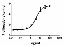
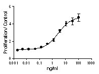
M-NFS-60 cell proliferation induced by mouse M-CSF. -

Recombinant mouse M-CSF induces proliferation of M-NFS-60 cells in a dose dependent manner. BioLegend’s protein was compared side-by-side to a competitor’s equivalent product.
M-CSF was first characterized as a glycoprotein that induces monocyte and macrophage colony formation from precursors in murine bone marrow cultures. M-CSF binds CD14+ monocytes and promotes the survival/proliferation of peripheral blood monocytes. In addition, M-CSF enhances inducible monocyte functions including phagocytic activity, microbial killing, and cytotoxicity for tumor cells as well as induces the synthesis of inflammatory cytokines such as IL-1, TNFα, and IFNγ in monocytes.
Multiple CSF1 mRNA species have been described that arise from alternative splicing in exon 6 and the alternative use of the 3’ end of exons 9 or 10. As a result, two distinct CSF1 protein products are encoded by these transcripts: a cell-surface or membrane-bound form of CSF1 (mCSF1) and a soluble form (sCSF1). Uterine sCSF1 is highly increased during pregnancy. On the contrary, uterine mCSF1 remains low during pregnancy. High levels of M-CSF have been associated to different pathologies such as pulmonary fibrosis and atherosclerosis.
M-CSF binds to its receptor M-CSFR, and this receptor is shared by a second ligand, IL-34. Mouse M-CSF and IL-34 exhibit cross-species specificity, both bind to the human and mouse M-CSF receptors. IL-34 can regulate myeloid development and substitute for CSF-1 in vivo. IL-34 has overlapping but not identical biological activities as M-CSF.
Product Details
- Source
- Mouse M-CSF, amino acids Lys33-Glu262 (Accession# NM_001113530.1) was expressed in 293E cells.
- Molecular Mass
- The 251 amino acid recombinant protein has a predicted molecular mass of approximately 28.2 kD. The DTT-reduced and non-reduced protein migrate at approximately 50 kD and 100 kD respectively by SDS-PAGE. The N-terminal contains a His9-(SGGG)2-IEGR-tag.
- Purity
- >98%, as determined by Coomassie stained SDS-PAGE.
- Formulation
- 0.22 µm filtered protein solution is in PBS.
- Endotoxin Level
- Less than 0.01 ng per µg cytokine as determined by the LAL method.
- Concentration
- 10 and 25 µg sizes are bottled at 200 µg/mL. 100 µg size and larger sizes are lot-specific and bottled at the concentration indicated on the vial. To obtain lot-specific concentration and expiration, please enter the lot number in our Certificate of Analysis online tool.
- Storage & Handling
- Unopened vial can be stored between 2°C and 8°C for up to 2 weeks, at -20°C for up to six months, or at -70°C or colder until the expiration date. For maximum results, quick spin vial prior to opening. The protein can be aliquoted and stored at -20°C or colder. Stock solutions can also be prepared at 50 - 100 µg/mL in appropriate sterile buffer, carrier protein such as 0.2 - 1% BSA or HSA can be added when preparing the stock solution. Aliquots can be stored between 2°C and 8°C for up to one week and stored at -20°C or colder for up to 3 months. Avoid repeated freeze/thaw cycles.
- Activity
- ED50 =2 - 6 ng/ml, corresponding to a specific activity of 1.6 - 5 x 10⁵ units/mg, as determined by M-NFS60 cell proliferation induced by mouse M-CSF in a dose dependent manner.
- Application
-
Bioassay
- Application Notes
-
BioLegend carrier-free recombinant proteins provided in liquid format are shipped on blue-ice. Our comparison testing data indicates that when handled and stored as recommended, the liquid format has equal or better stability and shelf-life compared to commercially available lyophilized proteins after reconstitution. Our liquid proteins are verified in-house to maintain activity after shipping on blue ice and are backed by our 100% satisfaction guarantee. If you have any concerns, contact us at tech@biolegend.com.
- Product Citations
-
Antigen Details
- Structure
- Disulfide-linked glycosylated homodimer.
- Distribution
-
M-CSF is broadly expressed in adult mouse tissues. M-CSF is released by fibroblasts, breast cancer cell lines, alveolar macrophages, stromal bone marrow cells, endothelial cells, and mesenchymal cells.
- Function
- M-CSF is the key regulator of the survival, proliferation, and differentiation of mononuclear phagocytes and plays a central role in the regulation of osteoclastogenesis. CSF-1 also regulates the development of Paneth cells, Langerhans cells, lamina propria dendritic cells, and microglia.
- Interaction
- Monocytes, macrophages, mononuclear phagocyte precursors, microglia, proliferating smooth muscle cells, umbilical vein endothelial cells, and breast cancer cell lines.
- Ligand/Receptor
- M-CSFR or CSF1R (CD115)
- Cell Type
- Embryonic Stem Cells, Hematopoietic stem and progenitors
- Biology Area
- Cell Biology, Cell Proliferation and Viability, Immunology, Stem Cells
- Molecular Family
- Cytokines/Chemokines, Growth Factors
- Antigen References
-
1. Kawasaki ES, et al. 1985. Science 230:291.
2. Wei S, et al. 2010. J. Leukocyte Biol. 88:495.
3. MacDonald KP, et al. 2010. Blood 116:3955.
4. Hodge JM, et al. 2011. Plos One 6:e21462.
5. Morandi et al. 2011. Plos One 6:e27450.
6. Erblich B, et al. 2011. Plos One 6:e26317. - Gene ID
- 12977 View all products for this Gene ID
- UniProt
- View information about M-CSF on UniProt.org
Related FAQs
- Why choose BioLegend recombinant proteins?
-
• Each lot of product is quality-tested for bioactivity as indicated on the data sheet.
• Greater than 95% Purity or higher, tested on every lot of product.
• 100% Satisfaction Guarantee for quality performance, stability, and consistency.
• Ready-to-use liquid format saves time and reduces challenges associated with reconstitution.
• Bulk and customization available. Contact us.
• Learn more about our Recombinant Proteins. - How does the activity of your recombinant proteins compare to competitors?
-
We quality control each and every lot of recombinant protein. Not only do we check its bioactivity, but we also compare it against other commercially available recombinant proteins. We make sure each recombinant protein’s activity is at least as good as or better than the competition’s. In order to provide you with the best possible product, we ensure that our testing process is rigorous and thorough. If you’re curious and eager to make the switch to BioLegend recombinants, contact your sales representative today!
- What is the specific activity or ED50 of my recombinant protein?
-
The specific activity range of the protein is indicated on the product datasheets. Because the exact activity values on a per unit basis can largely fluctuate depending on a number of factors, including the nature of the assay, cell density, age of cells/passage number, culture media used, and end user technique, the specific activity is best defined as a range and we guarantee the specific activity of all our lots will be within the range indicated on the datasheet. Please note this only applies to recombinants labeled for use in bioassays. ELISA standard recombinant proteins are not recommended for bioassay usage as they are not tested for these applications.
- Have your recombinants been tested for stability?
-
Our testing shows that the recombinant proteins are able to withstand room temperature for a week without losing activity. In addition the recombinant proteins were also found to withstand four cycles of freeze and thaw without losing activity.
- Does specific activity of a recombinant protein vary between lots?
-
Specific activity will vary for each lot and for the type of experiment that is done to validate it, but all passed lots will have activity within the established ED50 range for the product and we guarantee that our products will have lot-to-lot consistency. Please conduct an experiment-specific validation to find the optimal ED50 for your system.
- How do you convert activity as an ED50 in ng/ml to a specific activity in Units/mg?
-
Use formula Specific activity (Units/mg) = 10^6/ ED50 (ng/mL)
 Login / Register


Follow Us