
 Login / Register


- Clone
- PHF-6 (See other available formats)
- Regulatory Status
- RUO
- Other Names
- Microtubule-associated protein tau, PHF-tau, paired helical filament-tau, neurofibrillary tangle protein, microtubule-associated protein tau, isoform 4, G protein beta1/gamma2 subunit-interacting factor 1
- Previously
-
Covance Catalog# MMS-545P
- Isotype
- Mouse IgG1, κ
- Ave. Rating
- Submit a Review
- Product Citations
- 2 publications


| Cat # | Size | Price | Quantity Check Availability | Save | ||
|---|---|---|---|---|---|---|
| 828903 | 25 µL | 90€ | ||||
| 828901 | 100 µL | 286€ | ||||
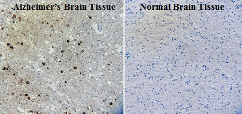
Paired helical filaments (PHFs) are the major building blocks of neurofibrillary lesions in Alzheimer's disease brains and are composed of hyperphosphorylated tau protein. Predominantly expressed in axons, alternatively spliced forms of tau comprise a family of microtubule-associated proteins that normally promote and stabilize the assembly of microtubules. PHF-tau differs from normal tau by its abnormal hyperphosphorylation, which results in decreased binding of tau to microtubules. The decreased affinity of PHF-tau for microtubules, coupled with reduced levels of normal tau, destabilizes microtubules leading to an impairment of axonal transport, neuronal death and the aggregation of PHFs. Therefore, hyperphosphorylation of tau is believed to be a key event in the pathogenesis of Alzheimer's disease.
Product DetailsProduct Details
- Verified Reactivity
- Human
- Antibody Type
- Monoclonal
- Host Species
- Mouse
- Immunogen
- The PHF-6 antibody was raised using PHF-tau preparations from human brain.
- Formulation
- Phosphate-buffered solution; no preservatives or carrier proteins.
- Preparation
- The antibody was purified by affinity chromatography.
- Concentration
- 1.0 mg/mL
- Storage & Handling
- This antibody should be handled aseptically as it is free of preservatives such as Sodium Azide. Store this antibody undiluted between 2°C and 8°C. Please note the storage condition for this antibody has been changed from -20°C to between 2°C and 8°C. You can also check the vial label or CoA to find the proper storage conditions.
- Application
-
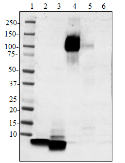
WB - Quality tested
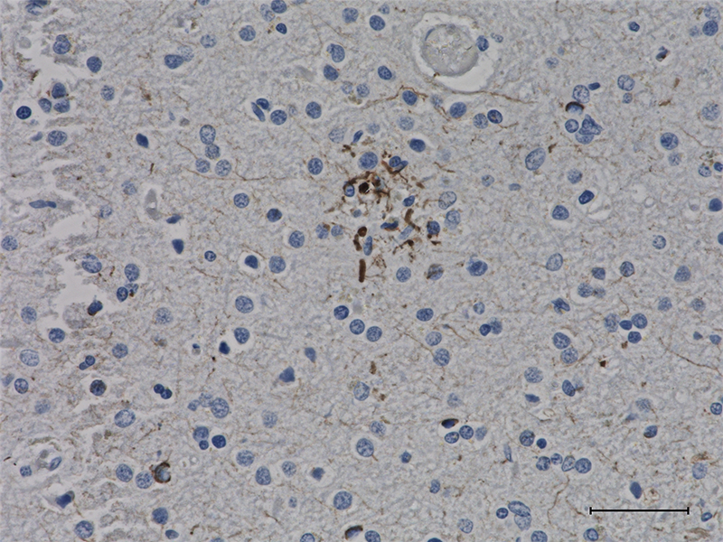
IHC-P - Verified - Recommended Usage
-
Each lot of this antibody is quality control tested by western blotting. For western blotting, the suggested use of this reagent is 0.5 - 1.0 µg/mL. For immunohistochemistry on formalin-fixed paraffin-embedded tissue sections, a concentration range of 2.5 - 5.0 µg/mL is suggested. It is recommended that the reagent be titrated for optimal performance for each application.
- Application Notes
-
This antibody is effective in immunoblotting (WB).
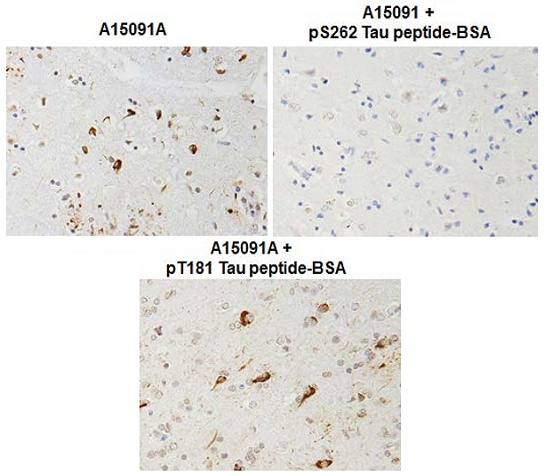
The PHF-6 antibody reacts with human tau phosphorylated at Thr 231.
Direct ELISA was done using phospho and non-phospho specific peptides, mainly to confirm specificity. Application has not been verified on tissue/cell lysates. -
Application References
(PubMed link indicates BioLegend citation) -
- Lasagna-Reeves CA, et al. 2012. FASEB J. 26:1946-1959.
- Mitchell TW, et al. 2000. J Histochem Cytochem. 48:1627-1637.
- Hong M, et al. 1997. J Biol Chem. 272:25326-25332.
- Bramblett GT, et al. 1993. Neuron. 10:1089-1099.
- Product Citations
-
- RRID
-
AB_2715849 (BioLegend Cat. No. 828903)
AB_2564924 (BioLegend Cat. No. 828901)
Antigen Details
- Biology Area
- Cell Biology, Neurodegeneration, Neuroscience, Protein Misfolding and Aggregation
- Molecular Family
- Phospho-Proteins, Tau
- Gene ID
- 4137 View all products for this Gene ID
- UniProt
- View information about Tau Phospho Thr231 on UniProt.org


Related FAQs
Other Formats
View All Tau Phospho (Thr231) Reagents Request Custom Conjugation| Description | Clone | Applications |
|---|---|---|
| Purified anti-Tau Phospho (Thr231) | PHF-6 | WB,IHC-P |
Customers Also Purchased
Compare Data Across All Formats
This data display is provided for general comparisons between formats.
Your actual data may vary due to variations in samples, target cells, instruments and their settings, staining conditions, and other factors.
If you need assistance with selecting the best format contact our expert technical support team.


Follow Us