- Clone
- JC70 (See other available formats)
- Regulatory Status
- RUO
- Other Names
- Platelet and endothelial cell adhesion molecule (PECAM-1), EndoCAM
- Isotype
- Mouse IgG1, κ
- Ave. Rating
- Submit a Review

| Cat # | Size | Price | Quantity Check Availability | Save | ||
|---|---|---|---|---|---|---|
| 623553 | 25 µg | 128€ | ||||
| 623554 | 100 µg | 324€ | ||||
CD31, widely known as “platelet and endothelial cell adhesion molecule” (PECAM), is a crucial transmembrane glycoprotein primarily found on the surface of endothelial cells and platelets. The quintessential endothelial cell marker, CD31 is vital for the maintenance of vascular integrity. It plays a pivotal role in cell-cell adhesion, promoting interactions between neighboring endothelial cells, and facilitates leukocyte diapedesis during the inflammatory response. CD31 also mediates platelet-endothelial interactions, contributing to platelet aggregation and thrombus formation at sites of vascular injury. Additionally, this multifunctional molecule is involved in signal transduction pathways that govern cell migration, proliferation, and survival, making it a key player in immune response regulation and angiogenesis.
Product DetailsProduct Details
- Verified Reactivity
- Human
- Antibody Type
- Monoclonal
- Host Species
- Mouse
- Immunogen
- Cell membrane preparation from the spleen of a patient with hairy cell leukemia
- Formulation
- Phosphate-buffered solution, pH 7.2, containing 0.09% sodium azide
- Preparation
- The antibody was purified by affinity chromatography and conjugated with Alexa Fluor® 647 under optimal conditions.
- Concentration
- 0.5 mg/mL
- Storage & Handling
- The antibody solution should be stored undiluted between 2°C and 8°C, and protected from prolonged exposure to light. Do not freeze.
- Application
-
IHC-P - Quality tested
ICC, ICFC, FC - Verified
- Recommended Usage
-
Each lot of this antibody is quality control tested by formalin-fixed paraffin-embedded immunohistochemical staining. For immunohistochemistry, a concentration range of 1 - 5 µg/mL is suggested. For immunocytochemistry, a concentration range of 1.25 - 2.5 μg/mL is recommended. For flow cytometric staining, the suggested use of this reagent is ≤ 0.25 µg per million cells in 100 µL volume. For intracellular flow cytometric staining, the suggested use of this reagent is ≤ 0.25 µg per million cells in 100 µL volume. It is recommended that the reagent be titrated for optimal performance for each application.
* Alexa Fluor® 647 has a maximum emission of 668 nm when it is excited at 633 nm / 635 nm.
Alexa Fluor® and Pacific Blue™ are trademarks of Life Technologies Corporation.
View full statement regarding label licenses - Excitation Laser
-
Red Laser (633 nm)
- Application Notes
-
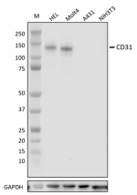
JC70 does not cross-react with mouse CD31 in Western Blot.
For ICC we recommend fixation/permeabilization with 100% ice-cold methanol, or 4% PFA followed by 100% ice-cold methanol. Fixation/permeabilization with 4% PFA followed by 0.5% Triton-X results in a significantly dimmer signal.
For IHC-P, we recommend antigen retrieval using Sodium Citrate, pH 6.0 (Cat. No. 420901). Product does not work with Tris-EDTA antigen retrieval. - Additional Product Notes
-
For use in immunohistochemistry on formalin-fixed paraffin-embedded tissue (IHC-P), it is recommended to perform antigen retrieval using Citrate Buffer, 10X (Cat. No. 420902) or Tris-EDTA pH 9.0 Antigen Retrieval Buffer (10X) (Cat. No. 422704).
For use in immunocytochemistry (ICC), it is recommended to fix/permeabilize with either of the following:- Fixation buffer (Cat. No. 420801) following by 100% ice-cold methanol
- 100% ice-cold methanol only
- RRID
-
AB_3675165 (BioLegend Cat. No. 623553)
AB_3675165 (BioLegend Cat. No. 623554)
Antigen Details
- Structure
- CD31 is a 738 amino acid protein with a predicted molecular weight of 82.5 kD.
- Distribution
-
Endothelial cells, Platelets, Granulocytes, Lymphocytes, Monocytes, Neutrophils, Cell surface
- Function
- Cell adhesion
- Interaction
- BDKRB2 and GNAQ
- Ligand/Receptor
- Forms homophillic interactions, also CD38, CD177, αV/β3 integrin
- Cell Type
- B cells, Dendritic cells, Endothelial cells, Granulocytes, Macrophages, Monocytes, Neutrophils, Platelets, T cells
- Biology Area
- Angiogenesis, Cell Adhesion, Cell Biology, Immunology, Neuroinflammation, Neuroscience
- Molecular Family
- Adhesion Molecules, CD Molecules
- Antigen References
-
- Barclay AN, et al. 1997. The Leukocyte Antigen FactsBook Academic Press.
- DeLisser HM, et al. 1994. Immunol. Today. 15:490-5.
- Newman PJ, et al. 1990. Science 247:1219-22.
- Lertkiatmongkol P, et al. 2016. Curr Opin Hematol. 23:253-9.
- Yeh JC, et al. 2008. Biochemistry. 47:9029-39.
- Gene ID
- 5175 View all products for this Gene ID
- UniProt
- View information about CD31 on UniProt.org
Related FAQs
Other Formats
View All CD31 Reagents Request Custom Conjugation| Description | Clone | Applications |
|---|---|---|
| Purified anti-CD31 (PECAM-1) | JC70 | WB,IHC-P,ICC,ICFC,FC |
| Alexa Fluor® 647 anti-CD31 (PECAM-1) Antibody | JC70 | IHC-P,FC,ICC,ICFC |
Compare Data Across All Formats
This data display is provided for general comparisons between formats.
Your actual data may vary due to variations in samples, target cells, instruments and their settings, staining conditions, and other factors.
If you need assistance with selecting the best format contact our expert technical support team.
-
Purified anti-CD31 (PECAM-1)

Whole cell extracts (15 µg total protein) from indicated cel... 
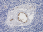
IHC staining of purified anti-CD31 (PECAM-1) (clone JC70) on... 
IHC staining of purified anti-CD31 (PECAM-1) (clone JC70) on... 
A431 cells (negative cell line) (panel A) and HEL cells (pos... 
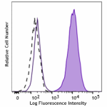
HEL cells (positive control) (filled histogram) and A431 cel... 
Human peripheral blood granulocytes were stained with either... -
Alexa Fluor® 647 anti-CD31 (PECAM-1) Antibody

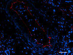
IHC staining with Alexa Fluor® 647 anti-CD31 (PECAM-1) (clon... 
HEL cells (positive cell line, panels A and C) and A-431 cel... 
HEL cells (positive cell line, filled histogram) and A-431 c... 
Human peripheral blood granulocytes were stained with either...

 Login / Register


Follow Us