- Clone
- 14.10 (See other available formats)
- Regulatory Status
- RUO
- Other Names
- CAML1, HSAS, HSAS1, MASA, MIC5, N-CAM-L1, N-CAML1, S10, SPG1, L1CAM, Neural cell adhesion molecule L1, antigen identified by monoclonal antibody R1
- Previously
-
Signet Catalog# 911-01
Covance Catalog# SIG-3911
- Isotype
- Mouse IgG1
- Ave. Rating
- Submit a Review
- Product Citations
- publications

-

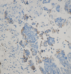
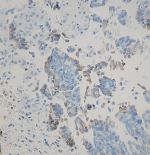
Staining of L1 (CD171), clone 14.10, on formalin fixed paraffin embedded human ovarian tumor.
| Cat # | Size | Price | Quantity Check Availability | Save | ||
|---|---|---|---|---|---|---|
| 826701 | 1 mL | 500€ | ||||
L1, also known as L1CAM, is a transmembrane protein; it is a neuronal cell adhesion molecule, member of the L1 protein family, of 200-220 kDa, and involved in axon guidance and cell migration with a strong implication in treatment-resistant cancers. L1CAM has also been designated CD171 (cluster of differentiation 171).
The protein encoded by this gene is an axonal glycoprotein belonging to the immunoglobulin superfamily of proteins. The ectodomain, consisting of several immunoglobulin-like domains and fibronectin-like repeats (type III), is linked via a single transmembrane sequence to a conserved cytoplasmic domain. This cell adhesion molecule plays an important role in nervous system development, including neuronal migration, and differentiation. Mutations in the gene cause three X-linked neurological syndromes known by the acronym CRASH (corpus callosum hypoplasia, retardation, aphasia, spastic paraplegia and hydrocephalus). Alternative splicing of a neuron-specific exon is thought to be functionally relevant. CD171 has been shown to function as a cell adhesion molecule mediating homotypic and heterotypic cell-cell interactions in neuronal myelination, neurite outgrowth and regeneration. Expression of CD171 is found on tetanus-toxin positive neurons, endothelial cells, certain epithelial cells, reticular fibroblasts and several malignant tumors including colon and breast carcinomas, colon melanoma, tumor cells of neuronal and mesothelial origin, where a role of CD171 in augmenting tumor growth has been demonstrated. Thus, CD171 is considered a novel marker for carcinoma progression.
CD171 displays binding to a number of ligands including homophilic interaction with itself and heterophilic binding to integrins, other cell adhesion molecules, extracellular matrix molecules and thus plays a vital role in cell adhesion and signal transduction. It is involved in the development of the nervous system and regulates processes such as neuron-neuron adhesion, myelination, axonal guidance, and neuronal migration.
Product Details
- Verified Reactivity
- Human
- Antibody Type
- Monoclonal
- Host Species
- Mouse
- Immunogen
- Clone 14.10 was raised to the ectodomain (amino acids M1 to E1123) of human L1 by using SKOV3ip cells as immunogen.
- Formulation
-
Phosphate-buffered solution with BSA + 0.09% NaN3.
Previous lots of this product may have been formulated with 0.1% or 0.05% NaN3 instead of 0.09% NaN3. For further information please contact BioLegend Technical Support or Customer Service. - Preparation
- The antibody was purified by affinity chromatography.
- Storage & Handling
- Store between 2°C and 8°C.
- Application
-
IHC-P - Quality tested
- Recommended Usage
-
Each lot of this antibody is quality control tested by immunohistochemical staining.
The optimal working dilution should be determined for each specific assay condition.
• IHC: ≥1:40 for Biotin based detection systems such as USA Ultra Streptavidin Detection (Cat. No. 929501).
Tissue Sections: Formalin-fixed, paraffin-embedded tissues, frozen sections
Pretreatment: Not required
Incubation: 20 minutes at room temperature - Application Notes
-
This antibody is effective in immunohistochemistry (IHC).
By Western blot analysis, the L1-14.10 epitope was mapped to the third Ig domain. Clone 14.10 recognizes L1, a 200-220 kD transmembrane glycoprotein that functions as an adhesion molecule. -
Application References
(PubMed link indicates BioLegend citation) -
- Wolterink, et al. Therapeutic Antibodies to Human L1CAM: Functional Characterization and Application in a Mouse Model for Ovarian Carcinoma. Cancer Res.; 70: 2504-2515, Mar 2010.
- Huszar M, et al. Expression profile analysis in multiple human tumors identifies L1 (CD17as a molecular marker for differential diagnosis and targeted therapy. Human Pathology , Vol. 37, Issue 8, p1000-1008, Jun 2006. (First publication listed, but needs to be updated.)
- Huszar M, Moldenhauer G, Gschwend V, Ben-Arie A, Altevogt P, Fogela, M. Expression profile analysis in multiple human tumors Identifies L1 (CD171)as a molecular marker for differential diagnosis and targeted therapy. Hum Pathol **Article in Press** accepted 24 March, 2006
- Kaifi JT, Heidtmann S, et al. Absence of L1 in pancreatic masses distinguishes adenocarcinomas from poorly differentiated neuroendocrine carcinomas. Anticancer Res 26(2A):1167-70, 2006.
- Kaifi JT, et al. L1(CD17is highly expressed in gastrointestinal stromal tumors. Mod Pathol 19(3):399-406, 2006.
- Meier F, et al. The adhesion molecule L1 (CD17promotes melanoma progression. Int J Cancer 119(3):549-55, 2006.
- Stoeck A, et al. A role for exosomes in the constitutive and stimulus-induced ectodomain cleavage of L1 and CD44. Biochem J 393:609-18, 2006.
- Gutwein P, et al. Cleavage of L1 in exosomes and apoptotic membrane vesicles released from ovarian carcinoma cells.
- Clin Cancer Res 11(7):2492-501, 2005.
- Product Citations
-
- RRID
-
AB_2564904 (BioLegend Cat. No. 826701)
Antigen Details
- Biology Area
- Cell Biology, Neuroscience, Synaptic Biology
- Molecular Family
- Adhesion Molecules
- Gene ID
- 3897 View all products for this Gene ID
- UniProt
- View information about CD171 on UniProt.org
Related Pages & Pathways
Pages
Related FAQs
Other Formats
View All CD171 Reagents Request Custom Conjugation| Description | Clone | Applications |
|---|---|---|
| Purified anti-CD171 (L1) | 14.10 | IHC-P |
Customers Also Purchased
Compare Data Across All Formats
This data display is provided for general comparisons between formats.
Your actual data may vary due to variations in samples, target cells, instruments and their settings, staining conditions, and other factors.
If you need assistance with selecting the best format contact our expert technical support team.
-
Purified anti-CD171 (L1)

Staining of L1 (CD171), clone 14.10, on formalin fixed paraf...
 Login / Register


Follow Us