 Login / Register


- Clone
- W15133B (See other available formats)
- Regulatory Status
- RUO
- Other Names
- Extracellular signal-regulated kinase 1/2, Mitogen-activated protein kinase MAPK3/MAPK1, p44ERK1/p42ERK2, p44-MAPK/p42MAPK, PRKM3/PRKM2, ERK
- Isotype
- Rat IgG1, κ
- Ave. Rating
- Submit a Review
- Product Citations
- publications

-

Total lysates (15µg protein) from 293T, 293T/ERK1 knockout (KO) and 293T/ERK2 knockdown (KD) cells were resolved by electrophoresis (4-12% Bis-Tris gel), transferred to nitrocellulose, and probed with 1:500 diluted (1µg/mL) purified anti-ERK1/2 antibody, clone W15133B (upper) or 1:3000 diluted anti-GAPDH (poly6314) antibody (lower). Proteins were visualized by chemiluminescence detection using a 1:3000 diluted goat anti-rat-IgG secondary antibody conjugated to HRP for the anti-ERK1/2 antibody, and a donkey anti-rabbit IgG Antibody conjugated to HRP for GAPDH. Lane M: Molecular weight ladder, M* indicates longer exposure. -

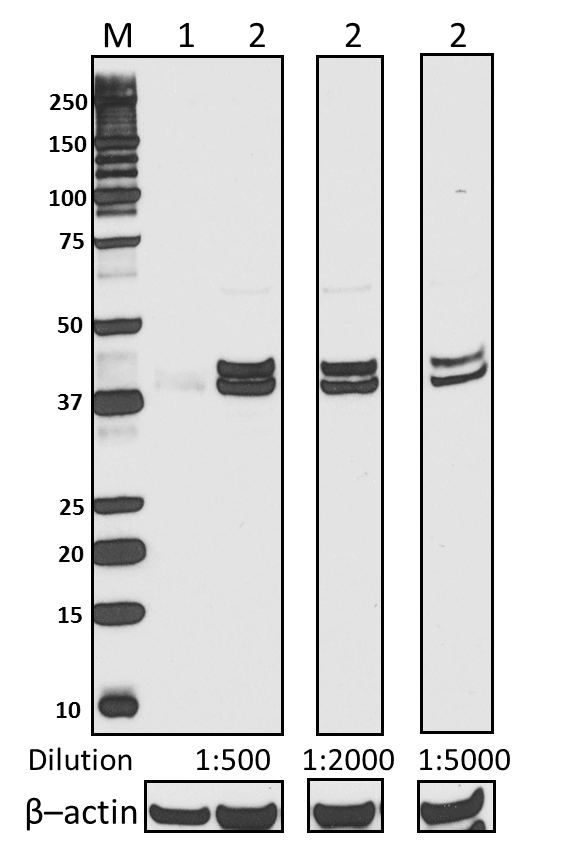
Total cell lysate from human HeLa cells (lane 1), Jurkat cells (lane 2), and mouse NIH-3T3 cells (lane 3) were resolved by electrophoresis, transferred to nitrocellulose, and probed with purified anti-ERK1/2 (clone W15133B) antibody. Proteins were visualized using an HRP goat anti-rat IgG antibody and chemiluminescence detection. -

HeLa cells were fixed with 4% paraformaldehyde (PFA) for 15 minutes, permeabilized with 0.5% Triton X-100 for three minutes, and blocked with 1% BSA for 60 minutes. Then the cells were intracellularly stained with 2.0 µg/ml purified anti-ERK1/2 (clone W15133B) antibody for one hour, followed by staining with Alexa Fluor® 594 conjugated anti-rat IgG antibody (red) and DAPI (blue) for one hour at room temperature. The image was captured with a 60X objective. -

Western blot of purified anti-ERK1/2 antibody (clone W15133B). Lane 1: Molecular weight marker; Lane 2: 20 µg of Drosophila head lysate; Lane 3: 20 µg of Drosophila S2 (embryonic) cell lysate. The blot was incubated with 0.5 µg/mL of the primary antibody overnight at 4°C, followed by incubation with HRP-labeled goat anti-rat IgG (Cat. No. 405405). Enhanced chemiluminescence was used as the detection system.
| Cat # | Size | Price | Quantity Check Availability | Save | ||
|---|---|---|---|---|---|---|
| 686901 | 25 µg | 90€ | ||||
| 686902 | 100 µg | 212€ | ||||
ERK1/2 are members of the mitogen-activated kinases (MAPKs) that are serine/threonine protein kinases. ERK1/2 can be activated by a range of extracellular stimuli, such as mitogen, growth factors, neurotransmitters, chemokines, and cytokines, through receptor tyrosine kinases (RTK), G protein-coupled receptors (GPCRs), or protein kinase C (PKC). Upon stimulation, ERK1/2 are phosphorylated by the upstream kinase MEK on residues Thr202 and Tyr204 and in turn phosphorylate many other downstream molecules that are involved in a range of cellular processes such as cell proliferation, differentiation, motility and cell death.
Product DetailsProduct Details
- Verified Reactivity
- Human, Mouse, Drosophila
- Antibody Type
- Monoclonal
- Host Species
- Rat
- Immunogen
- Partial human ERK1 recombinant protein (180-379 a.a.) expressed in E. coli.
- Formulation
- Phosphate-buffered solution, pH 7.2, containing 0.09% sodium azide.
- Preparation
- The antibody was purified by affinity chromatography.
- Concentration
- 0.5 mg/ml
- Storage & Handling
- The antibody solution should be stored undiluted between 2°C and 8°C.
- Application
-
WB - Quality tested
KO/KD-WB, ICC - Verified - Recommended Usage
-
Each lot of this antibody is quality control tested by Western blotting. For Western blotting, the suggested use of this reagent is 0.5 – 2.0 µg/ml in human, mouse, and Drosophila. For immunocytochemistry, a concentration range of 1.0 - 5.0 μg/ml is recommended. It is recommended that the reagent be titrated for optimal performance for each application.
- Application Notes
-
Reactivity to Drosophila was only verified with the purified format.
The drosophila homolog of mammalian ERK1/2 is the 43 kD mitogen-activated protein kinase ERK-A. - Product Citations
-
- RRID
-
AB_2861071 (BioLegend Cat. No. 686901)
AB_2629535 (BioLegend Cat. No. 686902)
Antigen Details
- Structure
- ERK1 and ERK2 encode 42 and 44 kD protein kinases and share 85% homology.
- Distribution
-
Nucleus and cytoplasm.
- Function
- ERK1/2 are broadly expressed protein kinases that are involved in proliferation, differentiation, motility, and cell death. In response to extracellular stimuli, ERK1/2 are phosphorylated at Thr202 and Tyr204 and in turn transduce signals to downstream substrates.
- Interaction
- Interacts with ADAM15, ARRB2, CANX, DAPK1, HSF4, IER3, MAP2K1/MEK1, MORG1, NISCH, SGK1, TPR, CDKN2AIP, PEA15, and MKNK2.
- Biology Area
- Apoptosis/Tumor Suppressors/Cell Death, Cell Biology, Signal Transduction
- Molecular Family
- Protein Kinases/Phosphatase
- Antigen References
-
1. Futran AS, et al. 2013. Curr. Biol. 23:R972.
2. Mendoza MC, et al. 2011. Trends Biochem. Sci. 36:320.
3. Chambard JC, et al. 2007. Biochim. Biophys. Acta. 1773:1299.
4. Roux PP, et al. 2004. Microbiol. Mol. Biol. Rev. 68:320.
5. Torii S, et al. 2004. J. Biochem. 136:557.
6. Clark EA, et al. 1996. J. Biol. Chem. 271:14814. - Gene ID
- 5595 View all products for this Gene ID 5594 View all products for this Gene ID
- UniProt
- View information about ERK1/2 on UniProt.org

Related FAQs
Other Formats
View All ERK1/2 Reagents Request Custom Conjugation| Description | Clone | Applications |
|---|---|---|
| Purified anti-ERK1/2 | W15133B | WB,KO/KD-WB,ICC |
| Alexa Fluor® 594 anti-ERK1/2 | W15133B | ICC |
Customers Also Purchased

Compare Data Across All Formats
This data display is provided for general comparisons between formats.
Your actual data may vary due to variations in samples, target cells, instruments and their settings, staining conditions, and other factors.
If you need assistance with selecting the best format contact our expert technical support team.
-
Purified anti-ERK1/2

Total lysates (15µg protein) from 293T, 293T/ERK1 knockout (... 
HeLa cells were fixed with 4% paraformaldehyde (PFA) for 15 ... 
Total cell lysate from human HeLa cells (lane 1), Jurkat cel... 
Western blot of purified anti-ERK1/2 antibody (clone W15133B... -
Alexa Fluor® 594 anti-ERK1/2

HeLa cells were fixed with 4% paraformaldehyde (PFA) for 15 ...


Follow Us