- Clone
- 359-81-11 (See other available formats)
- Regulatory Status
- RUO
- Other Names
- Lymphotoxin-α (LT- α), Tumor necrosis factor-β (TNF-β), Coley's toxin, Hemorrhagic factor, Necrosin, Natural killer cytotoxic factor (NKCF), Differentiation inducing factor (DIF), TNFSF-1
- Isotype
- Mouse IgG1, κ
- Ave. Rating
- Submit a Review
- Product Citations
- publications

-

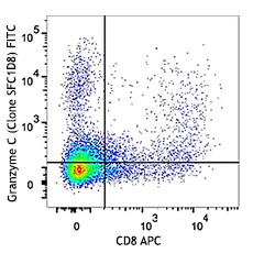
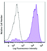
PMA+ionomycin-stimulated human T cells were surface stained with CD3 PE/Cyanine5 and intracellular stained with 359-81-11 PE
| Cat # | Size | Price | Quantity Check Availability | Save | ||
|---|---|---|---|---|---|---|
| 503105 | 25 µg | £89 | ||||
Lymphotoxin-α (LT-α), also known as tumor necrosis factor-beta (TNF-β), is a potent lymphoid factor that exerts cytotoxic effects on a wide range of tumor cells and certain other target cells. LT-α possesses a signal peptide sequence and is a secreted protein; however, LT-α is also present on the surface of activated T, B and LAK cells as a complex with LT-β. Bioactive LT-α exists as a homotrimer.
Product DetailsProduct Details
- Verified Reactivity
- Human
- Antibody Type
- Monoclonal
- Host Species
- Mouse
- Immunogen
- E. coli expressed, recombinant human LT-α.
- Formulation
- Phosphate-buffered solution, pH 7.2, containing 0.09% sodium azide.
- Preparation
- The antibody was purified by affinity chromatography, and conjugated with PE under optimal conditions.
- Concentration
- 0.2 mg/ml
- Storage & Handling
- The antibody solution should be stored undiluted between 2°C and 8°C, and protected from prolonged exposure to light. Do not freeze.
- Application
-
ICFC - Quality tested
- Recommended Usage
-
Each lot of this antibody is quality control tested by intracellular immunofluorescent staining with flow cytometric analysis. For flow cytometric staining, the suggested use of this reagent is ≤ 0.25 µg per 106 cells in 100 µl volume. It is recommended that the reagent be titrated for optimal performance for each application.
- Excitation Laser
-
Blue Laser (488 nm)
Green Laser (532 nm)/Yellow-Green Laser (561 nm)
- Application Notes
-
ELISA or ELISPOT Detection1,2: The biotinylated 359-81-11 antibody is useful as a detection antibody for a sandwich ELISA or ELISPOT assay, when used in conjunction with purified 359-238-8 antibody (Cat. No. 503002/503004) as the capture antibody.
Flow Cytometry3: The fluorochrome-labeled 359-81-11 antibody is useful for intracellular immunofluorescent staining and flow cytometric analysis to identify LT-α -producing cells within mixed cell populations. View intracellular cytokine staining protocol.
Neutralization1,2: The 359-81-11 antibody can neutralize the bioactivity of natural or recombinant LT-α. The LEAF™ purified antibody (Endotoxin <0.1 EU/μg, Azide-Free, 0.2 μm filtered) is recommended for neutralization of human LT-α bioactivity (Cat. No. 503108).
Additional reported applications (for the relevant formats) include: immunohistochemical staining of paraformaldehyde-fixed, saponin-treated frozen tissue sections, and immunocytochemistry. -
Application References
(PubMed link indicates BioLegend citation) -
- Meager A, et al. 1987. J. Immunol. Methods 104:31.
- Meager A, et al. 1987. Hybridoma. 6:305.
- Jason J, et al. 1999. Clin. Diagn. Lab Immunol. 6:73.
- RRID
-
AB_315275 (BioLegend Cat. No. 503105)
Antigen Details
- Structure
- TNF superfamily; trimer; 25 kD (Mammalian).
- Function
- Transformed cell cytotoxicity; mediator of inflammatory and immune functions; fibroblast synthesis of GM-CSF, G-CSF, IL-1, collagenase, prostaglandin E2; monocyte terminal differentiation, synthesis of G-CSF; neutrophil chemoattractant, production of reactive oxygen, fibroblast proliferation.
- Cell Sources
- Activated T and B cells, fibroblasts, astrocytes, myeloma, endothelial cells, epithelial cells.
- Cell Targets
- Monocytes, B cells, fibroblasts, neutrophils, osteoclasts, keratinocytes, endothelial cells.
- Receptors
- TNFRSF1A (TNF-R1, CD120a, TNFR-p60 Type β, p55); TNFRSF1B (TNF-R2, CD120b, TNFR-p80 Type A, p75).
- Biology Area
- Cell Biology, Immunology, Innate Immunity, Neuroinflammation, Neuroscience
- Molecular Family
- Cytokines/Chemokines
- Antigen References
-
1. Fitzgerald, K., et al. Eds. 2001. The Cytokine FactsBook. Academic Press, San Diego.
2. Aggarwal, B., et al.Eds. 1992. Tumor necrosis factors:structure, function, and mechanism of action. Marcel Dekker Inc.
3. Bonavida, B., et al.Eds. 1990. Tumor necrosis factor:structure, mechanisms of action, role in disease and therapy. Karger, Basel.
4. Paul, N., et al. 1987. Annu. Rev. Immunol. 6:407. - Regulation
- Type II integral membrane protein, forms heterotrimer with type II integral membrane protein LT-β either as LTα1β2 or LTα2β1; processed secreted form is trimeric.
- Gene ID
- 4049 View all products for this Gene ID
- UniProt
- View information about LT-alpha on UniProt.org
Related FAQs
- What type of PE do you use in your conjugates?
- We use R-PE in our conjugates.
Other Formats
View All LT-α Reagents Request Custom Conjugation| Description | Clone | Applications |
|---|---|---|
| Biotin anti-human LT-α (TNF-β) | 359-81-11 | ELISA Detection,ELISPOT Detection,ICFC |
| PE anti-human LT-α (TNF-β) | 359-81-11 | ICFC |
Customers Also Purchased
Compare Data Across All Formats
This data display is provided for general comparisons between formats.
Your actual data may vary due to variations in samples, target cells, instruments and their settings, staining conditions, and other factors.
If you need assistance with selecting the best format contact our expert technical support team.
-
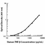
Biotin anti-human LT-α (TNF-β)
-
PE anti-human LT-α (TNF-β)

PMA+ionomycin-stimulated human T cells were surface stained ...
 Login / Register




Follow Us