- Clone
- 11A5C44 (See other available formats)
- Regulatory Status
- RUO
- Other Names
- Fibroblast growth factor 1 (FGF-1), Acidic fibroblast growth factor (aFGF), FGF-acidic, Endothelial cell growth factor (ECGF), Heparin-binding growth factor 1 (HBGF-1), FGF1, HBGF1
- Isotype
- Mouse IgG2b, λ
- Ave. Rating
- Submit a Review
- Product Citations
- publications

-

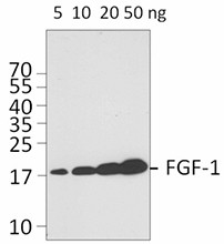
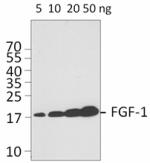
5, 10, 20, or 50 ng of human FGF-1 recombinant protein was resolved by electrophoresis, transferred to nitrocellulose, and probed with purified monoclonal anti-human FGF-1-acidic (clone 11A5C44) antibody. Proteins were visualized using a HRP conjugated anti-mouse-IgG secondary and chemiluminescence detection.
| Cat # | Size | Price | Quantity Check Availability | Save | ||
|---|---|---|---|---|---|---|
| 678602 | 100 µg | £157 | ||||
FGF-1, one of the most studied members of the fibroblast growth factor family, is a powerful mitogen exhibiting strong action on many different cell types. FGF-1 activity can be mediated not only by autocrine/paracrine pathways, but also by an intracrine pathway. FGF-1 lacks a secretion signal peptide and is exported through a non-classical pathway. Endogenous FGF-1 is found in the nucleus of most cell types. Nuclear localization is required for FGF-1 mitogenic activity. FGF-1 promotes tumor development by promoting cancer cell proliferation and survival. Increased FGF-1 expression in early stages of many different cancers have been reported. MCF-7 breast cancer cell lines that overexpress FGF-1 can form vascularized, metastatic tumors when injected into ovariectomized or tamoxifen-treated nude mice. FGF-1 also induces angiogenesis in vitro and in vivo. Thus, FGF-1 is an attractive candidate for cancer immunotargeting. FGF-1 is also involved in neuronal cell differentiation and survival. FGF-1 is highly expressed in motor neurons. In response to damage, motor neurons can release FGF-1, which results in astrocyte activation. Under oxidative stress, astrocytes can also release FGF-1, which stimulates ApoE/HDL generation in an autocrine manner for protection of the brain against oxidative stress. The involvement of FGF-1 in inflammation, cardioprotection, wound healing, adipocyte remodeling, and restenosis have also been reported.
Product DetailsProduct Details
- Verified Reactivity
- Human
- Antibody Type
- Monoclonal
- Host Species
- Mouse
- Immunogen
- Parital human human FGF-1 recombinant protein (16-155 a.a.) expressed in E. coli.
- Formulation
- Phosphate-buffered solution, pH 7.2, containing 0.09% sodium azide.
- Preparation
- The antibody was purified by affinity chromatography.
- Concentration
- 0.5 mg/ml
- Storage & Handling
- The antibody solution should be stored undiluted between 2°C and 8°C.
- Application
-
WB - Quality tested
- Recommended Usage
-
Each lot of this antibody is quality control tested by Western blotting. For Western blotting, the suggested use of this reagent is 0.25 - 1.0 µg per ml. It is recommended that the reagent be titrated for optimal performance for each application.
- RRID
-
AB_2565906 (BioLegend Cat. No. 678602)
Antigen Details
- Structure
- The 140 amino acid recombinant protein has a predicted molecular mass of approximately 16 kD. Growth factor.
- Distribution
- Secreted. FGF-1 is widely expressed in developing and adult tissues.
- Function
- FGF-1 plays an important role in many biological processes, including cell proliferation, inflammation, and angiogenesis. FGF-1 has a low stability and a very short half-life in vivo. Binding to heparin increases the stability of FGF-1 and is also important for the formation of active FGF-acidic/FGFR complex.
- Interaction
- Intracellular FGF-1 can interact directly with CK2, FIBP, mortalin, and the ribosome-binding protein p34/leucine-rich repeat containing 59 (LRRC59).
- Ligand/Receptor
- FGF-1 can signal through all known cell-surface FGFR isoforms (FGFR1b, 1c, 2b, 2c, 3b, 3c, and 4). FGF-1 also binds to heparin and heparin sulfate proteoglycans on the cell surface.
- Biology Area
- Angiogenesis, Cell Biology, Neuroscience, Synaptic Biology
- Molecular Family
- Adhesion Molecules, Growth Factors, Cytokines/Chemokines
- Antigen References
-
1. Presta M, et al. 2005. Cytokine Growth Factor Rev. 2:159.
2. Zakrzewska M, et al. 2008. Crit. Rev. Clin. Lab. Sci. 45:91.
3. Zhang L, et al. 1997. Oncogene. 15:2093.
4. Lin YZ, et al. 1996. J. Biol. Chem. 271:5305.
5. Culajay JF, et al. 2000. Biochemistry 39:7153.
6. Cassina P, et al. 2005. J. Neurochem. 93:38.
7. Pehar M, et al. 2005. Neurodegener. Dis. 2:139.
8. Jonker JW, et al. 2012. Nature 485:391.
9. Compagni A, et al. 2000. Cancer Res. 60:7163. - Gene ID
- 2246 View all products for this Gene ID
- UniProt
- View information about FGF-1-acidic on UniProt.org
Related Pages & Pathways
Pages
Related FAQs
Other Formats
View All FGF-1-acidic Reagents Request Custom Conjugation| Description | Clone | Applications |
|---|---|---|
| Purified anti-FGF-1-acidic | 11A5C44 | WB |
Compare Data Across All Formats
This data display is provided for general comparisons between formats.
Your actual data may vary due to variations in samples, target cells, instruments and their settings, staining conditions, and other factors.
If you need assistance with selecting the best format contact our expert technical support team.
-
Purified anti-FGF-1-acidic

5, 10, 20, or 50 ng of human FGF-1 recombinant protein was r...
 Login / Register


Follow Us