 Login / Register


- Clone
- 2F83 (See other available formats)
- Regulatory Status
- RUO
- Other Names
- Forkhead box protein A1, HNF3α, Hepatic nuclear factor 3 α
- Isotype
- Mouse IgG1, κ
- Ave. Rating
- Submit a Review
- Product Citations
- publications

-

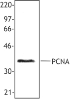
Whole cell extracts (15 µg protein) from HEL (negative control), HeLa (reduced expression control), HepG2, and NIH/3T3 cells were resolved on a 4-12% Bis-Tris gel, transferred to nitrocellulose and probed with 0.5 µg/mL (1:1000 dilution) of Purified anti-FOXA1 Antibody, clone 2F83, overnight at 4°C. Proteins were visualized by chemiluminescence detection using HRP Goat Anti-mouse IgG Antibody (Cat. No. 405306) at a 1:3000 dilution. Equal loading was confirmed by probing membranes with Direct Blot™ HRP anti-GAPDH Antibody (Cat. No. 607904) at a 1:50000 dilution. Lane M: Molecular Weight marker. Predicted expression data was obtained from Human Protein Atlas -

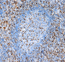
MCF-7 cells were fixed with 1% paraformaldehyde (PFA) for 10 minutes and permeabilized with 0.5% Triton X-100 buffer for 10 minutes at room temperature. Then blocked for 30 minutes at room temperature with 5% fetal bovine serum (FBS). The cells were stained with 5 µg/mL anti-FOXA1 (clone 2F83) purified at 4°C overnight. On the next day, cells were stained with Alexa Fluor® 594 anti-mouse IgG (clone Poly4053) for an hour at room temperature. Finally, cells were incubated with Flash Phalloidin™ Green 488 (green) for 20 mintues at room temperature. Nuclei were counterstained with DAPI (blue). Images were acquired with a 40X objective. -

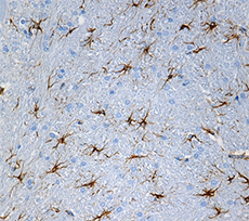
Human paraffin-embedded prostate tissue slices were prepared with a standard protocol of deparaffinization and rehydration. Antigen retrieval was done with Citrate buffer pH 6.0 1X at 95°C for 40 minutes. Tissue was washed with PBS/0.05% Tween 20 twice for five minutes, permealized with 0.5% TritonX-100 for 10 minutes and blocked with 5% FBS and 0.2% gelatin for 30 minutes. Then, the tissue was stained with 10 µg/mL of anti-human FOXA1 (clone 2F83) Purifed at 4°C overnight followed with one hour room temperature incubation of anti-mouse IgG Alexa Fluor® 594 (clone Poly4053, Red). Nuclei were counterstained with DAPI (Blue). The image was scanned with a 10X objective and stitched with Metamorph® software.
| Cat # | Size | Price | Quantity Check Availability | Save | ||
|---|---|---|---|---|---|---|
| 612001 | 25 µg | £70 | ||||
| 612002 | 100 µg | £174 | ||||
FOXA1 is a transcriptional activator that is required for tissue-specific differentiation during embryonic development. It also functions as a coactivator with nuclear hormone receptors, including ESR1, and has been shown to play a major role in driving estrogen receptor positive breast cancers.
Product DetailsProduct Details
- Verified Reactivity
- Human
- Antibody Type
- Monoclonal
- Host Species
- Mouse
- Immunogen
- Full length recombinant human FOXA1
- Formulation
- Phosphate-buffered solution, pH 7.2, containing 0.09% sodium azide.
- Preparation
- The antibody was purified by affinity chromatography.
- Concentration
- 0.5 mg/ml
- Storage & Handling
- The antibody solution should be stored undiluted between 2°C and 8°C.
- Application
-
WB - Quality tested
ICC, IHC-P - Verified - Recommended Usage
-
Each lot of this antibody is quality control tested by Western blotting. For Western blotting, the suggested use of this reagent is 0.1 - 1.0 µg per ml. For immunocytochemistry, a concentration range of 1.25 - 5.0 μg/ml is recommended. For immunohistochemistry on formalin-fixed paraffin-embedded tissue sections, a concentration range of 5.0 - 10 µg/ml is suggested. It is recommended that the reagent be titrated for optimal performance for each application.
- Application Notes
-
When using this clone for western blot, we highly recommend using lysate that has not been subjected to any freeze-thaw cycles.
-
Application References
(PubMed link indicates BioLegend citation) -
- Besenard V, et al. 2004. Gene Expr Patterns. 5:193-208. (IHC-P)
- RRID
-
AB_2801115 (BioLegend Cat. No. 612001)
AB_2801116 (BioLegend Cat. No. 612002)
Antigen Details
- Structure
- FOXA1 is a 472 amino acid protein with a predicted molecular weight of 49 kD.
- Distribution
-
Nucleus/Early endoderm derived cells
- Function
- Transcriptional activator
- Interaction
- FOXA2, NKX2-1, Histones H3 and H4
- Biology Area
- Cell Biology, Transcription Factors
- Molecular Family
- Nuclear Markers
- Gene ID
- 3169 View all products for this Gene ID
- UniProt
- View information about FOXA1 on UniProt.org

Related FAQs
Other Formats
View All FOXA1 Reagents Request Custom Conjugation| Description | Clone | Applications |
|---|---|---|
| Purified anti-FOXA1 | 2F83 | WB,ICC,IHC-P |
| Alexa Fluor® 647 anti-FOXA1 | 2F83 | ICFC |
| TotalSeq™-B1213 anti-FOXA1 Antibody | 2F83 | ICPG |
Customers Also Purchased
Compare Data Across All Formats
This data display is provided for general comparisons between formats.
Your actual data may vary due to variations in samples, target cells, instruments and their settings, staining conditions, and other factors.
If you need assistance with selecting the best format contact our expert technical support team.
-
Purified anti-FOXA1

Whole cell extracts (15 µg protein) from HEL (negative contr... 
MCF-7 cells were fixed with 1% paraformaldehyde (PFA) for 10... 
Human paraffin-embedded prostate tissue slices were prepared... -
Alexa Fluor® 647 anti-FOXA1

HEL cells were treated with True-Nuclear™ Transcription Fact... 
MCF-7 cells were treated with True-Nuclear™ Transcription Fa... -
TotalSeq™-B1213 anti-FOXA1 Antibody


Follow Us