- Clone
- 14D2C43 (See other available formats)
- Regulatory Status
- RUO
- Other Names
- ARG1, Liver Arginase, Type 1 Arginase, Arginase-1, Liver-Type Arginase
- Isotype
- Mouse IgG2b, κ
- Ave. Rating
- Submit a Review
- Product Citations
- publications
-

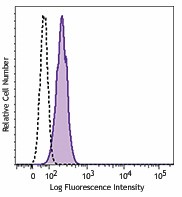
Human peripheral blood was surface stained with CD15 APC and CD16 Pacific Blue™, fixed, permeabilized, and then intracellularly stained with purified Arginase I (clone 14D2C43) (filled histogram) or mouse IgG2b, κ isotype control (open histogram), followed by anti-mouse IgG2b PE. Histogram was gated on the neutrophil (CD15+CD16+) population.
| Cat # | Size | Price | Quantity Check Availability | Save | ||
|---|---|---|---|---|---|---|
| 369701 | 25 µg | £70 | ||||
| 369702 | 100 µg | £174 | ||||
Arginase I, also known as ARG1, is a 34.7kD protein expressed by neutrophils and myeloid-derived suppressor cells (MDSCs). There are two isoforms that are differentiated based on their tissue distribution and subcellular localization. Arginase I converts L-arginine into L-ornithine and urea; it is the final enzyme in the urea cycle. While mostly found in the liver, it can also be expressed in cells lacking a comprehensive urea cycle. Also, it contributes to vasodilation and vascular function. Arginase I is also reported to be involved in MDSC mediated suppression of T cell proliferation. Arginase I has been implicated in hyperargininemia (decreased function of arginase I) and Q fever.
Product DetailsProduct Details
- Verified Reactivity
- Human
- Antibody Type
- Monoclonal
- Host Species
- Mouse
- Immunogen
- Full length recombinant protein expressed in E.coli.
- Formulation
- Phosphate-buffered solution, pH 7.2, containing 0.09% sodium azide.
- Preparation
- The antibody was purified by affinity chromatography.
- Concentration
- 0.5 mg/ml
- Storage & Handling
- The antibody solution should be stored undiluted between 2°C and 8°C.
- Application
-
ICFC - Quality tested
- Recommended Usage
-
Each lot of this antibody is quality control tested by intracellular immunofluorescent staining with flow cytometric analysis. For flow cytometric staining, the suggested use of this reagent is ≤0.5 µg per million cells in 100 µl volume. It is recommended that the reagent be titrated for optimal performance for each application.
- Product Citations
-
- RRID
-
AB_2571897 (BioLegend Cat. No. 369701)
AB_2571898 (BioLegend Cat. No. 369702)
Antigen Details
- Structure
- Belongs to the ureohydrolase family and has a molecular mass of 34.7 kD.
- Distribution
- Neutrophils and myeloid-derived suppressor cells (MDSC).
- Function
- Converts L-arginine into L-ornithine and urea.
- Interaction
- Arginine.
- Cell Type
- Neutrophils
- Biology Area
- Cell Biology, Immunology, Neuroinflammation, Neuroscience
- Molecular Family
- Enzymes and Regulators
- Antigen References
-
1. Munder M, et al. 2005. Blood 105:2549.
2. Luckner-Minden C, et al. 2010. JLB. 87:1125.
3. Holowatz L, et al. 2006. Journal of Physiology 574:573.
4. Sin YY, et al. 2013. PLoS One 8:11.
5. Benoit M, et al. 2008. Eur. J. Immunol. 4:1065. - Gene ID
- 383 View all products for this Gene ID
- UniProt
- View information about Arginase I on UniProt.org
Related Pages & Pathways
Pages
Related FAQs
Other Formats
View All Arginase I Reagents Request Custom Conjugation| Description | Clone | Applications |
|---|---|---|
| Purified anti-human Arginase I | 14D2C43 | ICFC |
| PE anti-human Arginase I | 14D2C43 | ICFC |
| APC anti-human Arginase I | 14D2C43 | ICFC |
| PE/Cyanine7 anti-human Arginase I | 14D2C43 | ICFC |
| PerCP/Cyanine5.5 anti-human Arginase 1 | 14D2C43 | ICFC |
Compare Data Across All Formats
This data display is provided for general comparisons between formats.
Your actual data may vary due to variations in samples, target cells, instruments and their settings, staining conditions, and other factors.
If you need assistance with selecting the best format contact our expert technical support team.
-
Purified anti-human Arginase I
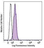
Human peripheral blood was surface stained with CD15 APC and... -
PE anti-human Arginase I
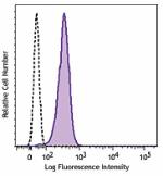
Human peripheral blood was surface stained with CD15 APC and... -
APC anti-human Arginase I
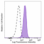
Human peripheral blood was surface stained with CD15 Pacific... -
PE/Cyanine7 anti-human Arginase I
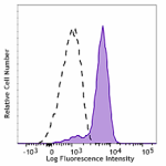
Human peripheral blood was surface stained with CD15 Pacific... -
PerCP/Cyanine5.5 anti-human Arginase 1
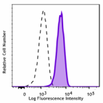
Human peripheral blood was surface stained with anti-human C...
 Login / Register


Follow Us