 Login / Register


- Clone
- NAT105 (See other available formats)
- Regulatory Status
- RUO
- Other Names
- PD1, PDCD1
- Isotype
- Mouse IgG1, κ
- Ave. Rating
- Submit a Review
- Product Citations
- publications

-

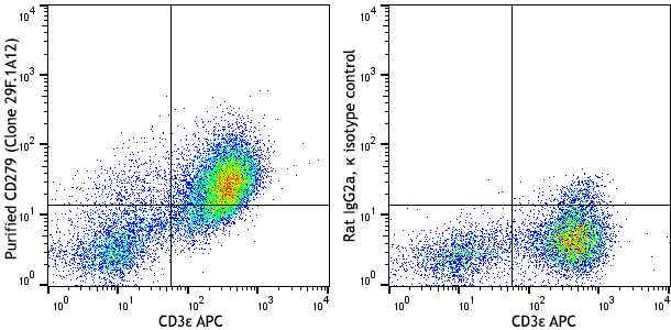
Human peripheral blood lymphocytes were stained with purified CD279 (clone NAT105), followed by anti-mouse IgG PE. The cells were then stained with anti-CD3 APC. -

Tissue imaged here is the lymphoid nodule/germinal center of a human tonsil. CD279 positive staining is prevalent within the nodule/germinal center. The antibody was used at 10 μg/ml here. Images shown at 10x and 40x magnification (inset). -

Human paraffin-embedded tonsil tissue slices were prepared with a standard protocol of deparaffinization and rehydration. Antigen retrieval was done with citrate buffered 1x (1.0M, pH 6.0) at 95°C for 40 minutes. Tissue was washed with PBS/0.05% Tween 20 twice for five minutes and blocked with 5% FBS and 0.2% gelatin for 30 minutes. Then, the tissue was stained with 10 µg/mL of purified CD279 (clone NAT105) antibody overnight at 4°C. On the next day, the tissue was washed twice with 1x PBS and stained with Alexa Fluor® 594 goat anti-mouse IgG (clone Poly4053, red) for an hour. Nuclei were counterstained with DAPI (green). The image was scanned with a 10X objective and stitched with MetaMorph® software. -

Western blot analysis of Jurkat (lane 1), PBMC untreated (lane 2) and PBMC activated with CD3 (clone OKT3) and CD28 (clone 28.2) antibodies for two days (lane 3) using anti-CD279 antibody (clone NAT105). Anti-β-actin antibody (poly6221) was used as a loading control. -

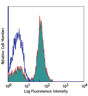
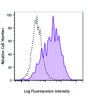
PHA-stimulated (three days) human PBMC were stained with purified CD279 (clone NAT105) (filled histogram) or purified mouse IgG1, κ isotype control, followed by anti-mouse IgG PE.
| Cat # | Size | Price | Quantity Check Availability | Save | ||
|---|---|---|---|---|---|---|
| 367402 | 100 µg | £77 | ||||
Programmed cell death 1 (PD-1), also known as CD279, is a 55 kD member of the immunoglobulin superfamily. CD279 contains the immunoreceptor tyrosine-based inhibitory motif (ITIM) in the cytoplasmic region and plays a key role in peripheral tolerance and autoimmune diseases. CD279 is expressed predominantly on activated T cells, B cells, and myeloid cells. PD-L1 (B7-H1, CD274) and PD-L2 (B7-DC, CD273) are ligands of CD279 (PD-1) and are members of the B7 gene family. Evidence suggests overlapping functions for these two PD-1 ligands and their constitutive expression on some normal tissues and upregulation on activated antigen-presenting cells. The interaction with CD279 ligands results in inhibition of T cell proliferation and cytokine secretion.
Product DetailsProduct Details
- Verified Reactivity
- Human
- Antibody Type
- Monoclonal
- Host Species
- Mouse
- Immunogen
- YT cell line.
- Formulation
- Phosphate-buffered solution, pH 7.2, containing 0.09% sodium azide.
- Preparation
- The antibody was purified by affinity chromatography.
- Concentration
- 0.5 mg/ml
- Storage & Handling
- The antibody solution should be stored undiluted between 2°C and 8°C.
- Application
-
FC - Quality tested
IHC-P, WB - Verified - Recommended Usage
-
Each lot of this antibody is quality control tested by immunofluorescent staining with flow cytometric analysis. For flow cytometric staining, the suggested use of this reagent is ≤ 0.5 µg per million cells in 100 µl volume. For immunohistochemistry of paraffin-embedded tissue, a concentration range of 0.1 - 10 µg/ml is suggested. For Western blotting, the suggested use of this reagent is 0.5 - 1.0 µg/ml. It is recommended that the reagent be titrated for optimal performance for each application.
Tissue Sections: Paraffin-embedded tissues.
Pretreatment: For optimal staining, the sections should be pretreated with an antigen unmasking solution such as Sodium Citrate H.I.E.R. Retrieval Solution (Cat. No. 928501, 928601).
Incubation: 24 hours at 2°C - 4 °C.
Positive Control: Human tonsil and spleen. - Application Notes
-
Additional reported applications (for the relevant formats of this clone) include: immunofluorescence staining.
We recommend using our Ultra Streptavidin (USA) HRP Detection Kit (Multi-Species, DAB) (Cat. No. 929501). -
Application References
(PubMed link indicates BioLegend citation) -
- Roncador G, et al. 2007. Haematologica 92:1059.
- Nam-Cha SH, et al. 2008. Am. J. Surg. Pathol. 32:1252.
- Rodriguez-Pinilla SM, et al. 2009. Am. J. Surg. Pathol. 33:81.
- Product Citations
-
- RRID
-
AB_2565782 (BioLegend Cat. No. 367402)
Antigen Details
- Structure
- 55 kD type I transmembrane protein.
- Distribution
-
Transiently expressed on CD4- and CD8- thymocytes, upregulated in thymocytes and splenic T and B lymphocytes, and is expressed on activated myeloid cells.
- Function
- Signaling, co-stimulation (co-inhibition), immunoglobulin superfamily.
- Interaction
- SHP-1 and SHP-2.
- Ligand/Receptor
- PD-L1 (CD274) and PD-L2 (CD273).
- Cell Type
- B cells, T cells, Thymocytes, Tregs
- Biology Area
- Cancer Biomarkers, Immunology, Inhibitory Molecules
- Molecular Family
- CD Molecules, Immune Checkpoint Receptors
- Antigen References
-
1. Francisco LM, Sage PT, and Sharpe AH. 2010. Immunological Rev. 236:219.
- Gene ID
- 5133 View all products for this Gene ID
- UniProt
- View information about CD279 on UniProt.org


Related FAQs
Other Formats
View All CD279 Reagents Request Custom ConjugationCustomers Also Purchased
Compare Data Across All Formats
This data display is provided for general comparisons between formats.
Your actual data may vary due to variations in samples, target cells, instruments and their settings, staining conditions, and other factors.
If you need assistance with selecting the best format contact our expert technical support team.
-
Purified anti-human CD279 (PD-1)

Human peripheral blood lymphocytes were stained with purifie... 
Tissue imaged here is the lymphoid nodule/germinal center of... 
Human paraffin-embedded tonsil tissue slices were prepared w... 
Western blot analysis of Jurkat (lane 1), PBMC untreated (la... 
PHA-stimulated (three days) human PBMC were stained with pur... -
PE anti-human CD279 (PD-1)

Human peripheral blood lymphocytes were stained with CD3 FIT... 
PHA-stimulated (three days) human PBMC were stained with CD2... 
Multiplexed IHC staining of PE anti-CD279 (clone NAT105) on ... -
APC anti-human CD279 (PD-1)

Human peripheral blood lymphocytes were stained with CD3 FIT... 
PHA-stimulated (three days) human PBMC were stained with CD2... -
Alexa Fluor® 488 anti-human CD279 (PD-1)

Human peripheral blood lymphocytes were stained with CD3 APC... 
PHA-stimulated (3-day) human peripheral blood lymphocytes we... -
PerCP/Cyanine5.5 anti-human CD279 (PD-1)

PHA-stimulated (3-day) human peripheral blood mononuclear ce... 
Human peripheral blood lymphocytes were stained with CD3 APC... -
PE/Cyanine7 anti-human CD279 (PD-1)

PHA-stimulated (3-day) human peripheral blood lymphocytes we... -
FITC anti-human CD279 (PD-1)

PHA-stimulated (3-day) human peripheral blood lymphocytes we... -
APC/Cyanine7 anti-human CD279 (PD-1)

PHA-stimulated (3-day) human peripheral blood lymphocytes we... -
Alexa Fluor® 647 anti-human CD279 (PD-1)

Human peripheral blood lymphocytes were stained with CD3 PE ... 

PHA-stimulated (3-day) human peripheral blood lymphocytes we... -
Biotin anti-human CD279 (PD-1)

Human peripheral blood lymphocytes were stained with CD3 APC... 
-
Brilliant Violet 510™ anti-human CD279 (PD-1)

Human peripheral blood lymphocytes were stained with CD3 PE ... 

PHA-stimulated (3-day) human peripheral blood lymphocytes we... -
Brilliant Violet 421™ anti-human CD279 (PD-1)

Human peripheral blood lymphocytes were stained with CD3 PE ... 

PHA-stimulated (3-day) human peripheral blood lymphocytes we... -
Brilliant Violet 711™ anti-human CD279 (PD-1)

Human peripheral blood lymphocytes were stained with CD3 FIT... 

PHA-stimulated (3-day) human peripheral blood lymphocytes we... -
Brilliant Violet 785™ anti-human CD279 (PD-1)

Human peripheral blood lymphocytes were stained with CD3 FIT... 

PHA-stimulated (3-day) human peripheral blood lymphocytes we... -
Brilliant Violet 605™ anti-human CD279 (PD-1)

Human peripheral blood lymphocytes were stained with CD3 FIT... 

PHA-stimulated (3-day) human peripheral blood lymphocytes we... -
Brilliant Violet 650™ anti-human CD279 (PD-1)

Human peripheral blood lymphocytes were stained with CD3 FIT... 
PHA-stimulated (3-day) human peripheral blood lymphocytes we... -
PE/Dazzle™ 594 anti-human CD279 (PD-1)

Human peripheral blood lymphocytes were stained with CD3 APC... -
TotalSeq™-Bn1303 anti-human CD279 (PD-1)
-
PE anti-human CD279

Typical results from PHA-stimulated human peripheral blood l... -
Spark Red™ 718 anti-human CD279 (PD-1) (Flexi-Fluor™)


Follow Us