- Clone
- O92H9 (See other available formats)
- Regulatory Status
- RUO
- Other Names
- Isocitrate Dehydrogenase 1 (NADP+), Soluble, Oxalosuccinate Decarboxylase, NADP(+)-Specific ICDH, PICD, IDH, IDP, Isocitrate Dehydrogenase [NADP] Cytoplasmic, Cytosolic NADP-Isocitrate Dehydrogenase, IDCD
- Isotype
- Mouse IgG1, κ
- Ave. Rating
- Submit a Review
- Product Citations
- publications

-

Total lysates (10µg protein) from HeLa and HeLa IDH1 CRISPR/Cas9 knockout (KO) cells were resolved by electrophoresis (4-20% Tris-glycine gel), transferred to nitrocellulose, and probed with 1:500 (1µg/ml) purified anti-IDH1 antibody , clone O92H9 (upper) or 1:3000 diluted anti-GAPDH (poly6314, cat#631401) antibody (lower). Proteins were visualized using chemiluminescence detection by incubation with HRP goat anti-mouse-IgG secondary antibody (cat#405306) for the anti-IDH1 antibody, and a donkey anti-rabbit IgG Antibody conjugated to HRP (Cat# 406401) for GAPDH. Lane M is the Molecular weight ladder. -
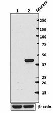
Total cell lysate from NIH3T3 cells (lane 1, 15 µg) and HeLa cells (lane 2, 15 µg) were resolved by electrophoresis (4-20% Tris-Glycine gel), transferred to nitrocellulose, and probed with purified anti-IDH1 antibody (Clone O92H9) antibody. Proteins were visualized using an HRP Goat anti-mouse IgG Antibody and chemiluminescence detection. Purified anti-β-actin antibody (poly6221) was used as a loading control. -

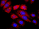
HeLa cells were fixed with ice cold methanol for five minutes, and blocked with 5% FBS for 30 minutes. Then, the cells were intracellularly stained with 2 µg/mL anti-human IDH1 (clone O92H9) for two hours followed by 2 µg/mL Alexa Fluor® 594 Goat anti-mouse IgG (minimal x-reactivity) Antibody for one hour at room temperature. Nuclei were counterstained with DAPI (blue). The image was captured with a 60X objective.
| Cat # | Size | Price | Quantity Check Availability | Save | ||
|---|---|---|---|---|---|---|
| 685302 | 100 µg | £182 | ||||
IDH1, also known as Isocitrate Dehydrogenase 1, enzyme plays and important role in lipid biosynthesis in different tissues such as the liver, adipose tissues, brain and tumors. IDH1 normally catalyze the decarboxylation of isocitrate to generate α-ketoglutarate (αKG), but recurrent mutations at Arg132 of IDH1 confer a neomorphic enzyme activity that catalyzes reduction of αKG into the putative oncometabolite D-2-hydroxyglutate (D2HG). D2HG inhibits αKG-dependent dioxygenases and is thought to create a cellular state permissive to malignant transformation by altering cellular epigenetics and blocking normal differentiation processes. To date, mutations in the IDH1 gene have been found in numerous cancers with the highest frequencies occurring in gliomas, chondrosarcomas or enchondromas, and cholangiocarcinomas. In addition, mutation of IDH1 is closely related to the progress and prognosis of certain tumors. Therefore, IDH1 is considered to be a promising biomarker for early diagnosis, prognosis and targeted therapy for tumors.
Product DetailsProduct Details
- Verified Reactivity
- Human
- Antibody Type
- Monoclonal
- Host Species
- Mouse
- Immunogen
- Purified full length recombinant IDH1 expressed in 293T cells.
- Formulation
- Phosphate-buffered solution, pH 7.2, containing 0.09% sodium azide.
- Preparation
- The antibody was purified by affinity chromatography.
- Concentration
- 0.5 mg/ml
- Storage & Handling
- The antibody solution should be stored undiluted between 2°C and 8°C.
- Application
-
WB - Quality tested
KO/KD-WB, ICC - Verified - Recommended Usage
-
Each lot of this antibody is quality control tested by Western blotting. For Western blotting, the suggested use of this reagent is 0.5 - 2.0 µg/ml. For immunocytochemistry, a concentration range of 1.0 - 5.0 μg/ml is recommended. It is recommended that the reagent be titrated for optimal performance for each application.
- Application Notes
-
Does not react with mouse IDH1.
For immunofluorescence microscopy, positive staining was observed in the cytoplasm of HeLa cells.
- RRID
-
AB_2616768 (BioLegend Cat. No. 685302)
Antigen Details
- Structure
- 414 amino acids with the predicted molecular weight of approximately 47 kD.
- Distribution
-
Cytoplasm and peroxisome.
- Function
- IDH1 enzyme plays an important role in lipid biosynthesis in different tissues such as the liver, adipose tissues, brain and tumors. Mutation of IDH is closely related to the progress and prognosis of certain tumors.
- Interaction
- Isocitrate
- Biology Area
- Cancer Biomarkers, Cell Biology
- Antigen References
-
1. De Quintana-Schmidt C, et al. 2015. Neurocirugia (Astur.) 26:276.
2. Waitkus MS, et al. 2016. Neuro. Oncol. 18:16.
3. Liu X, et al. 2015. Histol. Histopathol. 30:1155.
4. Fathi AT, et al. 2015. Semin. Hematol. 52:165.
5. Vigneswaran K, et al. 2015. Ann. Transl. Med. 3:95.
6. Bogdanovic E. 2015. Biochim. Biophys. Acta. 1850(9):1781-5.
7. Parker SJ, et al. 2015. Pharmacol. Ther. 152:54.
8. Krell D, et al. 2013. Future Oncol. 9:1923.
9. McKenney AS, et al. 2013. J. Clin. Invest. 123:3672. - Gene ID
- 3417 View all products for this Gene ID
- UniProt
- View information about IDH1 on UniProt.org
Related Pages & Pathways
Pages
Related FAQs
Other Formats
View All IDH1 Reagents Request Custom Conjugation| Description | Clone | Applications |
|---|---|---|
| Purified anti-IDH1 | O92H9 | WB,KO/KD-WB,ICC |
Compare Data Across All Formats
This data display is provided for general comparisons between formats.
Your actual data may vary due to variations in samples, target cells, instruments and their settings, staining conditions, and other factors.
If you need assistance with selecting the best format contact our expert technical support team.
 Login / Register


Follow Us