- Clone
- 1D6-E3 (See other available formats)
- Regulatory Status
- RUO
- Other Names
- Inosine Monophosphate Dehydrogenase 1, Inosine-5'-Monophosphate Dehydrogenase 1, IMP Dehydrogenase 1, IMPD1, IMP (Inosine Monophosphate) Dehydrogenase 1, Inosine Monophosphate Dehydrogenase Type 1, IMPDH 1
- Isotype
- Mouse IgG2b, κ
- Ave. Rating
- Submit a Review
- Product Citations
- publications

-

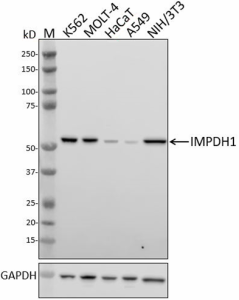
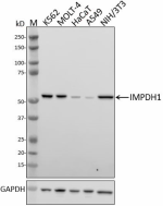
Whole cell extracts (15 µg total protein) from the indicated cell lines were resolved by 4-12% Bis-Tris gel electrophoresis, transferred to a PVDF membrane, and probed with 1.0 µg/mL of purified anti-IMPDH1 antibody (clone 1D6-E3) overnight at 4°C. Proteins were visualized by chemiluminescence detection using HRP goat anti-mouse IgG antibody (Cat. No. 405306) at a 1:3000 dilution. Direct-Blot™ HRP anti-GAPDH antibody (Cat. No. 607904) was used as a loading control at a 1:50000 dilution (lower). Lane M: Molecular weight marker. -

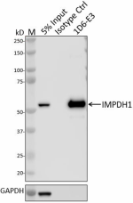
Whole cell extracts (250 µg total protein) prepared from MOLT4 cells were immunoprecipitated overnight with 2.5 µg of purified mouse IgG1, κ isotype ctrl antibody (Cat. No. 401402) or purified anti-IMPDH1 antibody (clone 1D6-E3). The resulting IP fractions and whole cell extract input (5%) were resolved by 4-12% Bis-Tris gel electrophoresis, transferred to a PVDF membrane and probed with a rabbit control antibody against a separate epitope of IMPDH. Lane M: Molecular weight marker. -

HeLa cells were fixed with 4% paraformaldehyde for 10 minutes, permeabilized with Triton X-100 for 10 minutes, and blocked with 5% FBS for 60 minutes. Cells were then intracellularly stained with a 1 µg/mL of either purified mouse IgG1, κ isotype ctrl antibody (Cat. No. 401402) (panel A) or purified anti-IMPDH1 antibody (clone 1D6-E3) (panel B) overnight at 4°C, followed by incubation with Alexa Fluor® 594 goat anti-mouse IgG antibody (Cat. No. 405326) at 2.0 µg/mL. Nuclei were counterstained with DAPI and the image was captured with a 60x objective.
| Cat # | Size | Price | Quantity Check Availability | Save | ||
|---|---|---|---|---|---|---|
| 935201 | 25 µg | £70 | ||||
| 935202 | 100 µg | £174 | ||||
Inosine-5'-monophosphate dehydrogenase 1 (IMPDH1) was initially identified as an oncogene in several human cancers. IMPDH1 catalyzes the conversion of inosine-5'-monophosphate (IMP) to xanthosine-5'-monophosphate (XMP) and is the first committed rate-limiting step in the de novo synthesis of GTP, and consequently it is involved in the regulation of cell growth. IMPDH1 is also part of the composition of the cytoplasmic rod and ring structures (RR) and is involved in maintaining cellular nucleotide pools needed for DNA and RNA synthesis.
Product DetailsProduct Details
- Verified Reactivity
- Human, Mouse, Hamster
- Antibody Type
- Monoclonal
- Host Species
- Mouse
- Immunogen
- Full length IMPDH purified from a Chinese hamster V79 cell variant
- Formulation
- Phosphate-buffered solution, pH 7.2, containing 0.09% sodium azide
- Preparation
- The antibody was purified by affinity chromatography.
- Concentration
- 0.5 mg/mL
- Storage & Handling
- The antibody solution should be stored undiluted between 2°C and 8°C.
- Application
-
WB - Quality tested
IP, ICC - Verified - Recommended Usage
-
Each lot of this antibody is quality control tested by western blotting. For western blotting, the suggested use of this reagent is 0.25 - 1.0 µg/mL. For immunoprecipitation, the suggested use of this reagent is 2.5 µg/test. For immunocytochemistry, a concentration range of 1.0 - 5.0 μg/mL is recommended. It is recommended that the reagent be titrated for optimal performance for each application.
- Application Notes
-
Clone recognizes human, mouse and hamster IMPDH1 and is predicted to recognize the rodent orthologs. Clone is likely to cross react with IMPDH2.
For ICC testing, HeLa cells were fixed in 4% PFA and permeabilized with either Triton X-100 or methanol; both methods produced strong staining at 1 µg/mL. - RRID
-
AB_2832890 (BioLegend Cat. No. 935201)
AB_2832890 (BioLegend Cat. No. 935202)
Antigen Details
- Structure
- IMPDH1 is an 514 aminoacid protein with a predicted molecular weight of 56 kD.
- Distribution
-
Localized to the cytosol filamentous rods and rings / Ubiquitously expressed
- Function
- Regulation of cell growth, and biosynthesis of de novo GTP
- Biology Area
- Cell Cycle/DNA Replication, Cell Proliferation and Viability, DNA Repair/Replication
- Antigen References
-
- Calise SJ, et al. 2015. Front Immunol. 6:41.
- Liao LX, et al. 2017. Proc Natl Acad Sci U S A. 29.
- Duan S, et al. 2018. J Exp Clin Cancer Res. 37:304.
- Zhao Y, et al. 2018. Biomarkers. 1:9.
- Carcamo WC, et al. 2011. PLoS One. 6.
- Xu Y, et al. 2017. Sci Rep. 7:745.
- Glesne DA, et al. 1991. Mol Cell Biol. 11:5417-25.
- Hager PW, et al. 1995. Biochem Pharmacol. 49:1323-9.
- Gene ID
- 3614 View all products for this Gene ID
- UniProt
- View information about IMPDH1 on UniProt.org
Related Pages & Pathways
Pages
Related FAQs
Other Formats
View All IMPDH1 Reagents Request Custom Conjugation| Description | Clone | Applications |
|---|---|---|
| Purified anti-IMPDH1 | 1D6-E3 | WB,IP,ICC |
Compare Data Across All Formats
This data display is provided for general comparisons between formats.
Your actual data may vary due to variations in samples, target cells, instruments and their settings, staining conditions, and other factors.
If you need assistance with selecting the best format contact our expert technical support team.

 Login / Register


Follow Us