 Login / Register


- Clone
- O4 (See other available formats)
- Regulatory Status
- RUO
- Other Names
- Galactocerebroside O4, Oligodendrocyte marker O4, Oligodendrocyte O4 antigen, Sulfated Galactocerebroside O4
- Isotype
- Mouse IgM, κ
- Ave. Rating
- Submit a Review
- Product Citations
- publications

-

IHC staining using purified anti-Oligodendrocyte marker O4 (clone O4) on formalin-fixed paraffin-embedded human cerebellum tissue. The tissue was incubated overnight at 4°C with 0.5 μg/mL of anti-Oligodendrocyte marker O4 (panels A and B) or without primary antibody (panels C and D), followed by incubation with 2.5 μg/mL of Alexa Fluor® 647 goat anti-mouse IgM (red) (Cat. No. 406526) for one hour at room temperature. Nuclei were counterstained with DAPI (blue) (Cat. No. 422801), and the slide was mounted with ProLong™ Gold Antifade mountant reagent. The images were captured with a 40X objective. Panels A and C are merged images of DAPI and Alexa Fluor® 647 channels, and panels B and D are images showing only Alexa Fluor® 647 channel. Scale bar = 50 μm -

Differentiated rat cortical stem cells were fixed with 4% paraformaldehyde and permeabilized with methanol. Cells were intracellularly stained with 5.0 µg/mL of purified anti-Oligodendrocyte marker O4 (clone O4) overnight at 4°C, followed by incubation with Alexa Fluor® 647 anti-mouse IgM (Cat. No. 406526) at 2.0 µg/mL. Nuclei were counterstained with DAPI and the image was captured with a 60X objective. Scale bar: 50 μm -

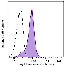
Differentiated rat cortical stem cells were fixed and permeabilized using Cyto-Fast™ Fix/Perm Buffer Set (Cat. No. 426803) and stained with purified anti-Oligodendrocyte marker O4 antigen (clone O4) (blue dot plot), or mouse IgM, κ isotype control (Cat. No. 401602) (red dot plot), followed by Alexa Fluor® 647 anti-mouse IgM (Cat. No. 406526). Left density plot (FSC-A vs. SSC-A) shows the gated population (stem cells and complex cells, and right dot plots (Alexa Fluor® 647 vs. FSC-A) shows stain intensity for each specific gated population.
| Cat # | Size | Price | Quantity Check Availability | Save | ||
|---|---|---|---|---|---|---|
| 609551 | 25 µg | £107 | ||||
| 609552 | 100 µg | £310 | ||||
Oligodendrocytes are the myelinating cells of the central nervous system (CNS). Similar to the Schwann cells in the peripheral nervous system (PNS), their function is to provide support and isolation to neuronal axons. Their importance has been denoted on de-myelinating diseases such as multiple sclerosis, amyotrophic lateral sclerosis, periventricular leukomalacia, and also, they have been associated with dementia, schizophrenia, cognitive impairment, and bipolar disorder. Oligodendrocytes develop during embryogenesis; however, a small proportion of progenitor stem cells are present in the adult brain. Thus, three stages of differentiation can be identified in the cerebral white matter, and specific markers have been associated which each particular stage. Sulfated Galactocerebroside O4 (marker O4) is a major galactosphingolipid component of oligodendrocyte plasma membrane. It is produced from early immature non-myelinating oligodendrocytes to mature myelinating oligodendrocytes, and together with expression of other markers, it has been widely used as characteristic marker for these specific differentiation stages.
Product DetailsProduct Details
- Verified Reactivity
- Human, Rat
- Antibody Type
- Monoclonal
- Host Species
- Mouse
- Immunogen
- White matter from bovine corpus callosum (from developer)
- Formulation
- Phosphate-buffered solution, pH 7.2, containing 0.09% sodium azide
- Preparation
- The antibody was purified by affinity chromatography.
- Concentration
- 0.5 mg/mL
- Storage & Handling
- The antibody solution should be stored undiluted between 2°C and 8°C.
- Application
-
IHC-P - Quality tested
ICC, ICFC - Verified - Recommended Usage
-
Each lot of this antibody is quality control tested by formalin-fixed paraffin-embedded immunohistochemical staining. For immunohistochemistry on testis samples, a concentration range of 1.25 - 5.0 µg/mL is suggested. For immunohistochemistry on cerebellum samples, a concentration range of 0.5 - 1.25 µg/mL is suggested. For immunocytochemistry, a concentration range of 2.5 - 5.0 μg/mL is recommended. For flow cytometric staining, the suggested use of this reagent is ≤ 0.5 µg per million cells in 100 µL volume. It is recommended that the reagent be titrated for optimal performance for each application.
- Application Notes
-
Clone O4 recognizes specifically O4 antigen by immunocytochemistry in oligodendrocytes derived from cortical stem cells using different fixation/permeabilization methods such as methanol, PFA/Triton X-100, and PFA/methanol, where the last method shows the highest signal intensity.
Following immunohistochemistry in paraffin sections, clone O4 showed specific high staining on cerebellum white matter, and very low to none staining in the molecular layer at the recommended concentration.
By flow cytometry staining on differentiated cortical stem cells, clone O4 stained at high intensity complex cells (oligodendrocytes) compared with low complexity cells (undifferentiated progenitor stem cells). - RRID
-
AB_2927990 (BioLegend Cat. No. 609551)
AB_2927990 (BioLegend Cat. No. 609552)
Antigen Details
- Structure
- It is a sulfated galactocerebroside (glycosphingolipid), composed of a single galactose linked to a ceramide
- Distribution
-
Present in the plasma membrane of early immature to mature oligodendrocytes
- Cell Type
- Neural Stem Cells, Oligodendrocytes
- Biology Area
- Neurodegeneration, Neuroscience, Neuroscience Cell Markers
- Antigen References
-
- Bansal R, et al. 1989. J Neurosci Res. 24:548-57.
- Bansal R, et al. 1999. J Neurosci. 19:7913-24.
- Ahmad I, et al. 2021. PloS One. 16:e0256155.
- Rasti BF, et al. 2020. Int J Nanomedicine. 15:3903-3920.
- Gene ID
- NA
- UniProt
- View information about Oligodendrocyte Marker O4 on UniProt.org

Related FAQs
Other Formats
View All Oligodendrocyte Marker O4 Reagents Request Custom Conjugation| Description | Clone | Applications |
|---|---|---|
| Purified anti-Oligodendrocyte Marker O4 | O4 | IHC-P,ICC,ICFC |
Customers Also Purchased

Compare Data Across All Formats
This data display is provided for general comparisons between formats.
Your actual data may vary due to variations in samples, target cells, instruments and their settings, staining conditions, and other factors.
If you need assistance with selecting the best format contact our expert technical support team.


Follow Us