- Clone
- W17231A (See other available formats)
- Regulatory Status
- RUO
- Other Names
- TPX2, Microtubule Nucleation Factor, Hepatocellular Carcinoma-Associated Antigen 519, Hepatocellular Carcinoma-Associated Antigen 90, P100, DIL2
- Isotype
- Rat IgG2a, λ
- Ave. Rating
- Submit a Review
- Product Citations
- publications

-

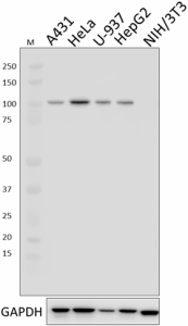
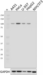
Whole cell extracts (15 µg protein) from the indicated cell lines were resolved by 4-12% Bis-Tris gel electrophoresis, transferred to a PVDF membrane, and probed with 1.0 µg/mL (1:500 dilution) of Purified anti-TPX2 Antibody, clone W17231A, overnight at 4°C. Proteins were visualized by chemiluminescence detection using HRP goat anti-rat IgG Antibody (Cat. No. 405405) at a 1:3000 dilution. Direct-Blot™ HRP anti-GAPDH Antibody (Cat. No. 607904) was used as a loading control at a 1:25000 dilution (lower). Lane M: Molecular Weight marker. -

HeLa cells were fixed with 4% paraformaldehyde for 10 minutes, permeabilized with ice-cold methanol for 10 minutes, and blocked with 5% FBS for 60 minutes. Cells were then intracellularly stained with a 1:500 dilution (1.0 µg/mL) of either Purified Rat IgG2ba, κ Isotype Control Antibody (Cat. No. 400502, panel A) or Purified anti-TPX2 Antibody (panel B) overnight at 4°C, followed by incubation with Alexa Fluor® 594 goat anti-rat IgG (Cat. No. 405422) at 2.0 µg/mL. Nuclei were counter-stained with DAPI, and the image was captured with a 60X objective. -

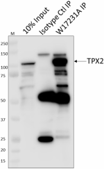
Whole cell extracts (250 μg total protein) prepared from HeLa cells were immunoprecipitated overnight with 2.5 µg of Purified rat IgG2a, κ Isotype Control Antibody (Cat. No. 400502) or Purified anti-TPX2 Antibody, clone W17231A. The resulting IP fractions and whole cell extract input (10%) were resolved by 4-12% Bis-Tris gel electrophoresis, transferred to a PVDF membrane, and probed with a 1.0 µg/mL (1:500 dilution) of W17231A. Lane M: Molecular Weight marker.
| Cat # | Size | Price | Quantity Check Availability | Save | ||
|---|---|---|---|---|---|---|
| 620601 | 25 µg | £61 | ||||
| 620602 | 100 µg | £157 | ||||
TPX2 protein, also known as targeting protein for Xklp2, is strictly associated with the spindle pole and mitotic spindle during mitosis. In the G2/S position of the cell cycle, TPX2 is diffusely distributed throughout the nucleus. TPX2 has been shown to be highly expressed in lung carcinoma cell lines, but not in normal lung tissues. TPX2 is thought to be required for the Ran-GTP dependent assembly of microtubules around chromosomes required to generate stable bipolar spindles with overlapping anti-parallel microtubule arrays and may also be involved in targeting Aurora-A kinase to the mitotic spindle. TPX2 has been shown to interact with a large number of proteins including serine/threonine protein kinase 6, ribosomal protein 6, Bop-1, α-tubulin, and nucleolin among others. TPX2 can be modified by phosphorylation on serine 738.
Product DetailsProduct Details
- Verified Reactivity
- Human
- Antibody Type
- Monoclonal
- Host Species
- Rat
- Immunogen
- Synthetic peptide corresponding to amino acid residues 413-429 of human TPX2 protein.
- Formulation
- Phosphate-buffered solution, pH 7.2, containing 0.09% sodium azide.
- Preparation
- The antibody was purified by affinity chromatography.
- Concentration
- 0.5 mg/mL
- Storage & Handling
- The antibody solution should be stored undiluted between 2°C and 8°C.
- Application
-
WB - Quality tested
ICC, IP - Verified - Recommended Usage
-
Each lot of this antibody is quality control tested by Western blotting. For Western blotting, the suggested use of this reagent is 1.0 µg per ml. For immunocytochemistry, a concentration range of 1.0 - 5.0 μg/ml is recommended. For immunoprecipitation, the suggested use of this reagent is 2.0 µg per ml. It is recommended that the reagent be titrated for optimal performance for each application.
- Application Notes
-
When tested for ICC, W17231A displayed heterogeneous nuclear localization in HeLa cells.
When tested for IP, W17231A detected minor, lower MW bands that were present in both input and IP fractions. These same bands were not observed in other WB experiments. We predict that these are minor degradation products due to slow lysis methods of extraction. - RRID
-
AB_2810681 (BioLegend Cat. No. 620601)
AB_2810681 (BioLegend Cat. No. 620602)
Antigen Details
- Structure
- TPX2 is a 747 amino acid protein with a predicted molecular weight of 86 kD.
- Distribution
-
Nuclear, during mitosis strictly associated with spindle pole and mitotic spindle. In G2/S cell cycle, diffusely distributed throughout nucleus. Highly expressed in lung carcinoma cell lines, but not in normal lung tissues.
- Function
- Microtubule assembly
- Biology Area
- Cell Biology, Cell Cycle/DNA Replication, Cell Motility/Cytoskeleton/Structure, Cell Proliferation and Viability
- Molecular Family
- Microtubules
- Antigen References
-
- Fung T, et al. 2007. Mol Biol Cell. 2.042361111.
- Bibby R, et al. 2009. J Biol Chem. 284:33177.
- Warner S, et al. 2009. Clin Cancer Res. 5.152083333.
- Eot-Houllier G, et al. 2010. J Biol Chem. 285:29556.
- Tagal V, et al. 2017. Nat Commun. 8:14098.
- Gene ID
- 22974 View all products for this Gene ID
- UniProt
- View information about TPX2 on UniProt.org
Related FAQs
Other Formats
View All TPX2 Reagents Request Custom Conjugation| Description | Clone | Applications |
|---|---|---|
| Purified anti-TPX2 | W17231A | WB,ICC,IP |
Compare Data Across All Formats
This data display is provided for general comparisons between formats.
Your actual data may vary due to variations in samples, target cells, instruments and their settings, staining conditions, and other factors.
If you need assistance with selecting the best format contact our expert technical support team.

 Login / Register


Follow Us