- Clone
- A16043B (See other available formats)
- Regulatory Status
- RUO
- Other Names
- Zeta chain of TCR associated protein kinase 70
- Isotype
- Mouse IgG1, κ
- Ave. Rating
- Submit a Review
- Product Citations
- publications
-

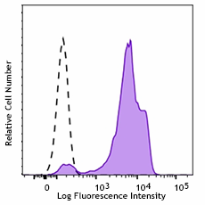
Human peripheral blood lymphocytes treated with True-Nuclear™ Transcription Factor Buffer Set, then stained with ZAP-70 (clone A16043B) Purified (filled histogram) or Purified Mouse IgG1, κ Isotype Ctrl (open histogram) followed by PE Goat anti-mouse IgG (minimal x-reactivity).
| Cat # | Size | Price | Quantity Check Availability | Save | ||
|---|---|---|---|---|---|---|
| 693502 | 100 µg | £141 | ||||
ZAP-70 is a 70kD protein expressed near the surface membrane of T-cells and NK cells. It is part of the intracellular signaling mechanism of the T-cell receptor, and plays integral role in T-cell signaling where it interacts with CD3-ζ to phosphorylate transmembrane protein LAT, which facilitates the binding of many signaling proteins. ZAP-70 plays a key role in the activation of T-cells, and their subsequent proliferation, differentiation, and production of cytokines in response to varied stimuli. It is used clinically as a prognostic marker helping differentiate the various types of chronic lymphocytic leukemia (CLL).
Product DetailsProduct Details
- Verified Reactivity
- Human, Mouse
- Antibody Type
- Monoclonal
- Host Species
- Mouse
- Immunogen
- Human ZAP 70 peptide phosphorylated at Y493. Complete Freund’s adjuvant.
- Formulation
- Phosphate-buffered solution, pH 7.2, containing 0.09% sodium azide.
- Preparation
- The antibody was purified by affinity chromatography.
- Concentration
- 0.5 mg/ml
- Storage & Handling
- The antibody solution should be stored undiluted between 2°C and 8°C.
- Application
-
ICFC - Quality tested
- Recommended Usage
-
Each lot of this antibody is quality control tested by intracellular immunofluorescent staining with flow cytometric analysis. For flow cytometric staining, the suggested use of this reagent is ≤ 0.125 µg per million cells in 100 µl volume. It is recommended that the reagent be titrated for optimal performance for each application.
- Application Notes
-
CD3, CD19, and CD56 co-staining indicates that this clone does not bind B-cells and will not bind Syk. Clone A16043B cross-reacts with mouse species.
This clone, A16043B, recognizes both non-phosphorylated ZAP-70 and phosphorylated forms of ZAP-70. - RRID
-
AB_2810698 (BioLegend Cat. No. 693502)
Antigen Details
- Structure
- Protein tyrosine kinase, contains two SH2 domains, 70 kD.
- Distribution
-
T cells, NK cells, and B-cell chronic lymphocytic leukemia (B-CLL).
- Function
- Signal transduction, associates with the T-cell antigen receptor zeta chain tyrosine-based activation motif when phosphorylated. Defects in ZAP70 causes selective T cell defect.
- Ligand/Receptor
- Binds to CD3 zeta chain, also associates with a variety of proteins including SLA, Fyn, RasGap, Lck, Vav1, and Shc.
- Cell Type
- Leukemia, NK cells, T cells
- Biology Area
- Cell Biology, Immunology, Signal Transduction
- Molecular Family
- TCRs
- Antigen References
-
1. Qian D, et al. 1997. J. Exp. Med. 185:1253. (ICFC)
2. Crespo M, et al. 2003. N. Engl. J. Med. 348:1764. (WB, IP)
3. Jamroziak K, et al. 2009. Cancer Epidemiol. Biomarkers Prev. 18:945. - Gene ID
- 7535 View all products for this Gene ID 22637 View all products for this Gene ID
- UniProt
- View information about ZAP-70 on UniProt.org
Related FAQs
Other Formats
View All ZAP-70 Reagents Request Custom Conjugation| Description | Clone | Applications |
|---|---|---|
| PE anti-ZAP-70 | A16043B | ICFC |
| Alexa Fluor® 488 anti-ZAP-70 | A16043B | ICFC |
| Purified anti-ZAP-70 | A16043B | ICFC |
| Alexa Fluor® 647 anti-ZAP-70 | A16043B | ICFC |
| TotalSeq™-C1490 anti-ZAP70 Antibody | A16043B | ICPG |
Compare Data Across All Formats
This data display is provided for general comparisons between formats.
Your actual data may vary due to variations in samples, target cells, instruments and their settings, staining conditions, and other factors.
If you need assistance with selecting the best format contact our expert technical support team.
-
PE anti-ZAP-70
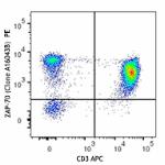
Human peripheral blood lymphocytes were surface stained with... 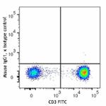
-
Alexa Fluor® 488 anti-ZAP-70
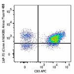
Human peripheral blood lymphocytes were surface stained with... 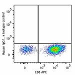
-
Purified anti-ZAP-70
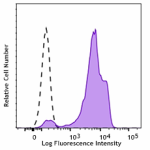
Human peripheral blood lymphocytes treated with True-Nuclear... -
Alexa Fluor® 647 anti-ZAP-70
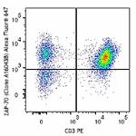
Human peripheral blood lymphocytes were surface stained with... 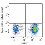
-
TotalSeq™-C1490 anti-ZAP70 Antibody
 Login / Register


Follow Us