IMPORTANT NOTICE: Our email addresses will soon change. Learn more >>

BioLegend offers extensive and diverse antibodies with one of the largest clone libraries in the industry. With two decades of antibody crafting experience, we offer high-quality antibodies validated for several applications including western blotting, flow cytometry, and microscopy. Within microscopy, immunohistochemistry can drive discovery in research fields including oncology, immunology, and neurology. With this in mind, we are continuously working to add further applications to existing products. Read below to learn about some of our newly validated antibodies for immunohistochemistry application.


View our IHC/ICC applications webpage for IHC protocols and a complete list of IHC validated antibodies.


Specificity Format Clone
anti-AMPKα Phospho (Thr172) A647 A20017A
anti-Avi-tag A488, A594, A647 W19258A
anti-CD31 (PECAM-1) PURE, A647 JC70
anti-CD340 (ErbB2/HER-2) PURE A20145B
anti-CD340 (ErbB2/HER-2) PURE W22266D
anti-c-Fos A647 7D6-A9-D4
anti-c-Fos Phospho (Ser32) PURE, A647 A22009C
anti-CREB1 Phospho (Ser133) PURE A22026B
anti-IBA1 PURE A19290E
anti-MDM2 PURE W23003F
anti-Nestin PURE Poly22A17
anti-Neurofilament H (NF-H), Nonphosphorylated SPKYG_570 SMI 32
anti-Neurofilament H (NF-H), Phosphorylated SPKYG_570 SMI 31
anti-NF-kB p65 PURE A21012A
anti-NF-κB p65 Phospho (Ser529) PURE A21012B
anti-NOS3 (eNOS) PURE W22110B
anti-Olig2 PURE W21023A
anti-Oligodendrocyte Marker O1 A488, A594, A647 O1
anti-Pan-Cytokeratin A488, A594, A647 AE-1/AE-3
anti-PTEN Phospho (Ser380) PURE, A488, A647 A22005D
anti-RBFOX3 (NeuN) A488, A647 W18230A
anti-SOX17 Recombinant PURE O92B6.Rec
anti-Tau Phospho (Thr231) PURE A23009A
anti-TMEM119 (Extracellular) A488, A594, A647 A16075D
anti-Vimentin A488 W16220A
anti-α Smooth Muscle Actin PURE, A488, A647 1A4
anti-β-Amyloid, 1-16 Recombinant PURE 6E10.Rec
anti-β-Amyloid, 17-28 PURE A18104E
anti-β-Catenin non-phospho (active) (S33/S37/T41) PURE W22009A
anti-γ-Catenin PURE W22004E


IHC staining of Alexa Fluor® 647 anti-Pan Cytokeratin (clone AE-1/AE-3) on formalin-fixed paraffin-embedded human tonsil tissue. Following antigen retrieval using 1X Tris-EDTA pH 9.0 Antigen Retrieval Buffer (Cat. No. 422704), the tissue was incubated either without (panel A) or with (panel B) Alexa Fluor® 647 anti-Pan Cytokeratin (clone AE-1/AE-3). Nuclei were counterstained with DAPI (Cat. No. 422801). Images were captured with a 40X objective and merged. Scale bar: 50 μm


IHC staining of Purified anti-CD340 (ErbB2/HER-2) (clone W22266D) (magenta) on formalin-fixed paraffin-embedded human breast cancer tissue. Following antigen retrieval using Sodium Citrate H.I.E.R. (Cat. No. 925501), the tissue was incubated without (panels A and C) and with (panels B and D) the antibody, followed by incubation with Alexa Fluor® 647 Goat anti-rat IgG for 1 hour at room temperature. Nuclei were counterstained with DAPI (blue) (panels A and B). Images were captured with a 40X objective. Scale bar: 50 µm


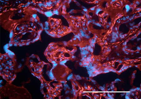
Formalin-fixed paraffin-embedded human placenta tissue was stained with Purified anti-human Podoplanin followed by Alexa Fluor® 647 goat anti-rat IgG antibody (red). Nuclei were counterstained with DAPI (blue). Scalebar = 50 µM.


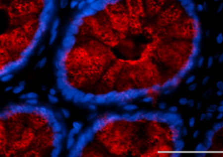
Formalin-fixed paraffin-embedded human colon tissue was stained with Purified anti-MUC1 antibody (clone 16A) followed by Alexa Fluor® 647 goat anti-mouse IgG antibody (red). Nuclei were counterstained with DAPI (blue). Scalebar = 50 µM.


ProductsHere

Login / Register
Remember me
Forgot your password? Reset password?
Create an Account