PerCP/Cyanine5.5 anti-mouse CD8a Antibody

Pricing & Availability
Clone
53-6.7 (See other available formats)
Regulatory Status
RUO
Other Names
T8, Lyt2, Ly-2
Isotype
Rat IgG2a, κ
Ave. Rating
Submit a Review
Product Citations
publications
53-6dot7_PerCPCyanine55_CD8a_Antibody_102318
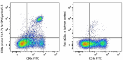
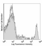
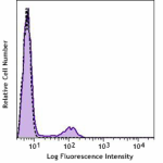
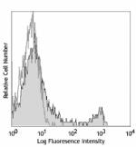
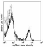
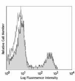
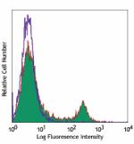
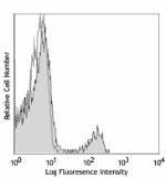
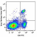
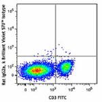
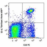
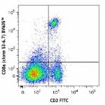
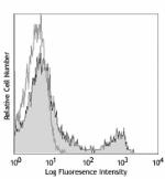
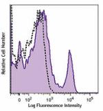
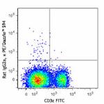
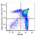
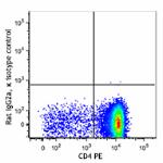
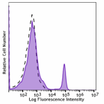
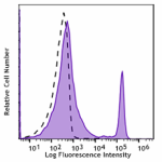
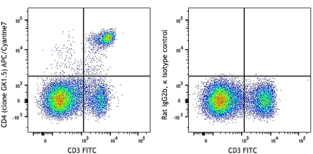
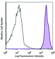
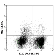
C57BL/6 mouse splenocytes were stained with CD3ε FITC and CD8a (clone 53-6.7) PerCP/Cyanine5.5, or Rat IgG2a, κ PerCP/Cyanine5.5 isotype control (bottom).
  • 53-6dot7_PerCPCyanine55_CD8a_Antibody_102318
    C57BL/6 mouse splenocytes were stained with CD3ε FITC and CD8a (clone 53-6.7) PerCP/Cyanine5.5, or Rat IgG2a, κ PerCP/Cyanine5.5 isotype control (bottom).
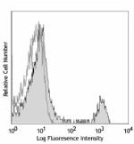
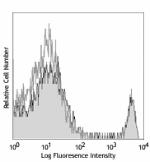
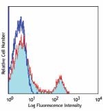
Compare all formats See PerCP/Cyanine5.5 spectral data
Cat # Size Price Quantity Check Availability Save
100733 25 µg 70€
Check Availability


Need larger quantities of this item?
Request Bulk Quote
100734 100 µg 190€
Check Availability


Need larger quantities of this item?
Request Bulk Quote
Description

CD8, also known as Lyt-2, Ly-2, or T8, consists of disulfide-linked α and β chains that form the α(CD8a)/β(CD8b) heterodimer and α/α homodimer. CD8a is a 34 kD protein that belongs to the immunoglobulin family. The CD8 α/β heterodimer is expressed on the surface of most thymocytes and a subset of mature TCR α/β T cells. CD8 expression on mature T cells is non-overlapping with CD4. The CD8 α/α homodimer is expressed on a subset of γ/δ TCR-bearing T cells, NK cells, intestinal intraepithelial lymphocytes, and lymphoid dendritic cells. CD8 is an antigen co-receptor on T cells that interacts with MHC class I on antigen-presenting cells or epithelial cells. CD8 promotes T cell activation through its association with the TCR complex and protein tyrosine kinase lck.

Product Details
Technical Data Sheet (pdf)

Product Details

Verified Reactivity
Mouse
Antibody Type
Monoclonal
Host Species
Rat
Immunogen
Mouse thymus or spleen
Formulation
Phosphate-buffered solution, pH 7.2, containing 0.09% sodium azide.
Preparation
The antibody was purified by affinity chromatography, and conjugated with PerCP/Cyanine5.5 under optimal conditions.
Concentration
0.2 mg/mL
Storage & Handling
The CD8a antibody solution should be stored undiluted between 2°C and 8°C, and protected from prolonged exposure to light. Do not freeze.
Application

FC - Quality tested

Recommended Usage

Each lot of this antibody is quality control tested by immunofluorescent staining with flow cytometric analysis. For flow cytometric staining, the suggested use of this reagent is < 1.0 µg per million cells in 100 µL volume. It is recommended that the reagent be titrated for optimal performance for each application.

* PerCP/Cyanine5.5 has a maximum absorption of 482 nm and a maximum emission of 690 nm.

Excitation Laser
Blue Laser (488 nm)
Green Laser (532 nm)/Yellow-Green Laser (561 nm)
Application Notes

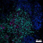
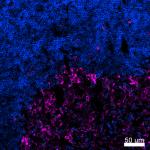
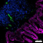
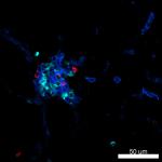
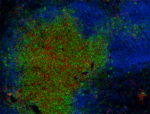
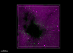
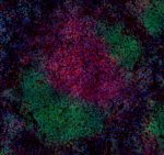
Clone 53-6.7 antibody competes with clone 5H10-1 antibody for binding to thymocytes3. The 53-6.7 antibody has been reported to block antigen presentation via MHC class I and inhibit T cell responses to IL-2. This antibody has also been used for depletion of CD8a+ cells. Additional reported applications (for the relevant formats) include: immunoprecipitation1,3, in vivo and in vitro cell depletion2,10,15, inhibition of CD8 T cell proliferation3, blocking of cytotoxicity3,4, immunohistochemical staining5,6 of acetone-fixed frozen sections and zinc-fixed paraffin-embedded sections, and spatial biology (IBEX)29,30. Clone 53-6.7 is not recommended for immunohistochemistry of formalin-fixed paraffin sections. The Ultra-LEAF™ purified antibody (Endotoxin < 0.01 EU/µg, Azide-Free, 0.2 µm filtered) is recommended for functional assays or in vivo studies (Cat No. 100746).

Application References
  1. Ledbetter JA, et al. 1979. Immunol. Rev. 47:63. (IHC, IP)
  2. Hathcock KS. 1991. Current Protocols in Immunology. 3.4.1. (Deplete)
  3. Takahashi K, et al. 1992. P. Natl. Acad. Sci. USA 89:5557. (Block, IP)
  4. Ledbetter JA, et al. 1981. J. Exp. Med. 153:1503. (Block)
  5. Hata H, et al. 2004. J. Clin. Invest. 114:582. (IHC)
  6. Fan WY, et al. 2001. Exp. Biol. Med. 226:1045. (IHC)
  7. Shih FF, et al. 2006. J. Immunol. 176:3438. (FC)
  8. Kamimura D, et al. 2006. J. Immunol. 177:306.
  9. Bouwer HGA, et al. 2006. P. Natl. Acad. Sci. USA 103:5102. (FC, Deplete)
  10. Kao C, et al. 2005. Int. Immunol. 17:1607. PubMed
  11. Ko SY, et al. 2005. J. Immunol. 175:3309. (FC) PubMed
  12. Rasmussen JW, et al. 2006. Infect. Immun. 74:6590. PubMed
  13. Lee CH, et al. 2009. Clin. Cancer Res. PubMed
  14. Geiben-Lynn R, et al. 2008. Blood 112:4585. (Deplete) PubMed
  15. Kingeter LM, et al. 2008. J. Immunol. 181:6244. PubMed
  16. Guo Y, et al. 2008. Blood 112:480. PubMed
  17. Andrews DM, et al. 2008. J. Virol. 82:4931. PubMed
  18. Britschqui MR, et al. 2008. J. Immunol. 181:7681. PubMed
  19. Kenna TJ, et al. 2008. Blood 111:2091. PubMed
  20. Jordan JM, et al. 2008. Infect. Immun. 76:3717. PubMed
  21. Todd DJ, et al. 2009. J. Exp. Med. 206:2151. PubMed
  22. Bankoti J, et al. 2010. Toxicol. Sci. 115:422. (FC) PubMed
  23. Medyouf H, et al. 2010. Blood 115:1175. PubMed
  24. Riedl P, et al. 2009. J. Immunol. 183:370. PubMed
  25. Apte SH, et al. 2010. J. Immunol. 185:998. PubMed
  26. Bankoti J, et al. 2010. Toxicol. Sci. 115:422. (FC) PubMed
  27. del Rio ML, et al. 2011. Transpl. Int. 24:501. (FC) PubMed
  28. Cui L, et al. 2015. J Control Release. 206:220. PubMed
  29. Radtke AJ, et al. 2020. Proc Natl Acad Sci U S A. 117:33455-65. (SB) PubMed
  30. Radtke AJ, et al. 2022. Nat Protoc. 17:378-401. (SB) PubMed
Product Citations
  1. Schuster M, et al. 2017. J Immunol. 199:920. PubMed
  2. Yoon J et al. 2018. J Vis Exp. -141 . PubMed
  3. Turecamo SE, et al. 2018. Matrix Biol. 67:01:00. PubMed
  4. Garber C, et al. 2019. Nat Neurosci. 1.802777778. PubMed
  5. Wang X, et al. 2019. Cell Res. 29:787. PubMed
  6. Zheng X, et al. 2019. PLoS Pathog. 15:e1008036. PubMed
  7. Lang J, et al. 2020. Nat Commun. 11:1338. PubMed
  8. Hudson WH, et al. 2020. Immunity. 51(6):1043-1058.e4.. PubMed
  9. Dai L, et al. 2020. Cell. 182(3):722-733.e11. PubMed
  10. Xing J, et al. 2021. Cell Reports. 35(12):109205. PubMed
  11. Zhang H, et al. 2021. Front Immunol. 12:644520. PubMed
  12. Vella JL, et al. 2021. Life Sci Alliance. 4:. PubMed
  13. Nesseler JP, et al. 2020. Cancers (Basel). 12: . PubMed
  14. Behr FM, et al. 2021. Eur J Immunol. 51:151. PubMed
  15. Denny L, et al. 2021. Clin Transl Immunology. 10:e1234. PubMed
  16. Pohlmeier L, et al. 2021. Allergy. 76:2030. PubMed
  17. Stone ET, et al. 2022. Cell Rep. 38:110388. PubMed
  18. Goncharov NV, et al. 2022. J Evol Biochem Physiol. 58:230. PubMed
  19. Wemlinger SM, et al. 2022. J Immunol. 208:1566. PubMed
  20. Parga-Vidal L, et al. 2022. Eur J Immunol. 52:1095. PubMed
  21. Wang S, et al. 2022. Sci Adv. 8:eabn3883. PubMed
  22. Piñeros AR, et al. 2022. Cell Rep. 39:111011. PubMed
  23. Silva Pereira S, et al. 2022. Elife. 11: . PubMed
  24. Hong Y, et al. 2022. J Leukoc Biol. 112:425. PubMed
  25. Balood M, et al. 2022. Nature. 611:405. PubMed
  26. Ge Y, et al. 2022. iScience. 25:105437. PubMed
  27. Wei M, et al. 2022. Front Immunol. 13:1017574. PubMed
  28. Zhu J, et al. 2022. Nat Commun. 13:7466. PubMed
  29. Koutník J, et al. 2022. Front Immunol. 13:1049033. PubMed
  30. Briukhovetska D, et al. 2023. Immunity. 56:143. PubMed
  31. Arimoto KI, et al. 2023. Nat Commun. 14:251. PubMed
  32. Ai L, et al. 2023. Cell Discov. 9:9. PubMed
  33. Chen L, et al. 2023. Bioeng Transl Med. 8:e10377. PubMed
  34. Teymournejad O, et al. 2023. NPJ Vaccines. 8:3. PubMed
  35. Marritt KL, et al. 2023. Front Immunol. 13:1087991. PubMed
  36. Wang R, et al. 2023. iScience. 26:105954. PubMed
  37. Kemna J, et al. 2023. Nat Immunol. 24:414. PubMed
  38. Zhu X, et al. 2023. Front Immunol. 14:1114802. PubMed
  39. Gon Y, et al. 2023. PLoS One. 18:e0279389. PubMed
  40. Scherer S, et al. 2023. Nat Immunol. 24:501. PubMed
  41. Ferapontov A, et al. 2023. Nat Commun. 14:976. PubMed
  42. Shi X, et al. 2023. iScience. 26:106143. PubMed
  43. Pattnaik A, et al. 2023. Vaccines (Basel). 11:. PubMed
  44. Abdelwahab T, et al. 2023. iScience. 26:106698. PubMed
  45. Milighetti M, et al. 2023. iScience. 26:106937. PubMed
  46. Hu Y, et al. 2023. JCI Insight. 8:. PubMed
  47. Murata A, et al. 2020. Front Immunol. 11:775. PubMed
  48. Lefebvre MN, et al. 2021. Cell Rep. 37:109956. PubMed
  49. Kim D, et al. 2021. Nanomicro Lett. 13:31. PubMed
  50. Platt DJ, et al. 2021. Cell Reports. 35(6):109113. PubMed
  51. Gern BH, et al. 2021. Cell Host Microbe. 29(4):594-606.e6. PubMed
  52. Cornax I, et al. 2021. Microorganisms. 9:00. PubMed
  53. Zhou J, et al. 2019. Immunity. 50:403. PubMed
  54. Zhang R, et al. 2019. Cell Rep. 28:2647. PubMed
  55. Barry KC, et al. 2018. Nat Med. 24:1178. PubMed
  56. Zhang P et al. 2019. Cell Rep. 27(11):3117-3123 . PubMed
  57. Kim CJ, et al. 2018. Immunity. 49:1034. PubMed
  58. Aldon Y, et al. 2020. J Immunol. 204:903. PubMed
  59. Van Den Eeckhout B, et al. 2020. NPJ Vaccines. 5:64. PubMed
  60. Yang C, et al. 2021. J Immunother Cancer. 9:. PubMed
  61. Sawada J, et al. 2021. Am J Pathol. 191:396. PubMed
  62. Alotaibi F, et al. 2021. Front Immunol. 11:584937. PubMed
  63. Pack AD, et al. 2021. Cell Reports. 36:109586. PubMed
  64. Delacher M, et al. 2021. Immunity. 54(4):702-720.e17. PubMed
  65. Chen L, et al. 2020. Cancer Immunol Res. 0.457638889. PubMed
  66. Chow MT et al. 2019. Immunity. 50(6):1498-1512 . PubMed
  67. Choi H et al. 2019. Cell Rep. 27(3):806-819 . PubMed
  68. Sultan H, et al. 2018. Cancer Immunol Res. 6:617. PubMed
  69. Yuzhu Hou et al. 2018. Immunity. 49(3):490-503 . PubMed
  70. Hayatsu N et al. 2017. Immunity. 47(2):268-283 . PubMed
  71. Wu J, et al. 2020. Sci Adv. 6:eaba3458. PubMed
  72. Brooks JF, et al. 2020. Cytotherapy. 22:436. PubMed
  73. Hu D, et al. 2021. J Cell Mol Med. 25:2900. PubMed
  74. Juul-Madsen K, et al. 2021. Proc Natl Acad Sci U S A. 118:. PubMed
  75. Xu X, et al. 2021. J Nanobiotechnology. 19:376. PubMed
  76. Sugita J, et al. 2021. Nat Commun. 12:1910. PubMed
  77. Paiva RS, et al. 2021. Eur J Immunol. 51:1968. PubMed
  78. Kienzl M, et al. 2020. Oncoimmunology. 9:1776059. PubMed
  79. Hong JY, et al. 2020. Cell. 180(5):847-861. PubMed
  80. Solanki A, et al. 2018. Development. 145. PubMed
  81. Stivala S, et al. 2019. J Clin Invest. 130:1596. PubMed
  82. Green DP, et al. 2019. Neuron. 101:412. PubMed
  83. Terashima Y, et al. 2020. Nat Commun. 11:609. PubMed
  84. Mizraji* G, et al. 2013. J Vis Exp. 77: 50388. PubMed
  85. Collinson-Pautz M, et al. 2016. PLoS One. 11:e0164547. PubMed
  86. Dos Santos Dias L, et al. 2021. PLoS Pathog. e1009324:17. PubMed
  87. Snyder LM, et al. 2022. Immunohorizons. 6:660. PubMed
  88. Smith AS, et al. 2022. J Immunother Cancer. 10:. PubMed
  89. Agrawal M, et al. 2021. Cells. 10:. PubMed
  90. Yin Q, et al. 2021. Proc Natl Acad Sci U S A. 118: . PubMed
  91. Chen D, et al. 2020. Cancer Immunol Res. 8:883. PubMed
  92. Ruhland MK, et al. 2020. Cancer Cell. 37(6):786-799.e5. PubMed
  93. JI B, et al. 2016. Cell Death Differ. 23:759-75. PubMed
  94. Holvoet B, et al. 2015. Stem Cell Reports. 5: 1183-1195. PubMed
  95. Dekhtiarenko I, et al. 2013. J Immunol. 190:3399. PubMed
  96. Chen X et al. 2017. Cell stem cell. 21(6):747-760 . PubMed
  97. Hönes J, et al. 2017. Sci Rep. . 10.1038/s41598-017-15866-9. PubMed
  98. Xu J et al. 2018. Cell. 173(3):762-775 . PubMed
  99. Kooreman NG et al. 2018. Cell stem cell. 22(4):501-513 . PubMed
  100. Ren J, et al. 2019. PLoS Biol. 17:e3000270. PubMed
  101. Hildebrand KM, et al. 2021. PLoS One. 16:e0253864. PubMed
  102. Shi R, et al. 2022. Theranostics. 12:875. PubMed
  103. Weaver JD, et al. 2022. Oncoimmunology. 11:2141007. PubMed
  104. Hu Y, et al. 2022. J Nanobiotechnology. 20:417. PubMed
  105. Fang E, et al. 2022. MedComm (2020). 3:e117. PubMed
  106. Tondini E, et al. 2022. NPJ Vaccines. 7:64. PubMed
  107. Brandi P, et al. 2022. Cell Rep. 38:110184. PubMed
  108. Chen X, et al. 2021. Cell Rep. 37:109991. PubMed
  109. Zuo W, et al. 2022. Cell Death Discov. 8:257. PubMed
  110. Zuo S, et al. 2021. J Immunother Cancer. 9:. PubMed
  111. Brigas HC, et al. 2021. Cell Reports. 36(9):109574. PubMed
  112. Barsoumian HB, et al. 2020. J Immunother Cancer. 8:00. PubMed
  113. Pilzecker B, et al. 2016. Nucleic Acids Res. 44: 4734 - 4744. PubMed
  114. Niknam S, et al. 2018. Clin Cancer Res. 24:5735. PubMed
  115. Novince CM, et al. 2017. Sci Rep. 7:5747. PubMed
  116. Place D, et al. 2017. PLoS One. 10.1371/journal.pone.0190384. PubMed
  117. Chung L et al. 2018. Cell host & microbe. 23(2):203-214 . PubMed
  118. Kemp V, et al. 2018. Cancer Gene Ther. 26:268. PubMed
  119. Wu J et al. 2019. Immunity. 50(5):1218-1231 . PubMed
  120. Kaur A, et al. 2019. Cancer Discov. 9:64. PubMed
  121. Dutow P, et al. 2014. J Infect Dis. 209:1269. PubMed
  122. White C, et al. 2015. J Immunol. 194:697. PubMed
  123. Uddback I, et al. 2016. Sci Rep. 6:20137. PubMed
  124. Liu W, et al. 2021. Cell Death Discov. 7:136. PubMed
  125. Carbone C, et al. 2021. J Immunother Cancer. 9:. PubMed
  126. Bourque J, et al. 2021. Heliyon. 7:e08311. PubMed
  127. Alameh MG, et al. 2021. Immunity. 54:2877. PubMed
  128. Bullard BL, et al. 2022. NPJ Vaccines. 7:65. PubMed
  129. Sun Y, et al. 2022. Mol Ther Oncolytics. 25:160. PubMed
  130. Frost JN, et al. 2021. Med (N Y). 2:164. PubMed
  131. Ohtsuka J, et al. 2021. iScience. Online ahead of print. PubMed
  132. Maulloo CD, et al. 2021. Front Immunol. 12:714842. PubMed
  133. Zhao D, et al. 2020. Cancer Discov. 10:1374. PubMed
  134. Dobson HE, et al. 2020. Mucosal Immunol. 0.901388889. PubMed
  135. Parayath NN, et al. 2020. Nat Commun. 4.680555556. PubMed
  136. Lee L, et al. 2016. PLoS One. 11:e0167693. PubMed
  137. Ying Zhang et al. 2017. Cancer cell. 32(3):377-391 . PubMed
  138. Karandikar SH, et al. 2019. Anal Chem. 91:3405. PubMed
  139. Wang Z et al. 2018. Immunity. 49(1):80-92 . PubMed
  140. Shi R, et al. 2019. J Cancer. 10:367. PubMed
  141. Karandikar SH, et al. 2019. JCI Insight. 5. PubMed
  142. Zhang W, et al. 2020. Nat Commun. 11:1187. PubMed
  143. Chandra J, et al. 2016. Immunol Cell Biol. 10.1038/icb.2016.83. PubMed
  144. Wuggenig P, et al. 2020. Commun Biol. 3:130. PubMed
  145. Yu H, et al. 2021. Front Oncol. 11:736882. PubMed
  146. Wüthrich M, et al. 2021. MBio. 12:e0201821. PubMed
  147. Ryan NM, et al. 2022. Front Immunol. 13:932742. PubMed
  148. Kohn EM, et al. 2022. J Immunol. 208:1417. PubMed
  149. Matsumura T, et al. 2022. Nat Commun. 13:7064. PubMed
  150. Ghorbani S, et al. 2022. Nat Commun. 13:2445. PubMed
  151. Yuan M, et al. 2022. Oxid Med Cell Longev. 2022:5479491. PubMed
  152. Wang Y, et al. 2020. Virol Sin. 36:122. PubMed
  153. Desai P, et al. 2021. Cell. 184(5):1214-1231.e16. PubMed
  154. Dekhtiarenko I, et al. 2016. PLoS Pathog. 12:e1006072. PubMed
  155. Tang Y, et al. 2019. Nat Commun. 10:4157. PubMed
  156. Lee SC et al. 2018. Cancer cell. 34(2):225-241 . PubMed
  157. Highton AJ, et al. 2019. Wellcome Open Res. 4:78. PubMed
  158. Liu YG, et al. 2019. FASEB J. 33:5018. PubMed
  159. Yao C, et al. 2018. Autophagy. 1.854861111. PubMed
  160. Hartwig S, et al. 2014. PLoS One. 9:90720. PubMed
  161. Younes AI, et al. 2021. Transl Oncol. 14:100983. PubMed
  162. Li E, et al. 2021. Front Immunol. 12:667177. PubMed
  163. Mezzapelle R, et al. 2021. EMBO Mol Med. 13:e12344. PubMed
  164. Wang D, et al. 2022. EMBO Rep. 23:e53691. PubMed
  165. Unsinger J, et al. 2021. J Leukoc Biol. 109:697. PubMed
  166. Jing Y, et al. 2021. Front Immunol. 12:651860. PubMed
  167. Woodring T, et al. 2022. iScience. 25:104934. PubMed
  168. Pan Y, et al. 2021. Br J Pharmacol. 178:4907. PubMed
  169. Shaikh H, et al. 2021. Front Immunol. 12:689896. PubMed
  170. Okada R, et al. 2021. EBioMedicine. 67:103345. PubMed
  171. MaruYama T, et al. 2015. J Leukoc Biol. 98: 385-393. PubMed
  172. Muri J, et al. 2020. Cell Reports. 29(9):2731-2744.e4.. PubMed
  173. Schuster M, et al. 2019. Front Immunol. 10:1583. PubMed
  174. Bennion BG, et al. 2019. J Virol. 93. PubMed
  175. Contreras NA, et al. 2019. PLoS Pathog. 15:e1007890. PubMed
  176. Fan MY et al. 2018. Cell reports. 25(5):1204-1213 . PubMed
  177. Zhao N, et al. 2018. J Clin Invest. 26:84. PubMed
  178. Borkner L, et al. 2017. J Immunol. 10.4049/jimmunol.1602115. PubMed
  179. Hendrikx S et al. 2019. Cell reports. 26(5):1227-1241 . PubMed
  180. Field CS, et al. 2020. Cell Metab. 31:422. PubMed
  181. Ireland L, et al. 2020. Front Immunol. 11:297. PubMed
  182. Van Den Eeckhout B, et al. 2020. NPJ Vaccines. 0.252777778. PubMed
  183. Cohen MA, et al. 2020. Cell Stem Cell. 26(4):579-592. PubMed
  184. Xu Y, et al. 2016. Nat Commun. 7:12073. PubMed
  185. Yin X, et al. 2020. Cell Rep. 33:108278. PubMed
  186. Xu W, et al. 2021. Immunity. 54(3):526-541.e7. PubMed
  187. Sheppard S, et al. 2021. Cell Reports. 35(9):109210. PubMed
  188. Di Pilato M, et al. 2021. Cell. 184(17):4512-4530.e22. PubMed
  189. Chao JL, et al. 2021. Cell Rep Med. 2:100399. PubMed
  190. Sun Y, et al. 2020. J Immunol. 205:2649. PubMed
  191. Zhang Z, et al. 2020. Nature. 579:415. PubMed
  192. Le QV, et al. 2022. Bioact Mater. 15:160. PubMed
  193. Sun Y, et al. 2022. iScience. 25:104846. PubMed
  194. Barsoumian HB, et al. 2022. Cancers (Basel). 14:. PubMed
  195. Teng F, et al. 2021. Cell Rep. 37:110051. PubMed
  196. Chen YL, et al. 2022. Front Neurosci. 16:876582. PubMed
  197. Wang J, et al. 2021. Am J Cancer Res. 11:2005. PubMed
  198. Kohn M, et al. 2021. Front Immunol. 626627:12. PubMed
  199. Guo X, et al. 2020. J Cell Mol Med. . PubMed
  200. Siolas D, et al. 2021. Cell Reports. 36:109578. PubMed
  201. Wan X, Thomas J, Unanue E 2016. J Exp Med. 213: 967 - 978. PubMed
  202. Kunishita Y, et al. 2020. Front Immunol. 11:98. PubMed
  203. Eckert EC, et al. 2020. Mol Ther Oncolytics. 0.710416667. PubMed
  204. Parks CA, et al. 2019. Proc Natl Acad Sci U S A. 116:3136. PubMed
  205. Säwen P et al. 2018. eLife. 7 pii: e41258. PubMed
  206. White JP et al. 2018. Cell. 175(5):1198-1212 . PubMed
  207. Li A, et al. 2018. Cancer Lett. 431:54. PubMed
  208. Caetano MS, et al. 2019. Clin Cancer Res. 25:7576. PubMed
  209. RL M, et al. 2015. Proc Natl Acad Sci U S A. 112:6506-6514. PubMed
  210. Gorman M, et al. 2016. J Virol. 90: 8212 - 8225. PubMed
  211. Goc J, et al. 2021. Cell. 184:5015. PubMed
  212. Hu Y, et al. 2021. J Nanobiotechnology. 19:416. PubMed
  213. Li CY, et al. 2022. Int J Mol Sci. 23:. PubMed
  214. Hodgson R, et al. 2022. Commun Biol. 5:1216. PubMed
  215. Chang MH, et al. 2021. Cell Rep. 37:109902. PubMed
  216. Dai B, et al. 2021. Cell Reports Medicine. 2(8):100381. PubMed
  217. Cortez MA, et al. 2020. Nat Commun. 3.819444444. PubMed
  218. Xiang W, et al. 2020. Signal Transduct Target Ther. 0.374305556. PubMed
  219. Harvey RE et al. 2017. Endocrinology. 158(7):2179-2189 . PubMed
  220. Strattan E, et al. 2020. Frontiers in Immunology. 2.131944444. PubMed
  221. Wolf Y, et al. 2019. Cell. 179:219. PubMed
  222. Mohammed RN, et al. 2019. Sci Rep. 4.185416667. PubMed
  223. Tan DQ, et al. 2019. Cell Rep. 26:2316. PubMed
RRID
AB_2075239 (BioLegend Cat. No. 100733)
AB_2075239 (BioLegend Cat. No. 100734)

Antigen Details

Structure
Ig superfamily, CD8α chain, 34 kD
Distribution

Most thymocytes, T cell subset, some NK cells, lymphoid dendritic cells

Function
Co-receptor for TCR
Ligand/Receptor
MHC class I molecule
Antigen References

1. Barclay A, et al. 1997. The Leukocyte Antigen FactsBook Academic Press.
2. Zamoyska R. 1994. Immunity 1:243.
3. Ellmeier W, et al. 1999. Annu. Rev. Immunol. 17:523.

Gene ID
12525 View all products for this Gene ID
UniProt
View information about CD8alpha on UniProt.org

Related FAQs

How stable is PerCP/Cyanine5.5 tandem as compared to PerCP alone?

PerCP/Cyanine5.5 is quite photostable and also better than PerCP alone in withstanding fixation.

Other Formats

View All CD8a Reagents Request Custom Conjugation
Description Clone Applications
APC anti-mouse CD8a 53-6.7 FC
Biotin anti-mouse CD8a 53-6.7 FC,IHC
FITC anti-mouse CD8a 53-6.7 FC
PE anti-mouse CD8a 53-6.7 FC
PE/Cyanine5 anti-mouse CD8a 53-6.7 FC
Purified anti-mouse CD8a 53-6.7 FC,CyTOF®,IHC-F,IP
PE/Cyanine7 anti-mouse CD8a 53-6.7 FC
APC/Cyanine7 anti-mouse CD8a 53-6.7 FC
Alexa Fluor® 488 anti-mouse CD8a 53-6.7 FC,3D IHC
Alexa Fluor® 647 anti-mouse CD8a 53-6.7 FC,IHC-F,3D IHC,SB
Pacific Blue™ anti-mouse CD8a 53-6.7 FC
Alexa Fluor® 700 anti-mouse CD8a 53-6.7 FC
PerCP/Cyanine5.5 anti-mouse CD8a 53-6.7 FC
PerCP anti-mouse CD8a 53-6.7 FC
Brilliant Violet 421™ anti-mouse CD8a 53-6.7 FC,IHC,SB
Brilliant Violet 570™ anti-mouse CD8a 53-6.7 FC
Brilliant Violet 650™ anti-mouse CD8a 53-6.7 FC
Brilliant Violet 605™ anti-mouse CD8a 53-6.7 FC
Ultra-LEAF™ Purified anti-mouse CD8a 53-6.7 FC,CyTOF®,IHC,IP,Depletion,Block
Brilliant Violet 711™ anti-mouse CD8a 53-6.7 FC
Brilliant Violet 785™ anti-mouse CD8a 53-6.7 FC
Brilliant Violet 510™ anti-mouse CD8a 53-6.7 FC
Purified anti-mouse CD8a (Maxpar® Ready) 53-6.7 FC,CyTOF®
Alexa Fluor® 594 anti-mouse CD8a 53-6.7 IHC-F,FC,3D IHC
PE/Dazzle™ 594 anti-mouse CD8a 53-6.7 FC
APC/Fire™ 750 anti-mouse CD8a 53-6.7 FC
GoInVivo™ Purified anti-mouse CD8a 53-6.7 FC
TotalSeq™-A0002 anti-mouse CD8a 53-6.7 PG
Spark Blue™ 550 anti-mouse CD8a 53-6.7 FC
Spark NIR™ 685 anti-mouse CD8a 53-6.7 FC
TotalSeq™-C0002 anti-mouse CD8a 53-6.7 PG
TotalSeq™-B0002 anti-mouse CD8a 53-6.7 PG
Spark YG™ 570 anti-mouse CD8a 53-6.7 IHC-F
PE/Fire™ 640 anti-mouse CD8a 53-6.7 FC
PE/Fire™ 700 anti-mouse CD8a 53-6.7 FC
Spark Blue™ 574 anti-mouse CD8a Antibody 53-6.7 FC
Spark Violet™ 423 anti-mouse CD8a Antibody 53-6.7 FC
Spark UV™ 387 anti-mouse CD8a 53-6.7 FC
Spark Blue™ 515 anti-mouse CD8a 53-6.7 FC
APC/Fire™ 810 anti-mouse CD8a 53-6.7 FC
Spark Red™ 718 anti-mouse CD8a (Flexi-Fluor™) 53-6.7 FC
PE/Fire™ 810 anti-mouse CD8a 53-6.7 FC
Spark PLUS UV395™ anti-mouse CD8a 53-6.7 FC
PerCP/Fire™ 780 anti-mouse CD8a 53-6.7 FC
Go To Top Version: 3    Revision Date: 10/23/2018

For Research Use Only. Not for diagnostic or therapeutic use.


This product is supplied subject to the terms and conditions, including the limited license, located at www.biolegend.com/terms) ("Terms") and may be used only as provided in the Terms. Without limiting the foregoing, BioLegend products may not be used for any Commercial Purpose as defined in the Terms, resold in any form, used in manufacturing, or reverse engineered, sequenced, or otherwise studied or used to learn its design or composition without express written approval of BioLegend. Regardless of the information given in this document, user is solely responsible for determining any license requirements necessary for user’s intended use and assumes all risk and liability arising from use of the product. BioLegend is not responsible for patent infringement or any other risks or liabilities whatsoever resulting from the use of its products.


BioLegend, the BioLegend logo, and all other trademarks are property of BioLegend, Inc. or their respective owners, and all rights are reserved.


8999 BioLegend Way, San Diego, CA 92121 www.biolegend.com
Toll-Free Phone: 1-877-Bio-Legend (246-5343) Phone: (858) 768-5800 Fax: (877) 455-9587

This data display is provided for general comparisons between formats.
Your actual data may vary due to variations in samples, target cells, instruments and their settings, staining conditions, and other factors.
If you need assistance with selecting the best format contact our expert technical support team.

ProductsHere

Login / Register
Remember me
Forgot your password? Reset password?
Create an Account