- Clone
- W21094A (See other available formats)
- Regulatory Status
- RUO
- Other Names
- Adapter protein CMS, AL024079, C78928, Cas ligand with multiple SH3 domains, CD2 associated protein, CMS, Mesenchyme to epithelium transition protein with SH3 domains 1, METS 1
- Isotype
- Rat IgG2a, κ
- Ave. Rating
- Submit a Review
- Product Citations
- publications

-

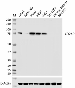
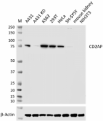
Whole cell extracts (15 µg total protein) from the indicated cell lines (lane 1 and 2 are A431 cells without and with CD2AP knockdown, respectively) were resolved on a 4-12% Bis-Tris gel, transferred to a PVDF membrane, and probed with the 1 µg/mL of Purified anti-CD2AP (clone W21094A) overnight at 4°C. Proteins were visualized by chemiluminescence detection using HRP Goat anti-rat IgG (Cat. No. 405405) at a 1:5000 dilution. Direct-Blot™ HRP anti-β-actin (Cat. No. 664804) was used as a loading control at a 1:25000 dilution. Western-Ready™ ECL Substrate Premium Kit (Cat. No. 426319) was used as a detection agent. Lane M: Molecular weight marker -

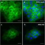
A431 cells without (panel A and B) and with (panel C and D) CD2AP knockdown were fixed with 4% paraformaldehyde for 10 minutes, permeabilized with 0.5% Triton X-100, and blocked with 5% FBS for 1 hour at room temperature. Cells were then intracellularly stained with 5.0 µg/mL of Purified anti-CD2AP (clone W21094A), followed by incubation with 2.5 µg/mL of Alexa Fluor® 488 Goat anti-rat IgG (Cat. No. 405418) for 1 hour at room temperature (panel A and panel C). Nuclei were counterstained with DAPI (Cat. No. 422801) (panel B and D), and the image was captured with a 40X objective. Scale bar: 25 µm -

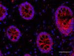
IHC staining using Purified anti-CD2AP (clone W21094A) on formalin-fixed paraffin-embedded human colon tissue. Following antigen retrieval using 1X Citrate buffer (Cat. No. 420901), the tissue was incubated with 5 µg/mL of Purified anti-CD2AP (clone W21094A) overnight at 4°C, followed by incubation with 2.5 µg/mL of Alexa Fluor® 647 Goat anti-rat IgG (red) (Cat. No. 405416) for 1 hour at room temperature. Nuclei were counterstained with DAPI (blue) (Cat. No. 422801). The image was captured with a 40X objective. Scale bar: 50 µm
| Cat # | Size | Price | Quantity Check Availability | Save | ||
|---|---|---|---|---|---|---|
| 625151 | 25 µg | 132€ | ||||
| 625152 | 100 µg | 328€ | ||||
CD2‐associated protein (CD2AP) is a scaffolding molecule connecting membrane proteins to the cytoskeleton. CD2AP also participates in the immunological synapse between CD2-expressing T cells and antigen-presenting cells. T cell activation induces cell adhesion through CD2-mediated binding of antigen-presenting cells to surface ligands. This enhances antigen-specific T cell activation, cell clustering, and induces cytoskeletal polarization. The interactions between CD2AP and other cytoskeletal proteins can regulate EGFR endocytosis. CD2AP is expressed at highest levels in liver, thymus, and spleen. Ablation of CD2AP disrupted the tissue architecture of the lamina propria cells of the gastric mucosa, indicating that CD2AP plays an important role in maintaining the normal morphology of the gastric mucosa. CD2AP-deficient mice develop a fatal congenital nephrotic syndrome, suggesting that CD2AP is also involved in maintaining renal glomerular integrity.
Product DetailsProduct Details
- Verified Reactivity
- Human
- Antibody Type
- Monoclonal
- Host Species
- Rat
- Immunogen
- Partial recombinant human CD2AP protein
- Formulation
- Phosphate-buffered solution, pH 7.2, containing 0.09% sodium azide
- Preparation
- The antibody was purified by affinity chromatography.
- Concentration
- 0.5 mg/mL
- Storage & Handling
- The antibody solution should be stored undiluted between 2°C and 8°C.
- Application
-
WB - Quality tested
ICC, IHC-P - Verified - Recommended Usage
-
Each lot of this antibody is quality control tested by western blotting. For western blotting, the suggested use of this reagent is 0.125 - 1.0 µg/mL. For immunocytochemistry, a concentration range of 1.0 - 5.0 μg/mL is recommended. For immunohistochemistry on formalin-fixed paraffin-embedded tissue sections, a concentration range of 1.0 - 10.0 µg/mL is suggested. It is recommended that the reagent be titrated for optimal performance for each application.
- Application Notes
-
The clone does not cross-react with mouse in western blotting (WB).
For immunocytochemistry (ICC), 4% PFA fixation followed by permeabilization with 0.5% Triton X-100 or ice-cold methanol is recommended.
For immunohistochemistry (IHC-P), antigen retrieval buffer Citrate Buffer (Cat. No. 420901) or Tris-EDTA pH 9.0 is recommended. - Additional Product Notes
-
This antibody has been tested in knockout/knockdown models for Western Blotting and Immunocytochemistry.
- RRID
-
AB_3097249 (BioLegend Cat. No. 625151)
AB_3097249 (BioLegend Cat. No. 625152)
Antigen Details
- Structure
- CD2AP is a 639 amino acid protein with a predicted molecular weight of 71 KD
- Distribution
-
Widely expressed in fetal and adult tissues
- Function
- Regulating signal transduction and cytoskeletal molecules
- Interaction
- F-actin, PKD2, NPHS1, NPHS2, WTIP, DDN, CBL, BCAR1/p130Cas, MVB12A, ARHGAP17, ANLN, CD2, CBLB, PDCD6IP, TSG101, RIN3, CGNL1 and SH3BP1
- Cell Type
- Neurons, T cells
- Biology Area
- Cell Biology, Cell Motility/Cytoskeleton/Structure, Immunology, Neuroscience
- Molecular Family
- Adaptor Proteins, APP/β-Amyloid
- Antigen References
-
- Kirsch KH, et al. 1999. EMBO J. 96:6211-16.
- Kirsch KH, et al. 2001. J Biol Chem. 276:4957-63.
- Lynch DK, et al. 2003. J. Biol. Chem. 278:21805-13.
- Hutchings NJ, et al. 2003. J. Biol. Chem. 278:22396-403.
- Shih NY. et al. 1999. Science. 286:312-15.
- Gene ID
- 23607 View all products for this Gene ID
- UniProt
- View information about CD2AP on UniProt.org
Related Pages & Pathways
Pages
Related FAQs
Other Formats
View All CD2AP Reagents Request Custom Conjugation| Description | Clone | Applications |
|---|---|---|
| Purified anti-CD2AP | W21094A | WB,IHC-P,ICC |
Compare Data Across All Formats
This data display is provided for general comparisons between formats.
Your actual data may vary due to variations in samples, target cells, instruments and their settings, staining conditions, and other factors.
If you need assistance with selecting the best format contact our expert technical support team.
 Login / Register


Follow Us