 Login / Register


- Clone
- Poly28294 (See other available formats)
- Regulatory Status
- RUO
- Other Names
- Glial fibrillary acidic protein
- Previously
-
Covance Catalog# PCK-591P
- Isotype
- Chicken IgY
- Ave. Rating
- Submit a Review
- Product Citations
- publications

-

IHC staining of purified anti-GFAP antibody (clone Poly28294) on formalin-fixed paraffin-embedded human brain tissue. The tissue was incubated with a 1:5,000 dilution of the primary antibody for 20 minutes at room temperature. For detection, a biotinylated anti-chicken secondary was followed by the HRP labeling reagent and DAB from BioLegend's Ultra Streptavidin (USA) HRP Detection Kit (Multi-Species, DAB, Cat. No. 929901) followed by hematoxylin counterstaining, according to the protocol provided. The image was captured with a 40X objective. Scale bar: 50 µm -

IHC staining of purified anti-GFAP antibody (clone Poly28294) on formalin-fixed paraffin-embedded mouse brain tissue. The tissue was incubated with a 1:5,000 dilution of the primary antibody for 20 minutes at room temperature. For detection, a biotinylated anti-chicken secondary was followed by the HRP labeling reagent and DAB from BioLegend's Ultra Streptavidin (USA) HRP Detection Kit (Multi-Species, DAB, Cat. No. 929901) followed by hematoxylin counterstaining, according to the protocol provided. The image was captured with a 40X objective. Scale bar: 50 µm -

IHC staining of purified anti-GFAP antibody (clone Poly28294) on formalin-fixed paraffin-embedded rat brain tissue. The tissue was incubated with a 1:5,000 dilution of the primary antibody for 20 minutes at room temperature. For detection, a biotinylated anti-chicken secondary was followed by the HRP labeling reagent and DAB from BioLegend's Ultra Streptavidin (USA) HRP Detection Kit (Multi-Species, DAB, Cat. No. 929901) followed by hematoxylin counterstaining, according to the protocol provided. The image was captured with a 40X objective. Scale bar: 50 µm -

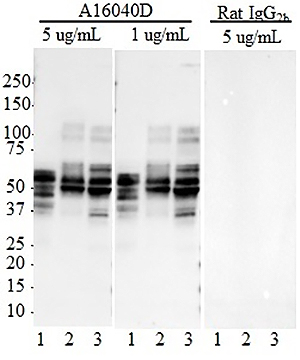
Western blot of purified anti-GFAP antibody (clone Poly28294). Lane 1: Molecular weight marker; Lane 2: 20 µg of human brain lysate; Lane 3: 20 µg of mouse brain lysate; Lane 4: 20 µg of rat brain lysate. The blot was incubated with a 1:10,000 dilution of the primary antibody overnight at 4°C, followed by incubation with HRP labeled goat anti-chicken. Enhanced chemiluminescence was used as the detection system.
| Cat # | Size | Price | Quantity Check Availability | Save | ||
|---|---|---|---|---|---|---|
| 829401 | 100 µL | 398€ | ||||
Glial fibrillary acidic protein (GFAP) is the 10nm or intermediate filament protein found specifically in astrocytic cells in the central nervous system. Antibodies to GFAP are therefore widely used to identify astrocytic cells in situ and in tissue culture. This antibody was developed using bovine spinal cord GFAP purified from a Triton X-100 extract of myelin associated material and further purified by centrifugation and ion exchange chromatography in 6m urea on DEAE cellulose.
Product DetailsProduct Details
- Verified Reactivity
- Human, Mouse, Rat
- Antibody Type
- Polyclonal
- Host Species
- Chicken
- Immunogen
- Native GFAP purified from bovine spinal cord.
- Formulation
- Phosphate-buffered solution, containing 0.2% sodium azide.
- Preparation
- The antibody was purified by affinity chromatography.
- Concentration
- Lot-specific (to obtain lot-specific concentration and expiration, please enter the lot number in our Certificate of Analysis online tool.)
- Storage & Handling
- The antibody solution should be stored undiluted between 2°C and 8°C. Please note the storage condition for this antibody has been changed from -20°C to between 2°C and 8°C. You can also check your vial or your CoA to find the most accurate storage condition for this antibody.
- Application
-
IHC-P - Quality tested
WB - Verified - Recommended Usage
-
Each lot of this antibody is quality control tested by formalin-fixed paraffin-embedded immunohistochemical staining. For immunohistochemistry, a dilution range of 1:1000 - 1:5000 is suggested. For Western blotting, the suggested use of this reagent is a 1:5000 - 1:10000 dilution. It is recommended that the reagent be titrated for optimal performance for each application.
- Application References
-
- Sosunov AA, et al. 2013. J. Neurosci. 33:7439-50. (IHC, WB) PubMed
- Product Citations
-
- RRID
-
AB_2564929 (BioLegend Cat. No. 829401)
Antigen Details
- Structure
- GFAP is a 432 amino acid protein with a molecular mass of ~50 kD.
- Distribution
-
Tissue distribution: GFAP is expressed by numerous cell types of the central nervous system (CNS) including astrocytes, ependymal cells, and Bergmann glia cells (protoplasmic astrocyte). GFAP is expressed in cells lacking fibronectin.
Cellular distribution: Cytoskeleton and cytosol Function
GFAP is a class-III intermediate filament and a structural constituent of the cytoskeleton. It is a cell-specific marker that is used to distinguish astrocytes from other glial cells during the development of the CNS. - Cell Type
- Astrocytes
- Biology Area
- Cell Biology, Neuroscience, Neuroscience Cell Markers
- Molecular Family
- Intermediate Filaments
- Gene ID
- 2670 View all products for this Gene ID
- UniProt
- View information about GFAP on UniProt.org

Related FAQs
Other Formats
View All GFAP Reagents Request Custom Conjugation| Description | Clone | Applications |
|---|---|---|
| Purified anti-GFAP | Poly28294 | IHC-P,WB |
Customers Also Purchased
Compare Data Across All Formats
This data display is provided for general comparisons between formats.
Your actual data may vary due to variations in samples, target cells, instruments and their settings, staining conditions, and other factors.
If you need assistance with selecting the best format contact our expert technical support team.


Follow Us