- Clone
- 13G8C67 (See other available formats)
- Regulatory Status
- RUO
- Other Names
- Histone Deacetylase 2, YY1-Associated Factor 1, HD2, Transcriptional Regulator Homolog RPD3, YAF1
- Isotype
- Mouse IgG1, κ
- Ave. Rating
- Submit a Review
- Product Citations
- publications
-

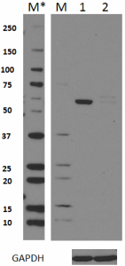
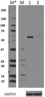
Total lysates (15 µg protein) from 293T (lane 1) and 293T/HDAC2 knockdown (KD) cells (lane 2) were resolved by electrophoresis (4-20% Tris-Glycine gel), transferred to nitrocellulose, and probed with 1:500 diluted (1 µg/mL) Purified anti-HDAC2 Antibody, clone 13G8C67 (upper) or 1:3000 diluted Purified anti-GAPDH Antibody, clone poly6314 (lower). Proteins were visualized by chemiluminescence detection using a 1:3000 diluted goat anti-mouse-IgG secondary antibody conjugated to HRP for the anti-HDAC2 Antibody, and a donkey anti-rabbit IgG Antibody conjugated to HRP for anti-GAPDH Antibody. Lane M: Molecular weight ladder, M* indicates longer exposure. -

Immunoprecipitation of HDAC2 from 293T cell extracts. Lane 1 is 5% input. Immunoprecipitation was performed using protein G resins only (lane 2), mouse IgG isotype control (lane 3), and anti-HDAC2 antibody (clone 13G8C67, lane 4). Western blot was performed using anti-HDAC2 antibody (clone 13G8C67). -

HepG2 cells were stained with purified anti-HDAC2 (clone 13G8C67) antibody, followed by staining with DyLight™ 488 conjugated goat anti-mouse IgG (green) antibodies, and then Alexa Fluor® 594 conjugated phalloidin (red). -

Chromatin Immunoprecipitation (ChIP) was performed using commercial Protein-G coated 96 well high-throughput ChIP assay kit by loading 3 µg of cross-linked chromatin samples from Jurkat cells with either A) 1:100 dilution of Go-ChIP-Grade™ Purified anti-HDAC2 Antibody (clone 13G8C67), B) equal amount of Purified Mouse IgG1, κ Isotype Control Antibody (clone MG1-45), or C) competitor's ChIP-grade Purified anti-HDAC2 Antibody and D) equal amount of matched Isotype Control Antibody as recommended by the manufacturer. The enriched DNA was purified and quantified by real-time qPCR using primers targeting human FOSL1 (Fos like 1) gene region, which is known to be bound to HDAC2. The amount of immunoprecipitated DNA in each sample is represented as signal relative to the 5% of total amount of input chromatin.
| Cat # | Size | Price | Quantity Check Availability | Save | ||
|---|---|---|---|---|---|---|
| 680101 | 25 µg | 90€ | ||||
| 680102 | 100 µg | 221€ | ||||
HDAC2 is a member of the class I histone deacetylase (HDACs) family, which modulates the chromatin structure by removing acetyl groups from the side chain of lysine residues on the N-terminal region of core histones. HDAC2 lacks DNA binding activity and executes its function by recruiting transcription factors and forming large transcriptional repressor complexes. In addition to histones, HDAC2 can deacetylate transcription factors and modify their transcriptional activity. HDAC2 is highly homologous to HDAC1 in regulating cell proliferation, differentiation, and apoptosis. HDAC2 has been shown to negatively regulate synaptic plasticity and memory formation. Elevated activity of HDAC2 has been associated with the loss of memory and neuronal degeneration.
Product DetailsProduct Details
- Verified Reactivity
- Human, Mouse
- Antibody Type
- Monoclonal
- Host Species
- Mouse
- Immunogen
- Human HDAC2 peptide (471-488 a.a.) conjugated to KLH.
- Formulation
- Phosphate-buffered solution, pH 7.2, containing 0.09% sodium azide.
- Preparation
- The antibody was purified by affinity chromatography.
- Concentration
- 0.5 mg/ml
- Storage & Handling
- The antibody solution should be stored undiluted between 2°C and 8°C.
- Application
-
WB - Quality tested
IP, ICC, KO/KD-WB, ChIP - Verified - Recommended Usage
-
Each lot of this antibody is quality control tested by Western blotting. For Western blotting, the suggested use of this reagent is 0.5 - 2.5 µg per ml. For immunoprecipitation, the suggested use of this reagent is 10 - 30 μg per ml. For immunocytochemistry, a concentration range of 0.1 - 10 μg/ml is recommended. For ChIP applications, the suggested dilution is 1:50-1:100 by volume. It is recommended that the reagent be titrated for optimal performance for each application.
- Application Notes
-
A minor non-specific band of ~70 kDa was observed by western blot during product development.
- RRID
-
AB_2566332 (BioLegend Cat. No. 680101)
AB_2566332 (BioLegend Cat. No. 680102)
Antigen Details
- Structure
- 488 amino acids with a predicted molecular weight of 55.4 kD. It contains a histone deacetylase domain.
- Distribution
-
Nucleus.
- Function
- HDAC2 is a histone deacetylase responsible for the deacetylation of ε-amino acid group of lysine residues on the N-terminal tail of core histones. Forms of complex with transcription factors and acts as a transcriptional co-repressor.
- Interaction
- Forms histone deacetylase complexes with HDAC1, RBBP4, and RBBP7. Interacts with GFI1, SNW1, HDAC7, PRDM6, SAP30, SETDB1, SUV39H1, H2AFY, ATR, CBFA2T3, BCL-6, DNMT1, MINT, HDAC10, HCFC1, NRIP1, KDM4A, and PELP1.
- Biology Area
- Cell Biology, Chromatin Remodeling/Epigenetics, Transcription Factors
- Antigen References
-
1. Winter M, et al. 2013. EMBO J. 32:3176.
2. Ma P, et al. 2013. PLoS Genet. 9:e1003377.
3. Kurita M, et al. 2012. Nat. Neurosci. 15:1245.
4. Segre CV, et al. 2011. J. Biomed. Biotechnol. 2011:690848.
5. Brunmeir R, et al. 2009. Int. J. Dev. Biol. 53:275.
6. Guan JS, et al. 2009. Nature 459:55. - Gene ID
- 3066 View all products for this Gene ID
- UniProt
- View information about HDAC2 on UniProt.org
Related Pages & Pathways
Pages
Related FAQs
Other Formats
View All HDAC2 Reagents Request Custom Conjugation| Description | Clone | Applications |
|---|---|---|
| Purified anti-HDAC2 | 13G8C67 | WB,IP,ICC,KO/KD-WB,ChIP |
| Direct-Blot™ HRP anti-HDAC2 | 13G8C67 | WB |
Compare Data Across All Formats
This data display is provided for general comparisons between formats.
Your actual data may vary due to variations in samples, target cells, instruments and their settings, staining conditions, and other factors.
If you need assistance with selecting the best format contact our expert technical support team.
-
Purified anti-HDAC2
Total lysates (15 µg protein) from 293T (lane 1) and 293T/HD... 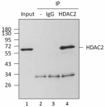
Immunoprecipitation of HDAC2 from 293T cell extracts. Lane 1... 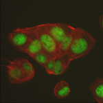
HepG2 cells were stained with purified anti-HDAC2 (clone 13G... 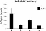
Chromatin Immunoprecipitation (ChIP) was performed using com... -
Direct-Blot™ HRP anti-HDAC2
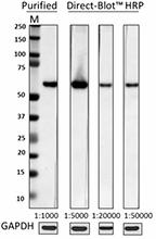
Total cell lysate (15µg protein) from HepG2 cells was resolv...
 Login / Register


Follow Us