 Login / Register


- Clone
- 1D4/CD66c (See other available formats)
- Regulatory Status
- RUO
- Other Names
- CEACAM 6
- Isotype
- Mouse IgG3, κ
- Ave. Rating
- Submit a Review
- Product Citations
- publications

-

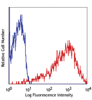
Human lysed washed granulocytes stained with purified anti-human CD66c (clone 1D4/CD66c) (filled histogram) or mouse IgG1, κ isotype control purified (open histogram) followed by anti-mouse IgG PE. -

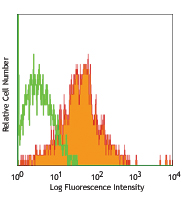
Human colon cancer cell line HT-29 stained with purified anti-human CD66c (clone 1D4/CD66c) (filled histogram) or mouse IgG1, κ isotype control purified (open histogram) followed by anti-mouse IgG PE. -

Human paraffin-embedded spleen tissue slices were prepared with a standard protocol of deparaffination and rehydration. Antigen retrieval was done with Citrate buffer 1X pH 6.0 at 95°C for 40 minutes. Tissue was washed with PBS/ 0.05% Tween-20 twice for five minutes, blocked with 5% FBS and 0.2% gelatin for 30 minutes. Then, the tissue was stained with 10 µg/mL of purified anti-human CD66c (clone 1D4/CD66c) at 4°C overnight. On the next day, the tissue was washed twice with PBS and stained with goat anti-mouse secondary antibody (clone Poly4053) Alexa Fluor® 594 (red) for two hours at room temperature. The nuclei were counter staining with DAPI (blue). The image was captured with a 10X objective. -

Human paraffin-embedded colon tissue slices were prepared with a standard protocol of deparaffination and rehydration. Antigen retrieval was done with Citrate buffer 1X pH 6.0 at 95°C for 40 minutes. Tissue was washed with PBS/ 0.05% Tween-20 twice for five minutes, blocked with 5% FBS and 0.2% gelatin for 30 minutes. Then, the tissue was stained with 10 µg/mL of purified anti-human CD66c (clone 1D4/CD66c) at 4°C overnight. On the next day, the tissue was washed twice with PBS and stained with goat anti-mouse secondary antibody (clone Poly4053) Alexa Fluor® 594 (red) for two hours at room temperature. The nuclei were counter staining with DAPI (blue). The image was captured with a 10X objective.
| Cat # | Size | Price | Quantity Check Availability | Save | ||
|---|---|---|---|---|---|---|
| 365002 | 100 µg | 118€ | ||||
CD66c, also known as CEACAM6, is one of the splice variants of CD66. It is expressed on endothelial, epithelial cells, neutrophils, monocytes, and myeloma and cancer cells, such as B-cell acute lymphoblastic lymphomas. CD66 expression level can be induced on T cells, B cells, and CD16-negative NK cells. Like the other CD66 splice variants, CD66c is involved in cell adhesion and growth, neutrophil activation, and signaling. It plays a role in hemophilic and heterophilic adhesion.
Product DetailsProduct Details
- Verified Reactivity
- Human
- Antibody Type
- Monoclonal
- Host Species
- Mouse
- Formulation
- Phosphate-buffered solution, pH 7.2, containing 0.09% sodium azide
- Preparation
- The antibody was purified by affinity chromatography.
- Concentration
- 0.5 mg/mL
- Storage & Handling
- The antibody solution should be stored undiluted between 2°C and 8°C.
- Application
-
FC - Quality tested
IHC-P - Verified - Recommended Usage
-
Each lot of this antibody is quality control tested by immunofluorescent staining with flow cytometric analysis. For flow cytometric staining, the suggested use of this reagent is ≤ 0.5 µg per million cells in 100 µL volume. For immunohistochemistry on formalin-fixed paraffin-embedded tissue sections, a concentration range of 5.0 - 10.0 µg/mL is suggested. It is recommended that the reagent be titrated for optimal performance for each application.
- RRID
-
AB_2904392 (BioLegend Cat. No. 365002)
Antigen Details
- Structure
- Member of the CEA (carcinoembryonic antigen) family of the Ig superfamily
- Distribution
-
Granulocytes, epithelial cells
- Function
- Homophilic and heterophilic adhesion
- Ligand/Receptor
- CD66a-e, CD62E and Galectins
- Cell Type
- Epithelial cells, Granulocytes
- Biology Area
- Immunology
- Molecular Family
- Adhesion Molecules, CD Molecules
- Antigen References
-
- Zola H, et al. 2007. Leukocyte and Stromal Cell Molecules:The CD Markers. Wiley-Liss. A John Wiley & Sons Inc. Publication
- Gene ID
- 4680 View all products for this Gene ID
- UniProt
- View information about CD66c on UniProt.org


Related FAQs
Other Formats
View All CD66c Reagents Request Custom Conjugation| Description | Clone | Applications |
|---|---|---|
| Purified anti-human CD66c | 1D4/CD66c | FC,IHC-P |
Customers Also Purchased

Compare Data Across All Formats
This data display is provided for general comparisons between formats.
Your actual data may vary due to variations in samples, target cells, instruments and their settings, staining conditions, and other factors.
If you need assistance with selecting the best format contact our expert technical support team.


Follow Us