- Clone
- TAp63-4.1 (See other available formats)
- Regulatory Status
- RUO
- Other Names
- TP63,Tumor Protein P63, P63, Transformation-Related Protein 63, Tumor Protein 63, P73L, Tumor Chronic Ulcerative Stomatitis Protein, Keratinocyte Transcription Factor KET, Protein P73-Like, Tumor Protein P53-Competing Protein
- Isotype
- Mouse IgG2a, κ
- Ave. Rating
- Submit a Review
- Product Citations
- publications
-

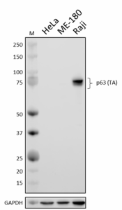
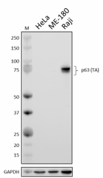
Whole cell extracts (15 µg protein) from HeLa (negative control), ME-180 (reduced expression control), and Raji (positive control) cells were resolved by 4-12% Bis-Tris gel electrophoresis, transferred to a PVDF membrane, and probed with 1.0 µg/mL (1:500 dilution) of purified anti-p63 (TA) antibody (clone TAp63-4.1) overnight at 4°C. Proteins were visualized by chemiluminescence detection using HRP goat anti-mouse IgG antibody (Cat. No. 405306) at a 1:3000 dilution. Direct-Blot™ HRP anti-GAPDH antibody (Cat. No. 607904) was used as a loading control at a 1:50000 dilution (lower). Lane M: Molecular weight marker. -

HeLa cells (negative control, panel A) and Raji cells (positive control, panel B) were fixed with 4% paraformaldehyde for 10 minutes, permeabilized with methanol for 10 minutes, and blocked with 5% FBS for 60 minutes. Cells were then intracellularly stained with 2.0 µg/mL of purified anti-p63 (TA) antibody followed by incubation with Alexa Fluor® 594 goat anti-mouse IgG antibody (Cat. No. 405326) at 2.0 µg/mL. Nuclei were counterstained with DAPI, and the image was captured with a 60X objective. -

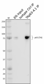
Whole cell extracts (250 µg total protein) prepared from Raji cells were immunoprecipitated overnight with 2.5 µg of purified mouse IgG2a, κ isotype ctrl antibody (Cat. No. 400302) or purified anti-p63 (TA) antibody, (clone TAp63-4.1). The resulting IP fractions and whole cell extract input (6%) were resolved by 4-12% Bis-Tris gel electrophoresis and transferred to a PVDF membrane. Lane M: Molecular weight marker.
| Cat # | Size | Price | Quantity Check Availability | Save | ||
|---|---|---|---|---|---|---|
| 938101 | 25 µg | 109€ | ||||
| 938102 | 100 µg | 278€ | ||||
p63 is a member of the p53 family of transcription factors. The TP63 gene encodes two major isoforms, p63 (TA) and p63 (ΔN) that are produced from alternative start sites through alternative promoter usage. p63 (TA) is able to induce expression from p53-dependent promoters, including those that control the expression of genes encoding the apoptotic response proteins Noxa and Puma. It has also been shown to play a role in energy metabolism by inducing genes encoding Sirt1, AMPKα2, and LKB1 proteins in response to metabolic stress. While p63 (ΔN) lacks the TA domain, it retains transactivation function and can induce expression from some promoters; it can also exert dominant-negative function by forming heterotetramers that sequester TAp63 and render it inactive, thereby promoting tumorigenesis.
Product DetailsProduct Details
- Verified Reactivity
- Human
- Antibody Type
- Monoclonal
- Host Species
- Mouse
- Immunogen
- Full-length recombinant human p63 (TA) protein
- Formulation
- Phosphate-buffered solution, pH 7.2, containing 0.09% sodium azide
- Preparation
- The antibody was purified by affinity chromatography.
- Concentration
- 0.5 mg/mL
- Storage & Handling
- The antibody solution should be stored undiluted between 2°C and 8°C.
- Application
-
WB - Quality tested
ICC, IP - Verified - Recommended Usage
-
Each lot of this antibody is quality control tested by western blotting. For western blotting, the suggested use of this reagent is 0.5 - 1 µg/mL. For immunocytochemistry, a concentration range of 1.0 - 5.0 μg/mL is recommended. For immunoprecipitation, the suggested use of this reagent is 2.5 µg/test. It is recommended that the reagent be titrated for optimal performance for each application.
- Application Notes
-
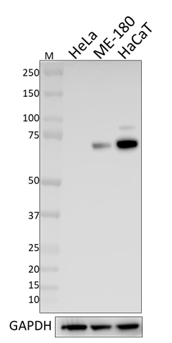
Epitope binding of TAp63-4.1 has been mapped to the LSDPxW motif at the C-terminal region of the p63 transactivation (TA) domain (Nekulova M, et al. 2013. Virchows Arch. 463:415). Because this sequence is absent in the p63 (?N isoform) and the p63 paralog p73, this antibody is not predicted to react with either protein. Lack of p63 (?N) reactivity was confirmed during product development testing.
This clone was ICC tested on Raji cells fixed with 4% PFA and permeabilized with either Triton X-100 or methanol. Both methods were compatible with p63 (TA) staining.
This clone is predicted to recognize all p63 (TA) isoforms.
This clone is predicted to recognize mouse p63 (TA) due to complete sequence conservation of the LSDPxW epitope between human and mouse p63 (TA) orthologs. - Product Citations
-
- RRID
-
AB_2876758 (BioLegend Cat. No. 938101)
AB_2876758 (BioLegend Cat. No. 938102)
Antigen Details
- Structure
- p63 (TA) is a 680 amino acid protein with a predicted molecular weight of 76 kD.
- Distribution
-
Nucleus
- Function
- Transcription factor
- Antigen References
-
- Yang A, et al. 1998. Mol Cell. 2:305-16.
- Ghioni P, et al. 2002. Mol Cell Biol. 22:8659.
- Waltermann A, et al. 2003. Oncogene. 22:5686.
- Nekulova M, et al. 2013. Virchows Arch. 463:415.
- Castellone M and Laukkanen M. 2017. Front Biosci. 9:31.
- Napoli M and Flores E. 2017. Br J Cancer. 116:149.
- Gene ID
- 8626 View all products for this Gene ID
- UniProt
- View information about p63 on UniProt.org
Related Pages & Pathways
Pages
Related FAQs
Other Formats
View All p63 (TA) Reagents Request Custom Conjugation| Description | Clone | Applications |
|---|---|---|
| Purified anti-p63 (TA) | TAp63-4.1 | WB,ICC,IP |
Customers Also Purchased
Compare Data Across All Formats
This data display is provided for general comparisons between formats.
Your actual data may vary due to variations in samples, target cells, instruments and their settings, staining conditions, and other factors.
If you need assistance with selecting the best format contact our expert technical support team.

 Login / Register






Follow Us