 Login / Register


- Clone
- 7C6B05 (See other available formats)
- Regulatory Status
- RUO
- Other Names
- Spleen focus forming virus (SFFV) proviral integration oncogene, Transcription factor PU.1
- Isotype
- Mouse IgG1, κ
- Ave. Rating
- Submit a Review
- Product Citations
- publications

-

Total cell lysates (15 µg protein) from THP1 (lane 1) and MCF-7 (lane 2) cells were resolved by electrophoresis (4-20% Tris-Glycine gel), transferred to nitrocellulose, and probed with 1:1000 diluted (0.5 µg/mL) Purified anti-SPI1 (PU.1) Antibody, clone 7C6B05 (upper panel). Proteins were visualized by chemiluminescence detection using a 1:3000 diluted goat anti-mouse-IgG secondary antibody conjugated with HRP for anti-SPI1 (PU.1) Antibody or 1:4000 Direct-Blot™ HRP anti-GAPDH antibody, clone W17079A (lower panel) as a loading control. Lane M: Molecular weight ladder. -

THP-1 cells were stained with purified anti-PU.1 (clone 7C6B05) antibody, followed by staining with DyLight™ 488 conjugated goat anti-mouse IgG (green) antibody. Nuclei were stained with DAPI (blue). -

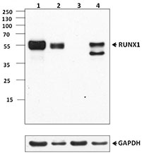
Immunoprecipitation of PU.1 from THP-1 cell extracts. Lane 1 is 5% input. Immunoprecipitation was performed using protein G resins only (lane 2), mouse IgG isotype control (lane 3), and anti-PU.1 antibody (clone 7C6B05, lane 4). Western blot was performed using anti-PU.1 antibody (clone 7C6B05). The astrostar indicates immunoglobulin heavy chain and light chain.
| Cat # | Size | Price | Quantity Check Availability | Save | ||
|---|---|---|---|---|---|---|
| 658002 | 100 µg | 216€ | ||||
SPI1 is a transcription factor belonging to the E26-transformation-specific (Ets) family and is exclusively expressed in hematopoietic cells. SPI1 regulates cell fate decisions during differentiation of hematopoietic stem cells, which is crucial for the development of lymphoid and myeloid cell lineages. SPI1-deficient mice lack macrophages, neutrophils, and B lymphocytes, and they die before or shortly after birth. Abnormally regulated expression of SPI1 can lead to developmental defects as well as occurring malignancy. Overexpression of SPI1 blocks erythroid differentiation and inhibits cell death. Mice carrying a mutant SPI1 allele show decreased SPI1 expression and develop acute myeloid leukaemia (AML), suggesting the role of SPI1 in oncogenesis.
Product DetailsProduct Details
- Verified Reactivity
- Human
- Antibody Type
- Monoclonal
- Host Species
- Mouse
- Immunogen
- Full length human SPI1 recombinant protein
- Formulation
- This antibody is provided in phosphate-buffered solution, pH 7.2, containing 0.09% sodium azide.
- Preparation
- Affinity purified.
- Concentration
- 0.5 mg/ml
- Storage & Handling
- Upon receipt, store undiluted between 2°C and 8°C.
- Application
-
WB - Quality tested
ICC, IP - Verified - Recommended Usage
-
Each lot of this antibody is quality control tested by Western blotting. For Western blotting, the suggested use of this reagent is 0.5 - 2.0 µg per ml. For immunocytochemistry, a concentration of 5.0 μg/ml is recommended. For immunoprecipitation, the suggested use of this reagent is 5.0 - 20 μg per ml. It is recommended that the reagent be titrated for optimal performance for each application.
- Application Notes
-
NOTE: For flow cytometric staining with this clone, True-Nuclear™ Transcription Factor Buffer Set (Cat. No. 424401) offers improved staining and is highly recommended.
- Product Citations
-
- RRID
-
AB_2562720 (BioLegend Cat. No. 658002)
Antigen Details
- Structure
- 270 amino acids, predicted molecular weight of 31 kD; contains a C-termianl ETS domain responsible for DNA binding.
- Distribution
-
Nucleus.
- Function
- SPI1 is a transcription factor required for the development of lymphoid and myeloid cells.
- Interaction
- SPI1 interacts with RUNX1, CEBPD, NONO, SPIB, and GFI1.
- Cell Type
- B cells, Dendritic cells, Neutrophils
- Biology Area
- Cell Biology, Immunology, Transcription Factors
- Molecular Family
- Nuclear Markers
- Antigen References
-
1. Hikami K, et al. 2011. Arthritis Rheum. 63:755.
2. Zakrzewska A, et al. 2010. Blood 116:e1.
3. Pham TH, et al. 2013. Nucleic Acids Res. 41:6391.
4. Pospisil V, et al. 2011. EMBO J. 30:4450.
5. Zarnegar MA, et al. 2010. Mol. Cell Biol. 30:4922.
6. Rimmelé P, et al. 2010. Cancer Res. 70:6757. - Gene ID
- 6688 View all products for this Gene ID
- UniProt
- View information about SPI1 on UniProt.org

Related FAQs
Other Formats
View All SPI1 Reagents Request Custom Conjugation| Description | Clone | Applications |
|---|---|---|
| Alexa Fluor® 647 anti-SPI1 (PU.1) | 7C6B05 | ICFC,ICC |
| Purified anti-SPI1 (PU.1) | 7C6B05 | WB,ICC,IP |
| Alexa Fluor® 594 anti-SPI1 (PU.1) | 7C6B05 | ICC,IHC-P |
| PE anti-SPI1 (PU.1) | 7C6B05 | ICFC |
| Alexa Fluor® 488 anti-SPI1 (PU.1) | 7C6B05 | ICFC |
| PE/Cyanine7 anti-SPI1 (PU.1) | 7C6B05 | ICFC |
| APC anti-SPI1 (PU.1) | 7C6B05 | ICFC |
Customers Also Purchased

Compare Data Across All Formats
This data display is provided for general comparisons between formats.
Your actual data may vary due to variations in samples, target cells, instruments and their settings, staining conditions, and other factors.
If you need assistance with selecting the best format contact our expert technical support team.
-
Alexa Fluor® 647 anti-SPI1 (PU.1)

Human peripheral blood lymphocytes were surface stained with... 

Human peripheral blood monocyte-derived macrophages were fix... -
Purified anti-SPI1 (PU.1)

Total cell lysates (15 µg protein) from THP1 (lane 1) and MC... 
THP-1 cells were stained with purified anti-PU.1 (clone 7C6B... 
Immunoprecipitation of PU.1 from THP-1 cell extracts. Lane 1... -
Alexa Fluor® 594 anti-SPI1 (PU.1)

Human peripheral blood monocyte-derived macrophages were fix... 
Human paraffin-embedded tonsil tissue slices were prepared w... -
PE anti-SPI1 (PU.1)

Human peripheral blood lymphocytes were surface stained with... 
-
Alexa Fluor® 488 anti-SPI1 (PU.1)

Human peripheral blood lymphocytes were surface stained with... 
-
PE/Cyanine7 anti-SPI1 (PU.1)

Human peripheral blood lymphocytes were surface stained with... -
APC anti-SPI1 (PU.1)

Human peripheral blood lymphocytes were surface stained with...


Follow Us