
 Login / Register


- Clone
- 4G4B45 (See other available formats)
- Regulatory Status
- RUO
- Other Names
- Signal transducer and activator of transcription 3, Acute-phase response factor (APRF), HIES, ADMIO
- Isotype
- Mouse IgG1, κ
- Ave. Rating
- Submit a Review
- Product Citations
- 6 publications


| Cat # | Size | Price | Quantity Check Availability | Save | ||
|---|---|---|---|---|---|---|
| 678001 | 25 µg | 90€ | ||||
| 678002 | 100 µg | 212€ | ||||
STAT3 is an 88 kD member of the STAT (signal transducer and activators of transcription) protein family that is phosphorylated in response to a cytokine receptor-associated kinase activity. Phosphorylation of STAT3 induces nuclear translocation to activate transcription. STAT3 forms both homo- and heterotrimers and is involved in the activation of genes required for cell growth and apoptosis. STAT3 is also involved in gp130 signaling and binds to IL-6 response elements in various acute phase protein promoters. STAT3 is phosphorylated by signaling from IFNs, EGF, FGF, IL-5, HGF, LIF, and BMP2. STAT3 activity is inhibited by PIAS3 and GRIM-19 and can also be regulated by the Rac1 protein.
Product DetailsProduct Details
- Verified Reactivity
- Human, Mouse
- Antibody Type
- Monoclonal
- Host Species
- Mouse
- Immunogen
- Partial human STAT3 recombinant protein (621-770 a.a.) expressed in E. coli.
- Formulation
- Phosphate-buffered solution, pH 7.2, containing 0.09% sodium azide.
- Preparation
- The antibody was purified by affinity chromatography.
- Concentration
- 0.5 mg/ml
- Storage & Handling
- The antibody solution should be stored undiluted between 2°C and 8°C.
- Application
-
WB - Quality tested
ICFC, ICC, IP, KO/KD-WB - Verified - Recommended Usage
-
Each lot of this antibody is quality control tested by Western blotting. For Western blotting, the suggested use of this reagent is 0.25 - 1.0 µg per ml. For intracellular flow cytometric staining, the suggested use of this reagent is ≤ 0.06 µg per million cells in 100 µl volume. For immunocytochemistry, a concentration range of 1.0 - 5.0 μg/ml is recommended. For immunoprecipitation, the suggested use of this reagent is 2.0 - 10 μg per ml. It is recommended that the reagent be titrated for optimal performance for each application.
- Application Notes
-
This clone is not recommended for ChIP (Chromatin Immunoprecipitation) assays (as determined by in-house testing).
- Product Citations
-
- RRID
-
AB_2861053 (BioLegend Cat. No. 678001)
AB_2861053 (BioLegend Cat. No. 678002)
Antigen Details
- Structure
- 770 amino acids with a predicted molecular weight of 88.1 kD that contains a C-terminal SH2 domain responsible for homo- or herterodimerization.
- Distribution
-
Cytoplasm.
- Function
- Phosphorylated in response to cytokine signaling by receptor-associated kinases, translocates to the nucleus to act as a transcriptional activator, activates a wide variety of genes involved in cell growth and apoptosis, and is involved in gp130-related signaling.
- Interaction
- STAT3 forms a homodimer or a heterodimer with other STAT family members and interacts with IL31RA, NCOA1, PELP1, SIPAR, SOCS7, STATIP1, CAV2, DAPK3, EIF2AK2, PIAS3, and TMF1.
- Cell Type
- Embryonic Stem Cells, Mesenchymal Stem Cells, Neural Stem Cells
- Biology Area
- Cell Biology, Neuroscience, Neuroscience Cell Markers, Stem Cells, Synaptic Biology, Transcription Factors
- Molecular Family
- Nuclear Markers
- Antigen References
-
1. Lufei C, et al. 2003. EMBO J. 22:1325.
2. Deo D, et al. 2002. J. Biol. Chem. 277:21237.
3. Pfeffer L, et al. 1997. Science 276:1418.
4. Akira S, et al. 1994. Cell 77:63. - Gene ID
- 6774 View all products for this Gene ID
- UniProt
- View information about STAT3 on UniProt.org

Related FAQs
Other Formats
View All STAT3 Reagents Request Custom Conjugation| Description | Clone | Applications |
|---|---|---|
| Purified anti-STAT3 | 4G4B45 | WB,ICFC,ICC,IP,KO/KD-WB |
| Alexa Fluor® 594 anti-STAT3 | 4G4B45 | ICC |
| PE anti-STAT3 | 4G4B45 | ICFC |
| PE/Cyanine7 anti-STAT3 | 4G4B45 | ICFC |
| Alexa Fluor® 647 anti-STAT3 | 4G4B45 | ICFC |
| APC anti-STAT3 | 4G4B45 | ICFC |
Customers Also Purchased
Compare Data Across All Formats
This data display is provided for general comparisons between formats.
Your actual data may vary due to variations in samples, target cells, instruments and their settings, staining conditions, and other factors.
If you need assistance with selecting the best format contact our expert technical support team.
-
Purified anti-STAT3

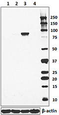
Total lysates (15 µg protein) from Jurkat (lane 1), PC3 (lan... 
Hela cells (filled histogram) or PC-3 cells (open histogram)... 
Total lysates (15 µg protein) from HeLa and HeLa STAT3 CRISP... 
HeLa cells were fixed with 2% paraformaldehyde (PFA) for 10 ... 
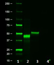
Immunoprecipitation of STAT3 from Hela cell extracts. Lane 1... -
Alexa Fluor® 594 anti-STAT3

HeLa cells were fixed with 2% paraformaldehyde (PFA) for ten... -
PE anti-STAT3

HeLa cells (filled histogram) or PC-3 cells (open histogram)... -
PE/Cyanine7 anti-STAT3

HeLa cells were fixed with Fixation Buffer, permeabilized wi... -
Alexa Fluor® 647 anti-STAT3

HeLa cells were fixed with Fixation Buffer, permeabilized wi... -
APC anti-STAT3

HeLa cells (filled histogram) or PC-3 cells (open histogram)...


Follow Us