- Clone
- W17211A (See other available formats)
- Regulatory Status
- RUO
- Other Names
- MKI67, Proliferation Marker Protein Ki-67, Antigen Ki67
- Isotype
- Rat IgG2a, κ
- Barcode Sequence
- CTGGTAGTTCCTGTA
- Ave. Rating
- Submit a Review
- Product Citations
- publications
| Cat # | Size | Price | Quantity Check Availability | Save | ||
|---|---|---|---|---|---|---|
| 398503 | 10 µg | 296€ | ||||
Antigen Ki-67 is a nuclear protein expressed as two isoforms with molecular weights of 395 and 345 kD. Both isoforms contain one forkhead-associated domain and 16 concatenated "Ki-67 repeats," each containing the epitope recognized by the mAb Ki-67. The antigen Ki-67 interacts with Hklp2, hNIFK, and chromobox protein homolog 1, 3, and 5. Ki-67 is required for cell proliferation and its expression is restricted to the phases G1, S, G2, and M of the cell cycle. This characteristic makes Ki-67 an excellent marker for proliferating cells and is commonly used as one of the prognostic factors in cancer studies. Ki-67 has also been used to study myocyte proliferation after myocardial infarction as well as lymphocyte proliferation during infection, and has been used in neurons of patients with different neuropathologies.
Product DetailsProduct Details
- Verified Reactivity
- Human
- Antibody Type
- Monoclonal
- Host Species
- Rat
- Immunogen
- Human Ki-67 recombinant protein
- Formulation
- Phosphate-buffered solution, pH 7.2, containing 0.09% sodium azide and EDTA
- Preparation
- The antibody was purified by chromatography and conjugated with TotalSeq™-Bn oligomer under optimal conditions.
- Concentration
- 0.5 mg/mL
- Storage & Handling
- The antibody solution should be stored undiluted between 2°C and 8°C. Do not freeze.
- Application
-
SB – Quality tested
- Recommended Usage
-
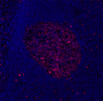
Each lot of this antibody is quality control tested by immunofluorescent staining in formalin-fixed, paraffin-embedded (FFPE) lymphoid tissue, and the oligomer sequence is confirmed by sequencing. TotalSeq™-Bn antibodies are compatible with the 10x Visium CytAssist Gene and Protein Expression Assay.
To maximize performance, it is strongly recommended that the reagent be titrated for each application, and that you centrifuge the antibody dilution at 14,000xg at 2 − 8°C for 10 minutes before use. Carefully pipette out the liquid avoiding the bottom of the tube when handling. To determine and optimize dilutions for the addition of Totalseq™-Bn antibodies into pre-designed antibody panels, refer to 10x Genomics Custom Add-on Antibody Optimization guide.
Buyer is solely responsible for determining whether Buyer has all intellectual property rights that are necessary for Buyer's intended uses of the BioLegend TotalSeq™ products. For example, for any technology platform Buyer uses with TotalSeq™, it is Buyer's sole responsibility to determine whether it has all necessary third party intellectual property rights to use that platform and TotalSeq™ with that platform. - Additional Product Notes
-
TotalSeq™-Bn reagents are designed to profile protein levels following an optimized protocol in spatial transcriptomics. Compatible spatial biology devices (e.g. Imaging System, 10x Genomics Visium Spatial CytAssist Gene and Protein Expression instruments and reagents) and sequencer (e.g. Illumina analyzers) are required. TotalSeq™-B reagents are not compatible with the 10x Genomics Visium system. The complete barcode sequence may be provided upon request. Please contact technical support for more information, or visit TotalSeq™-Bn Reagents for 10x Genomics Visium CytAssist Gene and Protein Assay.
- RRID
-
AB_3097316 (BioLegend Cat. No. 398503)
Antigen Details
- Structure
- Two isoforms with molecular weights of 395 and 345 kD, one forkhead-associated domain, 16 concatenated Ki-67 repeats, located in nucleus
- Distribution
-
Expressed in the phases G1, S, G2, and M of the cell cycle
- Function
- Required for cell proliferation
- Interaction
- Chromobox protein homolog 1, 3 and 5, Hklp2, and hNIFK
- Biology Area
- Cell Biology, Cell Cycle/DNA Replication, DNA Repair/Replication
- Molecular Family
- Nuclear Markers
- Antigen References
-
- Byeon IJ, et al. 2005. Nat. Struct. Mol. Biol. 12:987.
- Yerushalmi R, et al. 2010. Lancet. Oncol. 11:174.
- Drazen JM. et al. 2001. N. Engl. J. Med. 344:1750.
- Sachsenberg N, et al. 1998. J. Exp. Med. 187:1295.
- Nagy Z, et al. 1997. Acta. Neuropathol. 93:294.
- Gene ID
- 4288 View all products for this Gene ID
- UniProt
- View information about Ki-67 on UniProt.org
Related Pages & Pathways
Pages
Related FAQs
- If an antibody clone has been previously successfully used in IBEX in one fluorescent format, will other antibody formats work as well?
-
It’s likely that other fluorophore conjugates to the same antibody clone will also be compatible with IBEX using the same sample fixation procedure. Ultimately a directly conjugated antibody’s utility in fluorescent imaging and IBEX may be specific to the sample and microscope being used in the experiment. Some antibody clone conjugates may perform better than others due to performance differences in non-specific binding, fluorophore brightness, and other biochemical properties unique to that conjugate.
- Will antibodies my lab is already using for fluorescent or chromogenic IHC work in IBEX?
-
Fundamentally, IBEX as a technique that works much in the same way as single antibody panels or single marker IF/IHC. If you’re already successfully using an antibody clone on a sample of interest, it is likely that clone will have utility in IBEX. It is expected some optimization and testing of different antibody fluorophore conjugates will be required to find a suitable format; however, legacy microscopy techniques like chromogenic IHC on fixed or frozen tissue is an excellent place to start looking for useful antibodies.
- Are other fluorophores compatible with IBEX?
-
Over 18 fluorescent formats have been screened for use in IBEX, however, it is likely that other fluorophores are able to be rapidly bleached in IBEX. If a fluorophore format is already suitable for your imaging platform it can be tested for compatibility in IBEX.
- The same antibody works in one tissue type but not another. What is happening?
-
Differences in tissue properties may impact both the ability of an antibody to bind its target specifically and impact the ability of a specific fluorophore conjugate to overcome the background fluorescent signal in a given tissue. Secondary stains, as well as testing multiple fluorescent conjugates of the same clone, may help to troubleshoot challenging targets or tissues. Using a reference control tissue may also give confidence in the specificity of your staining.
- How can I be sure the staining I’m seeing in my tissue is real?
-
In general, best practices for validating an antibody in traditional chromogenic or fluorescent IHC are applicable to IBEX. Please reference the Nature Methods review on antibody based multiplexed imaging for resources on validating antibodies for IBEX.
Other Formats
View All Ki-67 Reagents Request Custom Conjugation| Description | Clone | Applications |
|---|---|---|
| Purified anti-human Ki-67 | W17211A | IHC-P |
| TotalSeq™-Bn1331 anti-human Ki-67 | W17211A | SB |
Compare Data Across All Formats
This data display is provided for general comparisons between formats.
Your actual data may vary due to variations in samples, target cells, instruments and their settings, staining conditions, and other factors.
If you need assistance with selecting the best format contact our expert technical support team.
-
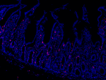
Purified anti-human Ki-67

Human paraffin-embedded intestine tissue slices were prepare... 
Human paraffin-embedded tonsil tissue slices were prepared w... -
TotalSeq™-Bn1331 anti-human Ki-67

 Login / Register


Follow Us