 Login/Register


3D IHC Data
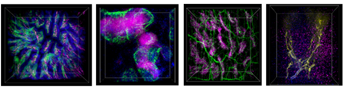
The images above are representative 3D IHC images of 500 μm-thick mouse samples. The sections were optically cleared after immunostaining, mounted in sample chambers, and imaged with a 10X or 20X objective.
From left to right:
- Mouse intestine: CD45 (green), CD326 (blue), Tubulin β (magenta)
- Mouse spleen: CD3ε (magenta), CD169 (green), I-A/I-E (blue)
- Mouse intestine: CD45 (magenta), Tubulin β (green)
- Mouse liver: DAPI (blue), CLEC4F (magenta), CD324 (yellow)
Watch dozens of our 3D imaging videos.
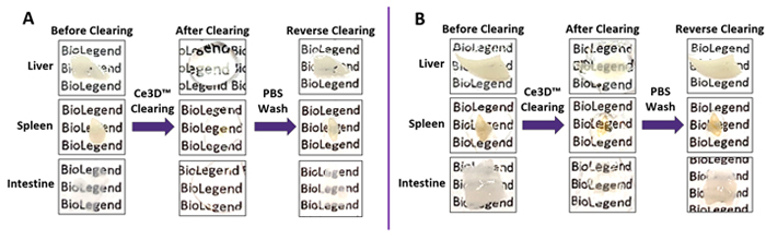
Optical Clearing Using Ce3D™ Tissue Clearing Solution
Representative 500 µm-thick mouse tissue sections before and after tissue clearing. Mice were perfused and organs were fixed with 1% (A) or 4% (B) PFA. Clearing was reversed by immersing tissues in PBS for one hour. 1% PFA is recommended for best clearing effect. 4% PFA is recommended for optimal epitope preservation and immunostaining.


Follow Us