Ab initio.

From the beginning.

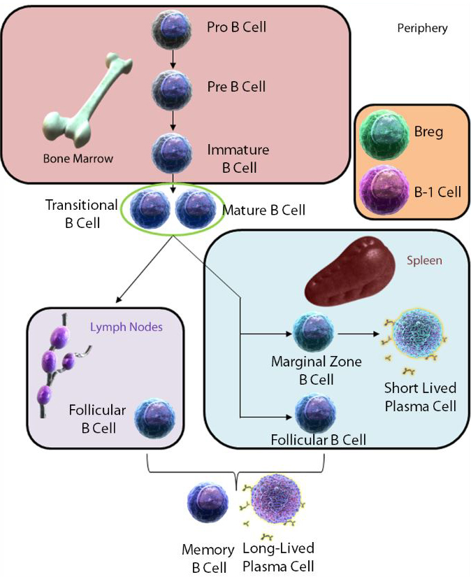
B cells are a lineage of lymphocytes originally discovered in the Bursa of Fabricus in birds (hence the B in B cells). B cell development in mammals was mainly found to occur in the bone marrow, as they do not have a Bursa of Fabricus. B cells are not only responsible for antibody production, but are also involved in several other areas of immunology. Click through the different sections of the shield to gain an understanding of everything B cells do in a mammalian system.

Want help designing your B Cell panel? Click here!

Nulla tenaci invia est via.

For the tenacious, no road is impassable.

Click on a cell below to learn more about it.

Pro B Cell

Developed from hematopoietic stem cells, the Pro B Cell is the earliest lineage stage of the B Cell. The main goal and checkpoint of the Pro B Cell stage is the proper construction of a heavy chain rearrangement, leading to the Pre B Cell stage.


Human Markers


Positive: CD10CD19CD24CD34CD38CD123CD127, EBF1, Pax5

Negative: c-kit lowIgM


Mouse Markers


Positive: B220CD19CD24CD43CD127, E2A, EBF1, Pax5

Negative: BP-1, c-kit lowFlt3IgM

Pre B Cell

Developed from Pro B cells, Pre B Cells express a proper heavy chain (µ) paired with surrogate light chains (VpreB and λ5) in their pre B Cell receptor. This step is crucial to development, as only B Cells expressing this pre B Cell receptor are allowed to progress to the next stage, the Immature B Cell.


Human Markers


Positive: CD10CD19CD20CD24CD38CD123CD124CD127, E2A, EBF1, Oct2, Pax5

Negative: CD34c-kitIgM


Mouse Markers


Positive: B220, BP-1, CD19CD24CD25+/-CD43 lowCD127, E2A, EBF1, Pax5, Siglec G

Negative: c-kitIgM

Immature B Cell

The Immature B Cell stage is characterized with the rearrangement of a proper light chain. This membrane bound IgM antibody is then paired with Ig-α and Ig-β to form a fully functioning B Cell receptor. Before progressing to a mature state, the cells must not react strongly to self. If they recognize self, they may have the chance to perform receptor editing to pass inspection.


Human Markers


Positive: CD10CD19CD20CD21CD24 hiCD38 hiCD40CD124IgM, EBF1, Pax5

Negative: CD27CD127,c-kitIgD


Mouse Markers


Positive: B220, BP-1, CD19CD24CD93, E2A, EBF1, IgM, Oct2, Pax5

Negative: CD23CD43IgD

Transitional B Cell

Transitional B Cells represent an almost intermediary state between Immature B Cells in the bone marrow and Mature B Cells in the periphery. Divided into T1 and T2 stages, these cells can be found in the bone marrow, blood, and spleen, giving rise to Marginal Zone and Follicular B Cells.


Human Markers


Positive: CD5CD19CD20CD21CD24 hiCD38 hi, E2A, EBF1, IgMOct2, Pax5

Negative: CD10 lowCD27IgD low


Mouse Markers


Positive: B220, BP-1, CD19CD21+/-CD23+/-CD24CD93, E2A, EBF1, IgM, Oct2, Pax5

Negative: IgD low

Mature B Cell (Activated)

Upon exiting the bone marrow, Mature B Cells will express both IgM and IgD on their cell surface. At this point, the cells will have acquired functional competence, hence their title of “mature”. They may be called naïve if they have not yet encountered their specific antigen.


Human Markers


Positive: CD19CD20CD25CD27CD30CD69CD80CD86CD135, EBF1, IgDIgMPax5


Mouse Markers


Positive: B220, BP-1, CD27CD69CD80Flt3IgDIgMMHC II hi, Pax5

Negative: CD138CXCR4

Breg Cell

Regulatory B Cells, or Bregs, are a subtype of B Cells capable of downregulating inflammation and inducing tolerance. However, these cells can be phenotypically difficult to identify, as they bear many common markers found on other B Cell types. The most well studied Bregs may be the B-10 and T2-MZP (Transitional 2 Marginal Zone Precursor) population. Many Breg subclasses execute their immunosuppression through IL-10.


Human Markers


Positive: CD1d hiCD5CD19CD21 hiCD24 hi, E2A, EBF1, IgD+/-IgM hiOct2Pax5

Negative: CD27+/-


Mouse Markers


Positive: CD1d hiCD5CD19CD24, E2A, EBF1, IgDIgM hi, Oct2, Pax5Tim-1

Negative: CD62LCD93+/-

B-1 Cell

B-1 Cells are considered an innate lymphocyte that appears early on in the development of the host. This has led to their being named “B-1,” while conventional B Cells that appear later on are termed “B-2”. B-1 Cells make up roughly 5% of mouse and human total B cell population. B-1 Cells arise from stem cell populations and exhibit the ability to self-renew. As they can also express a limited range of “natural”, lower-affinity antibodies, do not undergo affinity maturation, do not require T Cell help, and do not have a proclivity to develop into Memory B Cells. All of this lends credence to the theory they are mainly involved with quick, innate immune responses.

As seen below, phenotyping B-1 cells can be difficult. We recommend taking a look at this reference for more information.


Human Markers


Positive: CD5+/-CD11b+/-CD19CD20CD27CD43CD86+/-IgDIgM

Negative: CD70


Mouse Markers


Positive: CD5CD9+/-CD11bCD19CD21CD38CD43CD80CD273+/-IgM hiIL-5R

Negative: IgD low

Follicular B Cell

Follicular B Cells can be found within structures called follicles (which contain germinal centers) in secondary and tertiary lymphoid organs like the spleen, Peyer's patches, and lymph nodes. Unlike Marginal Zone B Cells, Follicular B Cells are capable of recirculating throughout the body. They are capable of becoming either Memory B Cells or Long Lived Plasma Cells. In most instances, these cells require T Cells for full activation.


Human Markers


Positive: CD19CD20CD21CD22CD23CD24, E2A, EBF1, IgDOct2, Pax5

Negative: CD10CD24 lowCD27CD38 lowIgM low


Mouse Markers


Positive: B220CD19CD22CD23, E2A, EBF1, CD38IgD, Oct2, Pax5

Negative: CD1d lowCD21/35 lowCD93IgM low

Marginal Zone B Cell

Marginal Zone (MZ) B Cells are a mature form of B Cells that home specifically to the MZ of the spleen. As such, they specifically guard against blood-borne pathogens they would be likely to encounter in the spleen. They typically have a lower threshold of activation compared to Follicular B Cells and can develop into Short Lived Plasma Cells, generating “natural antibodies” in a T Cell independent manner.


Human Markers


Positive: CD1cCD19CD20CD21 hiCD27+/-, E2A, EBF1, Oct2Pax5

Negative: IgD low


Mouse Markers


Positive: B220CD1dCD9CD21 hiCD22 hiCD35 hi, E2A, EBF1, IgM, Oct2, Pax5

Negative: CD23CD93IgD low

Short Lived Plasma Cell

Upon encountering antigen, Mature B Cells can develop into plasmablasts, which further differentiate into Plasma Cells. As indicated by their names, Short Lived Plasma Cells (SLPC) only persist for a few days, rapidly churning out antibody before undergoing apoptosis. SLPCs can typically be found in the red pulp of the spleen or in medullary chords of lymph nodes.


Human Markers


Positive: CD27CD138CD269

Negative: CD19 lowCD20MHC II low


Mouse Markers


Positive: Blimp-1CD93CD138CXCR4 hi

Negative: B220 lowCD19CD38 lowMHC II low

Memory B Cell

Memory B Cells develop after a primary infection within the host. After encountering an antigen, a subset of cells that developed from an individual cell can retain the experience and memory of its target. This leads to a quicker immune response should that antigen should ever reintroduce itself to the body.


Human Markers


Positive: CD19CD20CD27+/-CD40CXCR4CXCR5CXCR7Pax5, SPI-B

Negative: CD23 lowCD38


Mouse Markers


Positive: B220, BP-1, CD38+/-CD62L+/-CD80+/-IgAIgEIgMPax5, SPI-B

Negative: CD95 lowIgD

Long Lived Plasma Cell

Upon encountering antigen, Mature B Cells can develop into plasmablasts, which further differentiate into Plasma Cells. Plasma Cells are, quite frankly, powerhouses of antibody production. They generate a single class of immunoglobulin designated for a specific antigen. Antibody production can continue for days or months until the target is neutralized.


Human Markers


Positive: CD27CD38 hiCD138CD269CXCR4IRF4XBP1

Negative: CD19 lowCD20MHC IISurface Immunoglobulins


Mouse Markers


Positive: CD138CXCR4 hiIRF4XBP1

Negative: B220 lowCD19CD38 lowMHC IISurface Immunoglobulins

Incepto ne desistam

May I not shrink from my purpose!

E pluribus unum

Out of many, one.


Germinal centers (GC) can be found in secondary lymphoid organs (like spleen and lymph nodes). GCs are vital for B cells, as it allows them to proliferate upon recognition of their target antigen on follicular dendritic cells. In addition, B cells may require CD4+ T cells to provide costimulatory signals and to recognize the processed antigen in its MHC II (via the TCR). Also, the B cells can mutate their antibody genes in a process known as affinity maturation (check out the video below). This assures that only B cells with the highest affinity antibodies survive this process. Antibody class switching can also occur in GCs, where the constant region of the heavy chain changes. This generates a new isotype (i.e., IgM can become IgG). Cells with Fc receptors can have different affinities for different constant regions. The constant region also determines how easily an antibody can get through mucosal epithelial membranes, diffusion into tissue, polymerization (which increases avidity), and antibody stability. The variable portion, which recognizes antigen, remains unaffected.


Tile scan of spleen section from immunized mouse at day 7 stained with anti-IgD (red), anti-Bcl-6 (green), anti-CD4 (blue) and anti-Foxp3 (white). Picture represents the germinal centers at various locations which are identified by IgD staining the follicular B cells that surround the Bcl-6+ germinal center with an adjacent CD4+ T cell zone. Image provided courtesy of Dr. Michelle Linterman and Devina Divekar of the Babraham Institute.


Iuncta iuvant.

Together, they strive.


B cells are capable of reacting to antigen in two different ways:

TI (Thymus, T cell Independent) Antigens, as their name implies, do not require T cells to fully activate a B cell. An antigen will bind to the B cell receptor (BCR) either in a specific or non-specific manner. A second signal is obtained when that antigen is then recognized by another cell surface receptor. For example, LPS might be bound to the BCR and then to TLR4, generating the activation signals the B cell requires without any T cell help. The secondary signal can also be provided if several BCRs are clustered together and cross-linked.
 

On the other hand, TD (Thymus, T cell Dependent) Antigens require the presence of T helper cells for full activation. With TD antigens, the first activation signal comes from antigen cross-linking surface BCRs. The second set of signals comes from a T cell and its costimulatory molecules. T cells will also secrete cytokines to help stimulate the B cell.


B cells that recognize TD Antigens require additional signals and help from T cells.

Boni pastoris est tondere pecus non deglubere

It is a good shepherd's job to shear his flock, not to flay them.


Due to the ability of B cells to generate a variety of cytokines, they are capable of modulating the response of several cell types. This includes the ability to shift the Th1/Th2 balance and modify dendritic cells. B cell deficient mice show increased IL-12 production by dendritic cells, leading to an enhanced Th1 response. In addition, mice that lack B cells during development?display deficiencies in spleen dendritic cell and T cell compartments, and a lack of Peyer patch organogenesis, follicular dendritic cell networks, and marginal zone and metallophilic macrophages (these macrophages also have decreased chemokine expression). Beyond this, B regulatory cells are thought to contribute to immune cell regulation. Pinpointing the exact phenotype of this cell type has been difficult, as many of its markers are shared by other B cell phenotypes. Many of them (including the most well studied Breg, B-10 cells) are thought to exact their function through the anti-inflammatory cytokine, IL-10. While some exist naturally in the periphery, others can be induced by particular stimuli (i.e, TLR2, 4, 9, costimulation of CD40, CD80, and CD86, and BAFF). Similar to Tregs, human Bregs have recently been proposed to be classified into Br1 (B10) or Br3 cells based on their expression of IL-10 and TGF-β, respectively. Some Bregs are also capable of suppression via cell contact-dependent mechanisms.


Species Phenotype Organ of Origin Major Effector Disease State
Mouse B10 Spleen IL-10 CHS
  T2 MZ Spleen IL-10 Arthritis
  MZ Spleen IL-10 CIA
  B1 Peritoneum IL-10 CHS
  CD1dhi Spleen IL-10 AAI, IBD, Anaphylaxis
  CD23+ Mes. LN ? AAI, EAE
Human Immature Trans. B Blood IL-10, CD80/CD86 SLE
  B10 (Br1) Blood IL-10 Healthy and Autoimmune
  CD1dhi Blood IL-10 -
  Br3 Blood TGF-β CLL
  Breg Blood Foxp3 Healthy


CHS: contact hypersensitivity, T2 MZ: transitional 2 marginal zone, CIA: collagen induced arthritis, AAI: allergic airway inflammation, IBD: inflammatory bowel disease, Mes. LN: mesenteric lymph nodes, EAE: experimental autoimmune encephalomyelitis, SLE: systemic lupus erythematosus, CLL: chronic lymphocytic leukemia.


  1. Kalampokis, I. et al. 2013. Arthritis Res. Ther. 15 (Suppl 1): S1.
  2. Mauri, C. and Blair, P.A. 2010. Nat. Rev. Rheumatol. 6:636.
  3. Morva, A. et al. 2012. Blood. 119:106.
  4. Susanne Sattler, et al. 2012. Regulatory B Cells - Implications in Autoimmune and Allergic Disorders, Recent Advances in Immunology to Target Cancer, Inflammation and Infections. Dr. Jagat Kanwar (Ed.), ISBN: 978-953-51-0592-3, InTech, DOI: 10.5772/38319. Direct link.

Datum perficiemus munus

We shall accomplish the mission assigned


There have been conflicting reports for the role of B cells in cancer. Some animal models with B cell knockouts show enhanced protection from tumor development. B cells are also capable of exacerbating inflammation. However, B cells can also promote anti-tumor responses. They are required for optimal T cell activation and costimulation. In particular, their antigen presentation abilities can induce tumor-specific cytotoxic T cell activation. Antibody production can also lead to opsonization of target antigens in tumors.


More research is being conducted into the role of B cells in transplant rejection. Like in cancer, B cells can either positively or negatively regulate graft rejection depending on the type of allograft. While antibodies may contribute to rejection, regulatory B cells may be expanded to promote tolerance.


Previous studies have revealed that B cells and even antibodies are present within wound tissue. In a mouse model, CD19 (a common mature B cell marker) deficiency inhibited wound healing, infiltration of neutrophils and macrophages, and cytokine expression. On the other hand, CD19 overexpression enhanced wound healing and cytokine expression. B cells have also been shown to recognize Hyaluronanin wounds, which is an endogenous ligand for TLR4, stimulating them to make IL-6 and TGF-β in a CD19-dependent manner.


References:

1. Candolfi, M. et al. 2011. Neoplasia. 13:947. 
2. Dalloul, A. 2013. Front. Immunol. 4:444. 
3. DiLillo, D.J. et al. 2010. J. Immunol. 184:4006. 
4. DiLillo, D.J. et al. 2011. J. Immunol. 186:2643. 
5. Iwata, Y. et al. 2009. Am. J. Pathol. 175:649. 


Ense petit placidam sub libertate quietem.

By the sword, she seeks a serene repose under liberty.


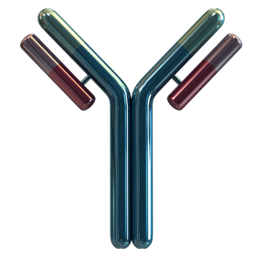
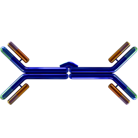
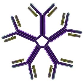
Antibodies are one of the chief products of B cells and plasma cells. Antibodies are also known as immunoglobulins, which is why they begin with the abbreviation Ig. In mammals, the letters A, D, E, G, and M are assigned based on the particular heavy chain (α, δ, ε, γ, µ respectively). While the basic components of antibodies are the same (2 light chains, 2 heavy chains), they can serve different functions. Click on each isotype to learn more about each type.


IgG and its four subclasses are the most abundant antibody class found in serum. They can also cross the placenta to provide passive immunity for the developing fetus. Many cells possess IgG Fc receptors, which are important for opsonization. Most IgGs also activate the classical pathway of the complement system.


IgD is typically co-expressed with IgM on B cells when they have become mature B cells. However, IgD is not absolutely required for B cell development, as δ-chain knockout mice do not develop any B cell abnormalities. Recently, research has suggested IgD may bind basophils and mast cells, aiding in allergic responses and anti-microbial factor production.


IgE is primarily associated with allergic reactions, parasite defense, and asthma. Upon binding allergen, this antibody is capable of triggering basophils and mast cells to release histamine in a process called degranulation.

While rarer in serum, IgA is the most abundant immunoglobulin of bodily secretions like breast milk, saliva, tears, and the mucosa of bronchial, digestive, and urogenital tracts. Because it is so heavily present in the mucosa, it provides defense against pathogens that might seek to cross this barrier. In serum, IgA is most often found in a monomer form. However, dimers, trimers, and even tetramers of IgA have been observed. The subunits are linked together by the J-chain or joining chain.

IgM is the first immunoglobulin expressed by developing B cells. Later on, plasma cells can secrete this antibody in a pentamer form. Similar to IgA, pentameric IgM also contains a J-chain, which seems to be necessary for its polymerization. IgM can also be secreted in external secretions, albeit at lower levels compared to IgA. IgM is the first antibody generated in response to antigen. Its pentameric form provides ten potential binding spots, which increases its antigen binding capabilities.

Crescat scientia vita excolatur.

Let knowledge grow, let life be enriched.


To learn more about B Cells, check out the following pages:

Maturation Markers

How to Make an Antibody

Basic Immunology

Cell Markers


Login/Register
Remember me
Forgot your password? Reset Password
Request an Account
My Cart (0 items)