- Clone
- RS38E (See other available formats)
- Regulatory Status
- RUO
- Workshop
- VIII
- Other Names
- BST2, Tetherin, HM1.24, Bone marrow stromal antigen 2
- Isotype
- Mouse IgG1, κ
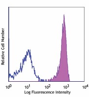
-

Human peripheral blood monocytes stained with RS38E APC
| Cat # | Size | Price | Quantity Check Availability | ||
|---|---|---|---|---|---|
| 348410 | 100 tests | $362.00 | |||
CD317, also known as BST2, Tetherin, and HM1.24, is a type II transmembrane GPI-protein with a molecular weight of about 29-33 kD. It is an interferon-induced protein expressed on dendritic cells, plasma cells, B lymphoblast cells, monocytes, granulocytes, T cells, NK cells, stromal cells, and some non-hematopoietic cells. BST2 inhibits cytokine production through interaction with ILT7 (CD85g). It is also involved in the regulation of B cell growth. More importantly, BST2 has been found to restrict the release of a number of viruses from infected cells, including all tested retroviruses (such as HIV-1) and some arenaviruses and filoviruses. In HIV-1 studies, it has been reported that BST2 retains the nascent virons on the surface of infected cells by incorporation of the protein into HIV-1 particles. HIV-1 Vpu is able to induce BST2 degradation.
Product Details
- Verified Reactivity
- Human, Cynomolgus, Rhesus
- Reported Reactivity
- African Green, Baboon, Pigtailed Macaque
- Antibody Type
- Monoclonal
- Host Species
- Mouse
- Formulation
- Phosphate-buffered solution, pH 7.2, containing 0.09% sodium azide and BSA (origin USA)
- Preparation
- The antibody was purified by affinity chromatography and conjugated with APC under optimal conditions.
- Concentration
- Lot-specific (to obtain lot-specific concentration, please enter the lot number in our Concentration and Expiration Lookup or Certificate of Analysis online tools.)
- Storage & Handling
- The antibody solution should be stored undiluted between 2°C and 8°C, and protected from prolonged exposure to light. Do not freeze.
- Application
-
FC - Quality tested
- Recommended Usage
-
Each lot of this antibody is quality control tested by immunofluorescent staining with flow cytometric analysis. For flow cytometric staining, the suggested use of this reagent is 5 µl per million cells in 100 µl staining volume or 5 µl per 100 µl of whole blood.
- Excitation Laser
-
Red Laser (633 nm)
-
Application References
(PubMed link indicates BioLegend citation) - Product Citations
-
- RRID
-
AB_2067121 (BioLegend Cat. No. 348410)
- Disclaimer
-
This product is manufactured and sold under a license and covered by a number of issued patents. Users who intend to file a patent relating to the use of this product or who intend to develop therapeutic use of this antibody are required to report to Biolegend in advance.
Antigen Details
- Structure
- Type II transmembrane, GPI-anchored protein, 29-33 kD
- Distribution
-
Dendritic cells, plasma cells, B lymphoblast cells, monocytes, granulocytes, T cells, NK cells, stromal cells
- Function
- Regulate B cell growth, inhibit cytokine production, retain progeny virons on the surface of infected cells
- Ligand/Receptor
- ILT7 (CD85g)
- Cell Type
- B cells, Dendritic cells, Granulocytes, Monocytes, NK cells, Plasma cells, T cells
- Biology Area
- Costimulatory Molecules, Immunology, Innate Immunity
- Molecular Family
- Adhesion Molecules, CD Molecules
- Antigen References
-
1. Sugamata OT, et al. 1999. Biochem. Bioph. Res. Co. 258:583.
2. Neil SJ, et al. 2008. Nature 451:425.
3. Fitzpatrick K, et al. 2010. PLoS Pathog. 6:e1000701.
4. Azuma KS, et al. 2008. Cancer Sci. 99:2461.
5. Cao W, et al. 2009. J. Exp. Med. 206:1603.
6. Zola H, et al. 2007. Leukocyte and Stromal Cell Molecules:The CD Markers. - Gene ID
- 684 View all products for this Gene ID
- UniProt
- View information about CD317 on UniProt.org
Other Formats
View All CD317 Reagents Request Custom Conjugation| Description | Clone | Applications |
|---|---|---|
| Purified anti-human CD317 (BST2, Tetherin) | RS38E | FC |
| Alexa Fluor® 647 anti-human CD317 (BST2, Tetherin) | RS38E | FC |
| PE anti-human CD317 (BST2, Tetherin) | RS38E | FC |
| APC anti-human CD317 (BST2, Tetherin) | RS38E | FC |
| PerCP/Cyanine5.5 anti-human CD317 (BST2, Tetherin) | RS38E | FC |
| PE/Cyanine7 anti-human CD317 (BST2, Tetherin) | RS38E | FC |
| TotalSeq™-A0936 anti-human CD317 (BST2, Tetherin) | RS38E | PG |
Compare Data Across All Formats
This data display is provided for general comparisons between formats.
Your actual data may vary due to variations in samples, target cells, instruments and their settings, staining conditions, and other factors.
If you need assistance with selecting the best format contact our expert technical support team.
-
Purified anti-human CD317 (BST2, Tetherin)
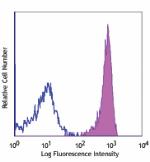
Human peripheral blood monocytes stained with purified RS38E... -
Alexa Fluor® 647 anti-human CD317 (BST2, Tetherin)
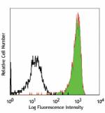
Human peripheral blood monocytes stained with RS38E Alexa Fl... -
PE anti-human CD317 (BST2, Tetherin)
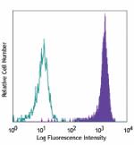
Human peripheral blood monocytes stained with RS38E PE -
APC anti-human CD317 (BST2, Tetherin)

Human peripheral blood monocytes stained with RS38E APC -
PerCP/Cyanine5.5 anti-human CD317 (BST2, Tetherin)
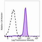
Human peripheral blood cells were stained with CD317 (clone ... -
PE/Cyanine7 anti-human CD317 (BST2, Tetherin)
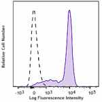
Human peripheral blood was incubated with True-Stain Monocyt... -
TotalSeq™-A0936 anti-human CD317 (BST2, Tetherin)
