DAPI (4',6-Diamidino-2-Phenylindole, Dilactate)

Pricing & Availability
Regulatory Status
RUO
Other Names
4',6-Diamidino-2-Phenylindole, Dilactate
1_Ankita_Patil.jpg
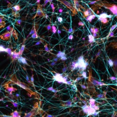
Methanol fixed superior cervical ganglion neurons were stained with anti-tubulin β3 (pink) and anti-TRIM46 (aqua), then stained with secondary antibodies, phalloidin (orange) and DAPI (blue). Image generously submitted to the 2017 Cell Life Imaging Competition by Ankita Patil from Drexel University.
  • 1_Ankita_Patil.jpg
    Methanol fixed superior cervical ganglion neurons were stained with anti-tubulin β3 (pink) and anti-TRIM46 (aqua), then stained with secondary antibodies, phalloidin (orange) and DAPI (blue). Image generously submitted to the 2017 Cell Life Imaging Competition by Ankita Patil from Drexel University.
  • 2_Kathleen-Ignatoski
    Frozen mouse intestine stained with purified anti-Ankyrin-B (green), E-Cadherin (red) and DAPI (blue) followed by anti-goat Alexa Fluor® 488 and anti-rabbit Alexa Fluor® 568. Image generously submitted to the 2017 Cell Life Imaging Competition by Kathleen Ignatoski from University of Michigan.
  • 3_KifLiakath.jpg
    Whole mount mouse tail epidermis was stained with purified anti-Keratin 14 (green, clone Poly19053), purified anti-Keratin 15 (orange, clone Poly18339) and DAPI (blue). Image generously submitted to the 2017 Cell Life Imaging Competition by Kif Liakath.
  • 4_DAPI_IF_080712
    Human cervical cancer cell line, HeLa, was fixed with 4% paraformaldehyde and stained with 300 nM DAPI.
  • 5_DAPI_080712
    Human cervical cancer cell line, HeLa, was fixed with 4% paraformaldehyde and stained with 300 nM DAPI.
See DAPI (4',6-Diamidino-2-Phenylindole, Dilactate) spectral data
Cat # Size Price Quantity Check Availability
422801 10 mg $101.00
Check Availability


Need larger quantities of this item?
Request Bulk Quote
Description

DAPI (4',6-Diamidino-2-Phenylindole, Dilactate), a blue fluorescent nucleic acid stain, preferentially binds dsDNA and associates with the minor groove AT clusters. The molecular weight for DAPI is 457.5.  Association of DAPI with dsDNA increases fluorescence approximately 20-fold, with an emission maximum of 460 nm. The increase in fluorescence is believed to be due to displacement of water molecules from the minor groove and DAPI. DAPI can also bind RNA and emits at a longer wavelength of 500 nm, with only a 20% increase in fluorescence.

DAPI is a popular nuclear stain for identification of cell cycle and specifically stains nuclei but not cytoplasm. The dilactate form is more water soluble compared to the dihydrochloride form.

DAPI can be excited by a UV laser, xenon, or a mercury-arc lamp. 

Technical data sheet

Product Details

Formulation
Lyophilized
Preparation
To make 5 mg/ml (10.9 mM) solution, dissolve entire contents of vial in 2 ml of deionized water.
Storage & Handling
Store between 2°C and 8°C, protected from light. For long term storage, aliquot and store at -20°C.
Application

ICFC, ICC

Recommended Usage

This product can be used at 3 µM for flow cytometry and 300 nM for immunofluorescence.  It is recommended to titrate the dye for optimal performance. 

Excitation Laser
Ultraviolet Laser (355 nm)
Application Notes

Protocols for Counterstaining with DAPI:

Immunofluorescence Staining
Note: Cells may be fixed by method of choice, such as with 4% paraformaldehyde. DAPI staining is done after staining for other markers. Fixation and permeabilization of cells is not necessary to counterstain with DAPI.

1. Fix cells using method of choice.
2. Incubate the cells with phosphate buffered saline (PBS) for 15 minutes.
3. Dilute the DAPI solution (we recommend to 300 nM) with PBS. Remove excess PBS from the slide and cover with DAPI solution, making sure the cells are completely covered.
4. Incubate for up to 5 minutes. Rinse the slide several times to remove all free DAPI. It is recommended to use a mounting medium with antifade reagent to reduce fluorescence quenching. The slide is ready to view under the microscope with appropriate filters.


Nuclear Staining for Flow Cytometry
Note: Cells may be fixed with 4% paraformadehyde or absolute ethanol.

1. Fix cells with absolute ethanol or 4% paraformaldehyde.
2. Centrifuge, discard the supernatant, and add 5 ml of PBS, allowing cells to rehydrate for 15 minutes.
3. Dilute the DAPI to 3 µM (recommended) in staining buffer (such as BioLegend's FOXP3 Perm Buffer, Cat. No. 421402).
4. Centrifuge cell suspension from step 3 above, discard supernatant, and add 1 ml of DAPI solution; then incubate at room temperature for 15 minutes.
5. Analyze by flow cytometry using an instrument with appropriate lasers.

Additional Product Notes

View more applications data for this product in our Scientific Poster Library.

Application References

(PubMed link indicates BioLegend citation)
  1. Solomonov I, et al. 2014. Sci Rep. 4:5987. PubMed
  2. Sodhi CP, et al. 2015. J Immunol. 194:4931. PubMed
Product Citations
  1. Davila ML, et al. 2022. iScience. 25:104426. PubMed
  2. DeDreu J, et al. 2021. Matrix Biol. 96:18. PubMed
  3. Duraiswamy J, et al. 2021. Cancer Cell. 39:1623. PubMed
  4. McCubbrey AL, et al. 2022. Cell Rep. 38:110222. PubMed
  5. Xiang J, et al. 2022. J Virol. 96:e0005722. PubMed
  6. Matta B, et al. 2022. Front Immunol. 13:951254. PubMed
  7. Kimmerling RJ, et al. 2022. Commun Biol. 5:1295. PubMed
  8. Reemst K, et al. 2022. Transl Psychiatry. 12:507. PubMed
  9. Kaneko K, et al. 2022. Cancers (Basel). 14: . PubMed
  10. Shahid AM, et al. 2022. PLoS One. 17:e0278209. PubMed
  11. Sountoulidis A, et al. 2023. Nat Cell Biol. 25:351. PubMed
  12. Wang CQ, et al. 2023. Cells. 12: . PubMed
  13. Jeon EY, et al. 2023. Bioeng Transl Med. 8:e10362. PubMed
  14. Xiong E, et al. 2023. Front Immunol. 13:1083119. PubMed
  15. Aru B, et al. 2023. Balkan Med J. 40:117. PubMed
  16. González-Terán B, et al. 2022. Cell. 185:794. PubMed
  17. Grubwieser P, et al. 2022. Front Cell Infect Microbiol. 12:875543. PubMed
  18. Sierro F, et al. 2017. Immunity. 47:374. PubMed
  19. Wiedemann SJ, et al. 2022. Cell Metab. 34:991. PubMed
  20. Michalek S, et al. 2022. EMBO Rep. 23:e54195. PubMed
  21. Cherkaoui S, et al. 2022. Mol Syst Biol. 18:e11033. PubMed
  22. Chang L, et al. 2023. Life Sci Alliance. 6:. PubMed
  23. Kozlovski S, et al. 2023. Front Immunol. 13:1041552. PubMed
  24. Vanneste D, et al. 2023. Nat Immunol. 24:827. PubMed
  25. Flinn MA, et al. 2023. Front Cardiovasc Med. 10:1142612. PubMed
  26. Ramalingam P, et al. 2023. Nat Commun. 14:2018. PubMed
  27. Huber M, et al. 2023. Nat Commun. 14:2143. PubMed
  28. Kerkering J, et al. 2023. J Clin Invest. . PubMed
  29. Basta MD, et al. 2023. iScience. 26:106570. PubMed
  30. Arora S, et al. 2023. PLoS One. 18:e0286211. PubMed
  31. Gómez-Pinedo U, et al. 2023. Cells. 12:. PubMed
  32. Hoover MY, et al. 2023. Nat Protoc. . PubMed
  33. Rodrigues M, et al. 2022. Biochem J. 479:1891. PubMed
  34. Gabriely G, et al. 2021. iScience. 24:103347. PubMed
  35. Effern M, et al. 2022. STAR Protoc. 3:101038. PubMed
  36. Sountoulidis A, et al. 2020. PLoS Biol. 18:e3000675. PubMed
  37. Mester B, et al. 2015. PLoS One. 10: e0140432. PubMed
  38. Wu N, et al. 2020. Cell Rep. 30:1129. PubMed
  39. Velilla J, et al. 2019. Neurol Genet. 5:e312. PubMed
  40. Jiao Q, et al. 2017. Clin Exp Immunol. 188:420. PubMed
  41. Anguiano T, et al. 2020. Toxicol Sci. 177:494. PubMed
  42. Afshar Y, et al. 2021. Nat Commun. 12:4408. PubMed
  43. Goodnough LH, et al. 2022. Front Endocrinol (Lausanne). 13:924927. PubMed
  44. Nguyen DT, et al. 2022. Cells. 11:. PubMed
  45. Baldominos P, et al. 2022. STAR Protoc. 3:101795. PubMed
  46. Wilson K, et al. 2016. Cell Death Dis. 7: e2197. PubMed
  47. Kobayashi T, et al. 2019. Cell. 176:982. PubMed
  48. Miedema A, et al. 2022. Acta Neuropathol Commun. 10:8. PubMed
  49. Emmons TR, et al. 2021. Cancer Immunol Res. 9:790. PubMed
  50. Valkov N, et al. 2021. Life Sci Alliance. 4:. PubMed
  51. Gamboa CM, et al. 2021. STAR Protoc. 100345:2. PubMed
  52. Sugawara E, et al. 2020. Autophagy. 1.323611111. PubMed
  53. Elias A, et al. 2016. Sci Rep. 6: 24971. PubMed
  54. Bannard O, et al. 2016. J Exp Med. 13: 993 - 1009. PubMed
  55. Sumitomo Y, et al. 2016. Blood. 128: 1614 - 1624. PubMed
  56. Paris AJ, et al. 2020. Nat Cell Biol. 1.747916667. PubMed
  57. Brunner JS, et al. 2020. Nat Commun. 0.757638889. PubMed
  58. Fukushima T, et al. 2019. Cell Rep. 29:4144. PubMed
  59. Alexander MS, et al. 2021. Muscle Nerve. 63:928. PubMed
  60. Hendricks A, et al. 2021. Front Oncol. 11:646885. PubMed
  61. Hiramatsu-Asano S, et al. 2021. Front Immunol. 11:616141. PubMed
  62. Ambrosi TH, et al. 2021. Elife. 10: . PubMed
  63. Hopp AK, et al. 2021. Mol Cell. 81:340. PubMed
  64. Zhang M, et al. 2017. Oncol Lett. 3.069444444. PubMed
  65. Greenblatt SM, et al. 2018. Cancer Cell. 33:1111. PubMed
  66. Wilson AS, et al. 2022. Nat Commun. 13:528. PubMed
  67. Deng J, et al. 2021. Breast Cancer Res Treat. 185:85. PubMed
  68. DeDreu J, et al. 2020. FASEB J. 34:9316. PubMed
  69. Allam O, et al. 2018. PLoS One. 13:e0194185. PubMed
  70. Fujiwara H, et al. 2021. Nat Immunol. 22:1440. PubMed
  71. Ravi A, et al. 2021. Dev Cell. 56:1661. PubMed
  72. Wu CL, et al. 2021. Nat Commun. 12:362. PubMed
  73. Koh P, et al. 2016. Sci Data. 3:160109. PubMed
  74. Sodhi C, et al. 2015. J Immunol. 194:4931. PubMed
  75. Kanda K, et al. 2018. JCI Insight. 3:e91316. PubMed
  76. So EY, et al. 2021. Am J Physiol Cell Physiol. 321:C569. PubMed
  77. Poulos M, et al. 2016. Nat Commun. 7:13829. PubMed
  78. Noah AC, et al. 2020. J Appl Physiol (1985). 473:128. PubMed
  79. Wang ECE, et al. 2019. Cell Stem Cell. 24:654. PubMed
  80. Wennerberg E, et al. 2022. Sci Transl Med. 14:eabe8195. PubMed
  81. Kumar A, et al. 2021. STAR Protoc. 2:100874. PubMed
  82. Hendricks A, et al. 2020. Cancers (Basel). 12:00. PubMed
  83. Aronovich A, et al. 2021. J Invest Dermatol. 141:619. PubMed
  84. Teo YV, et al. 2019. Cell Rep. 27:997. PubMed
  85. Hayama Y, et al. 2018. J Immunol. 200:3790. PubMed
  86. Fredriksson-Lidman K, et al. 2017. PLoS One. 10.1371/journal.pone.0185509. PubMed
  87. Caeser R, et al. 2021. Nat Protoc. 16:2499. PubMed
  88. Faisal S, et al. 2016. Sci Rep. 6:39530. PubMed
  89. Zhang X, et al. 2022. JCI Insight. 7:. PubMed
  90. Zhu M, et al. 2021. Eneuro. 8:. PubMed
  91. Flores-Pliego A, et al. 2021. Cells. 10:. PubMed
  92. Shani O, et al. 2021. Elife. 10: . PubMed
  93. Thompson M, et al. 2015. J Immunol. 195: 5637 - 5647. PubMed
  94. Kundu K, et al. 2019. Cancer Immunol Res. 1.069444444. PubMed
  95. Pagan JD, et al. 2018. Cell. 172:564. PubMed
  96. Heath O, et al. 2021. Cancer Immunol Res. 9:665. PubMed
  97. Dasinger JH, et al. 2021. Pregnancy Hypertens. 24:126. PubMed
  98. Ren X, et al. 2022. STAR Protoc. 3:101818. PubMed
  99. Moser J, et al. 2016. Sci Rep. 6:35387. PubMed
  100. Mould KJ, et al. 2019. JCI Insight. 4:00. PubMed
  101. Pascual-Pasto G, et al. 2022. J Immunother Cancer. 10:. PubMed
  102. Lehrke MJ, et al. 2021. Elife. 10:. PubMed
  103. Wang Y, et al. 2021. EMBO Mol Med. 13:e14089. PubMed
  104. Correa A, et al. 2015. PLoS One. 10: 0143391. PubMed
  105. Yanting C, et al. 2018. J Dairy Sci. 101:1601. PubMed
  106. Kurena B, et al. 2017. Front Immunol. 1.490972222. PubMed
  107. Pinho–Ribeiro FA, et al. 2018. Cell. 173:1083. PubMed
  108. Aziz M, et al. 2017. J Immunol. 10.4049/jimmunol.1602056. PubMed
  109. Valdespino-Vázquez MY, et al. 2021. J Med Virol. 93:4480. PubMed
  110. Kissiov DU, et al. 2022. Elife. 11:. PubMed
  111. Isobe T, et al. 2022. Nat Commun. 13:4501. PubMed
  112. Chiang M, et al. 2019. Cell Rep. 28:3212. PubMed
  113. Canals Hernaez D, et al. 2022. Front Oncol. 12:856424. PubMed
  114. Mascharak S, et al. 2022. Cell Stem Cell. 29:315. PubMed
  115. Rodriguez A, et al. 2021. Cell Stem Cell. 28:33. PubMed
  116. Pang J, et al. 2021. STAR Protoc. 2:100879. PubMed
  117. Haschak M, et al. 2021. Aging (Albany NY). 13:16938. PubMed
  118. Phan TS, et al. 2021. Sci Adv. 7:. PubMed
  119. Stancil IT, et al. 2021. Nat Commun. 12:4566. PubMed
  120. Margaroli C, et al. 2021. Cell Rep Med. :100242. PubMed
  121. Caporarello N, et al. 2020. Aging Cell. 19:e13196. PubMed
  122. Vergoni B, et al. 2016. Diabetes. 65: 3062 - 3074. PubMed
  123. Dent JR, et al. 2019. J Appl Physiol (1985). 127:1117. PubMed
  124. Schmidt A, et al. 2017. Toxicol Lett. 10.1016/j.toxlet.2017.08.008. PubMed
  125. Burns F, et al. 2015. Aquatic Toxicology. 164: 52-60. PubMed
  126. Stevens M, et al. 2016. Nat Biotechnol. 34:1161-1167. PubMed
  127. Shilts J, et al. 2021. Sci Rep. 0.745138889. PubMed
  128. Zheng C, et al. 2022. Acta Pharm Sin B. 12:3726. PubMed
  129. Anisul M, et al. 2021. Elife. 10:. PubMed
  130. DeDreu J, et al. 2022. FASEB J. 36:e21995. PubMed
  131. Pelham-Webb B, et al. 2021. Mol Cell. 81:1732. PubMed
  132. Kohn M, et al. 2021. Front Immunol. 626627:12. PubMed
  133. Jain A, et al. 2020. Nat Immunol. 0.920138889. PubMed
  134. Aimi F, et al. 2021. Mod Pathol. 34:1468. PubMed
  135. Barcia Durán JG, et al. 2021. Commun Biol. 4:406. PubMed
  136. Karimzadeh A, et al. 2021. Front Cell Dev Biol. 9:734176. PubMed
  137. Reeves KM, et al. 2021. Clin Cancer Res. Online ahead of print. PubMed
  138. Chmielewski M and Abken H 2017. Cell Rep.. 10.1016/j.celrep.2017.11.063. PubMed
  139. Hassanein W, et al. 2017. Organogenesis. 13:16. PubMed
  140. Guo P, et al. 2022. Nat Cell Biol. 24:99. PubMed
  141. Tominaga K, et al. 2022. iScience. 25:103667. PubMed
  142. Chen HY, et al. 2020. J Immunol. 1158:204. PubMed
  143. Gosztyla C, et al. 2020. Tissue Eng Part C Methods. 26:67. PubMed
  144. Köhler VK, et al. 2020. Int J Mol Sci. 21:00. PubMed
  145. Solomonov I, et al. 2014. Sci Rep. 4:5987. PubMed
  146. Srijiwangsa P, et al. 2018. BMC Pharmacol Toxicol. 19:32. PubMed
  147. Ladd MR, et al. 2018. Tissue Eng Part C Methods. 24:331. PubMed
  148. Bennett AH, et al. 2018. PLoS Genet. 14:e1007226. PubMed
  149. Doorley L, et al. 2017. Immunobiology.. 10.1016/j.imbio.2017.08.002. PubMed

Antigen Details

Biology Area
Cell Biology, Cell Cycle/DNA Replication, Neuroscience
Antigen References

1. Langlois RG, et al. 1980. Chromosoma. 77:229.
2. Kubista M, et al. 1987. Biochemistry 26:4545.

Gene ID
NA
Go To Top Version: 1    Revision Date: 11/30/2012

8999 BioLegend Way, San Diego, CA 92121 www.biolegend.com
Toll-Free Phone: 1-877-Bio-Legend (246-5343) Phone: (858) 768-5800 Fax: (877) 455-9587

Login/Register
Forgot your password? Reset Password
Request an Account