- Regulatory Status
- RUO
- Other Names
- Phalloidin

-

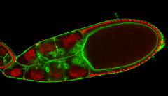
Drosophila ovaries were fixed with paraformaldehyde, incubated with Flash Phalloidin™ Green 488 (green) to visualize F-actin and mounted in VECTASHIELD containing DAPI (red) to visualize the nucleus. Image was then collected using a Leica TCS SP5 II (Leica Microsystems) confocal microscope. Image provided courtesy of Dr. Eurico Morais-de-Sá at the Instituto de Investigação e Inovação em Saúde, Portugal. -

C57BL/6 mouse frozen cerebellum section was fixed with 4% paraformaldehyde (PFA) for ten minutes at room temperature then permeabilized with 0.5% Triton X-100 for ten minutes and blocked with 5% FBS for one hour at room temperature. Then the section was stained with 5 µg/mL anti-GFAP (clone 2E1.E9) Alexa Fluor® 647 (red) at 4°C overnight. The following day the section was stained with Flash Phalloidin™ Green 488 (green, 25 µL of the stock solution -

HeLa cells were fixed with 1% paraformaldehyde (PFA) for ten minutes then permeabilized with 0.5% Triton X-100 for ten minutes and blocked by 5% FBS for 30 minutes. Then, the cells were stained with anti-Cytokeratin Alexa Fluor® 647 (red) at 4°C overnight. The following day, the cells were stained with Flash Phalloidin™ Green 488 (green, 25 µL of the stock solution in 1 mL of PBS) for 30 minutes at 4°C in the dark. The nuclei were then counterstained with DAPI (blue). The image was captured by 40X objective. -

Frozen bone marrow sections with osteoclasts expressing tdTomato (yellow) were stained with Flash Phalloidin™ Green 488 (green) and counterstained with DAPI (pink). Image generously submitted to the 2017 Cell Life Imaging Competition by Andres Garcia-Garcia from University of Cambridge.
| Cat # | Size | Price | Quantity Check Availability | ||
|---|---|---|---|---|---|
| 424201 | 300 units | $347.00 | |||
Molecular Mass 1582.65 g/mol-1. Flash Phalloidin™ Green 488 excites maximally at 488 nm and emits maximally at 520 nm.
Phalloidin is a bicyclic peptide that can be found naturally in the death cap mushroom. This molecule is considered to bind so tightly to F-actin that when ingested by an organism, it will prevent the depolymerization of the actin polymeric filaments which leads to cellular toxicity. In cell imaging, this is a very useful probe for imaging and stabilizing filamentous F-actin in fixed and permeabilized cells, providing structural and volumetric context to the cell. Phallotoxins are conjugated to a wide array of fluorophores to enable their use in multicolor microscopy.
Product Details
- Verified Reactivity
- Human, Mouse, Rat, All Species
- Preparation
- Flash Phalloidin™ Green 488 is lyophilized. Reconstitute with 1.5mL of methanol to make a stock solution of 300 units.
- Storage & Handling
- Store Flash Phalloidin™ Green 488 at -20°C, protected from light.
- Application
-
ICC - Quality tested
IHC-F - Verified - Recommended Usage
-
Reconstitute the Flash Phalloidin™ Green 488 with 1.5mL of methanol to make 300 units. Then prepare a working concentration by diluting 1:20 - 1:100 of stock in 1X PBS. It is recommended that the reagent be titrated for optimal performance for each application.
- Application Notes
-
- Prior to reconstitution, spin down the vial of lyophilized reagent in a microcentrofuge to ensure the reagent is at the bottom of the vial.
- Fix cultured cells with 1% - 4% paraformaldehyde (PFA) for 10 minutes at room temperature.
- Wash the cells two times with 1X PBS.
- Permeabilize the cells with 0.5% Triton X-100 for 10 minutes at room temperature or at 4°C.
- Wash the cells two times with 1X PBS.
- Block cells with 5% fetal bovine serum for 30 minutes at room temperature.
- Prepare the working solution by diluting Flash Phalloidin™ Green 488 1:20 - 1:100 in 1X PBS.
- Stain the cells with diluted solution for 20 minutes at room temperature in the dark protected from light.
- Mount the slides with an antifade mounting media and image the slides.
- Product Citations
-
Antigen Details
- Distribution
-
Cytoskeleton.
- Biology Area
- Cell Biology, Cell Motility/Cytoskeleton/Structure, Neuroscience
- Gene ID
- NA
