- Clone
- 1518CT337.123.86.269.232 (See other available formats)
- Regulatory Status
- RUO
- Other Names
- Apurinic/Apyrimidinic Endodeoxyribonuclease 1, REF1, HAP1, APEN, APE, APX, AP Endonuclease 1
- Isotype
- Mouse IgG1, κ

-

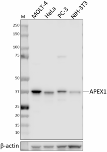
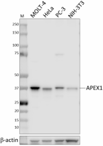
Whole cell extracts (15 µg total protein) from the indicated cells were resolved by 4-12% Bis-Tris gel electrophoresis, transferred to a PVDF membrane, and probed with 1.0 μg/mL of purified anti-APEX1 (clone 1518CT337.123.86.269.232) overnight at 4°C. Proteins were visualized by chemiluminescence detection using HRP goat anti-mouse IgG (Cat. No. 405306) at a 1:3000 dilution. Direct-Blot™ HRP anti-β-actin (Cat. No. 643807) was used as a loading control at a 1:10000 dilution (lower). Western-Ready™ ECL Substrate Premium Kit (Cat. No. 426319) was used as a detection agent. Lane M: Molecular weight marker -

IHC staining of purified anti-APEX1 (clone 1518CT337.123.86.269.232) on formalin-fixed paraffin-embedded colon tissue. Following antigen retrieval using Sodium Citrate H.I.E.R. (Cat. No. 928502), the tissue was incubated with 5.0 μg/mL of purified mouse IgG1 isotype control (Cat. No. 400102) (panel A) or 5.0 μg/mL purified anti-APEX1 (panel B) overnight at 4°C. BioLegend’s Ultra Streptavidin HRP Kit (Multi-Species, DAB, Cat. No. 929501) was used for detection followed by hematoxylin counterstaining, according to the protocol provided. The images were captured with a 20X objective. Scale bar: 50 μm -

ICC staining of purified anti-APEX1 (clone 1518CT337.123.86.269.232) on HeLa cells. The cells were fixed and permeabilized with 100% methanol, and blocked with 5% FBS for 1 hour. The cells were then stained with 10 µg/mL of purified mouse IgG1, κ isotype control (Cat. No. 400102) (panel A) or 10 µg/mL of anti-APEX1 antibody (panel B), followed by incubation with 2.5 µg/mL of Alexa Fluor® 647 anti-goat anti-mouse IgG (Cat. No. 405322) for 1 hour at room temperature. Nuclei were counterstained with DAPI, and the images were captured with a 40X objective. Scale bar: 50 μm
| Cat # | Size | Price | Quantity Check Availability | ||
|---|---|---|---|---|---|
| 605051 | 25 µg | $118.00 | |||
| 605052 | 100 µg | $293.00 | |||
Apurinic-apyridimic endonuclease-1, or APEX1, is a multifunctional enzyme required for the repair of DNA damaged by both endogenous and exogenous agents. It also exerts oncogenic function as a redox signaling protein by reductively stimulating the DNA-binding activity of transcription factors that drive cancer progression, including c-Jun, AP-1, NF-κB, p53, and HIF1α. APEX1 expression level and subcellular localization are positively correlated with cancer progression, and elevated APEX1 expression may also confer resistance to DNA-damaging chemotherapeutic agents.
Product Details
- Verified Reactivity
- Human, Mouse
- Antibody Type
- Monoclonal
- Host Species
- Mouse
- Immunogen
- Full-length recombinant human APEX1 protein
- Formulation
- Phosphate-buffered solution, pH 7.2, containing 0.09% sodium azide
- Preparation
- The antibody was purified by affinity chromatography.
- Concentration
- 0.5 mg/mL
- Storage & Handling
- The antibody solution should be stored undiluted between 2°C and 8°C.
- Application
-
WB - Quality tested
IHC-P, ICC - Verified - Recommended Usage
-
Each lot of this antibody is quality control tested by western blotting. For western blotting, the suggested use of this reagent is 0.25 - 1.0 µg/mL. For immunohistochemistry on formalin-fixed paraffin-embedded tissue sections, a concentration of 5.0 µg/mL is suggested. For immunocytochemistry, a concentration range of 5.0 - 10.0 μg/mL is recommended. It is recommended that the reagent be titrated for optimal performance for each application.
- Application Notes
-
When using this clone for ICC, we recommend using PFA + methanol or methanol only fix-perm conditions. We do not recommend using PFA + Triton X-100 due to non-specific staining.
When testing this clone for ICC, this antibody produced filamentous, extranuclear staining in some cells. This pattern was also observed with a control antibody generated using a different APEX1 immunogen, consistent with this staining being APEX1 dependent. - RRID
-
AB_2910484 (BioLegend Cat. No. 605051)
AB_2910484 (BioLegend Cat. No. 605052)
Antigen Details
- Structure
- APEX1 is a 318 amino acid protein with a predicted molecular weight of ~36 kD.
- Distribution
-
Nucleus/Ubiquitously expressed
- Function
- DNA repair enzyme
- Biology Area
- Cell Biology, DNA Repair/Replication
- Molecular Family
- Enzymes and Regulators
- Antigen References
-
- Fishel ML, et al. 2008. DNA Repair. 7:177-86.
- Kim MH, et al. 2013. J Clin Invest. 123:3211-3230.
- Thakur, et al. 2014. Exp Mol Med. 46:e106.
- Gene ID
- 328 View all products for this Gene ID
- UniProt
- View information about APEX1 on UniProt.org
Other Formats
View All APEX1 Reagents Request Custom Conjugation| Description | Clone | Applications |
|---|---|---|
| Purified anti-APEX1 | 1518CT337.123.86.269.232 | WB,ICC,IHC-P |
Compare Data Across All Formats
This data display is provided for general comparisons between formats.
Your actual data may vary due to variations in samples, target cells, instruments and their settings, staining conditions, and other factors.
If you need assistance with selecting the best format contact our expert technical support team.


