- Clone
- W22009A (See other available formats)
- Regulatory Status
- RUO
- Other Names
- Catenin beta-1, β-catenin, EVR7, CTNNB1, MRD19, NEDSDV, armadillo
- Isotype
- Rat IgG2b, κ
-

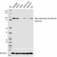
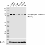
Whole cell extracts (15 µg total protein per lane) from the indicated cell lines (lane 3 and 4 are 293T cells untreated and treated 20 mM LiCl for 20 hours, respectively) were resolved on a 4-12% Bis-Tris gel, transferred to a PVDF membrane, and probed with the 1 µg/mL of Purified anti-β-catenin non-phospho (active) (clone W22009A) overnight at 4°C. Proteins were visualized by chemiluminescence detection using HRP Goat anti-rat IgG (Cat. No. 405405) at a 1:5000 dilution. Direct-Blot™ HRP anti-β-actin (Cat. No. 664804) was used as a loading control at a 1:25000 dilution. Western-Ready™ ECL Substrate Premium Kit (Cat. No. 426319) was used as a detection agent. Lane M: Molecular weight marker -

Whole cell extracts (15 µg total protein per lane) from the indicated cell lines were resolved on a 4-12% Bis-Tris gel and transferred to a PVDF membrane. For peptide blocking, 1 µg/mL of Purified anti-β-catenin non-phospho (active) (clone W22009A) was pre-incubated without (left blot) and with 1 µg/mL of phosphorylated (middle blot) or non-phosphorylated (S33/S37/T41) peptide (right blot) at a 1:1 ratio. Then the PVDF membrane was probed overnight at 4°C. Proteins were visualized by chemiluminescence detection using HRP Goat anti-rat IgG (Cat. No. 405405) at a 1:5000 dilution. Western-Ready™ ECL Substrate Premium Kit (Cat. No. 426319) was used as a detection agent. Lane M: Molecular weight marker -

A431 cells were fixed with ice-cold methanol, and then blocked with 5% FBS for 1 hour at room temperature. Cells were then intracellularly stained with 5 µg/mL of Purified anti-β-catenin non-phospho (active) (clone W22009A) (panel A and B), followed by incubation with 2.5 µg/mL of Alexa Fluor® 488 Goat anti-rat IgG (Cat. No. 405418) for 1 hour at room temperature. For peptide blocking, 5 µg/mL of Purified anti-β-catenin non-phospho (active) (clone W22009A) was pre-incubated with 5 µg/mL of the indicated peptide (1:1 ratio) (panels C and D: non-phospho peptide; panels E and F: phosphorylated peptide). Cells were then stained. Nuclei were counterstained with DAPI (Cat. No. 422801) (blue). The image was captured with a 40X objective. Scar bar: 25 µm -

IHC staining using Purified anti-β-catenin non-phospho (active) (clone W22009A) on formalin-fixed paraffin embedded human colon tissue. Following antigen retrieval using 1X Citrate buffer (Cat. No. 420901), the tissue was incubated with 5 µg/mL of Purified anti-β-catenin non-phospho (active) (clone W22009A) overnight at 4°C, followed by incubation with 2.5 µg/mL of Alexa Fluor® 594 Goat anti-rat IgG (red) (Cat. No. 405422) for 1 hour at room temperature. Nuclei were counterstained with DAPI (Cat. No. 422801) (blue). The image was captured with a 40X objective. Scar bar: 50 µm -

IHC staining using Purified anti-β-catenin non-phospho (active) (clone W22009A) on formalin-fixed paraffin embedded human colon tissue. For peptide blocking, 2 µg/mL of Purified anti-β-catenin non-phospho (active) (clone W22009A) was pre-incubated without (negative control, panel A) or with 2 µg/mL of phosphorylated (panel B) or non-phosphorylated (S33/S37/T41) peptide (panel C) at a 1:1 ratio. Following antigen retrieval using 1X Citrate buffer (Cat. No. 420901), the tissue sections were stained overnight at 4°C, followed by incubation with 2.5 µg/mL of Alexa Fluor® 594 Goat anti-rat IgG (red) (Cat. No. 405422) for 1 hour at room temperature. Nuclei were counterstained with DAPI (Cat. No. 422801) (blue). The image was captured with a 40X objective. Scar bar: 50 µm
| Cat # | Size | Price | Quantity Check Availability | ||
|---|---|---|---|---|---|
| 631851 | 25 µg | $180.00 | |||
| 631852 | 100 µg | $480.00 | |||
β-catenin is part of a complex of proteins that constitute adherens junctions (AJs). AJs are necessary for the creation and maintenance of epithelial cell layers by regulating cell growth and adhesion between cells. The encoded protein also anchors the actin cytoskeleton and may be responsible for transmitting the contact inhibition signal that causes cells to stop dividing once the epithelial sheet is complete. β-catenin also plays a key role in Wnt signaling pathways and thus is involved in neural differentiation, synaptic plasticity, neurodegenerative disease, and prevention of apoptosis. GSK-3β destabilizes β-catenin by phosphorylating it at Ser33, Ser37, and Thr41. Mutations at these sites lead to stabilization of β-catenin protein levels and have been found in many tumor cell lines. This protein binds to the product of the adenomatous polyposis coli (APC) gene, which is mutated in the adenomatous polyposis of the colon. Mutations in this gene are a cause of colorectal cancer (CRC), pilomatrixoma (PTR), medulloblastoma (MDB), and ovarian cancer.
Product Details
- Verified Reactivity
- Human, Mouse, Rat
- Antibody Type
- Monoclonal
- Host Species
- Rat
- Immunogen
- Recombinant human β-Catenin protein containing residues Serine 33 to Threonine 41.
- Formulation
- Phosphate-buffered solution, pH 7.2, containing 0.09% sodium azide
- Preparation
- The antibody was purified by affinity chromatography.
- Concentration
- 0.5 mg/mL
- Storage & Handling
- The antibody solution should be stored undiluted between 2°C and 8°C.
- Application
-
WB - Quality tested
ICC, IHC-P - Verified - Recommended Usage
-
Each lot of this antibody is quality control tested by western blotting. For western blotting, the suggested use of this reagent is 0.125 - 1.0 µg/mL. For immunocytochemistry, a concentration range of 1.0 - 5.0 μg/mL is recommended. For immunohistochemistry, a concentration range of 0.5 - 5.0 µg/mL is suggested. It is recommended that the reagent be titrated for optimal performance for each application.
- Application Notes
-
This antibody (clone W22009A) cross-reacts with mouse and rat non-phospho β-catenin (active) in western blot (WB).
For immunocytochemistry (ICC), 4% PFA fixation followed by permeabilization with 0.5% Triton X-100 or ice-cold methanol or fixation with ice-cold methanol is recommended.
For Immunohistochemistry - Paraffin (IHC-P), Citrate Buffer (Cat. No. 420901) or Tris-EDTA pH 9.0 is recommended for antigen retrieval. - RRID
-
AB_3097567 (BioLegend Cat. No. 631851)
AB_3097567 (BioLegend Cat. No. 631852)
Antigen Details
- Structure
- β-catenin is a 781 amino acid protein with a predicted molecular weight of 85 kD.
- Distribution
-
Expressed in several hair follicle cell types: basal and peripheral matrix cells, and cells of the outer and inner root sheaths. Expressed in colon, cortical neurons and breast cancer tissues.
- Function
- Wnt signaling pathway, regulation of cell-cell adhesion, neurogenesis, transcription, transcription regulation, early embryonic patterning, asymmetric cell division, stem cell renewal, and the epithelial to mesenchymal transition
- Interaction
- Wnt signaling pathway, apoptotic cleavage of cellular proteins, and EphBEphrinB signaling
- Biology Area
- Cell Adhesion, Cell Biology, Neuroscience, Signal Transduction, Synaptic Biology
- Molecular Family
- Adhesion Molecules
- Antigen References
-
- Hirano S, et al. 2012. Physiol Rev. 92:597.
- Yost C, et al. 1996. Genes Dev. 10:1443-54.
- Morin PJ, et al. 1997. Science. 275:1787-90.
- Gene ID
- 1499 View all products for this Gene ID
- UniProt
- View information about beta-Catenin on UniProt.org
Other Formats
View All Reagents Request Custom Conjugation| Description | Clone | Applications |
|---|---|---|
| Purified anti-β-Catenin non-phospho (active) (S33/S37/T41) | W22009A | WB,IHC-P,ICC |
Compare Data Across All Formats
This data display is provided for general comparisons between formats.
Your actual data may vary due to variations in samples, target cells, instruments and their settings, staining conditions, and other factors.
If you need assistance with selecting the best format contact our expert technical support team.
-
Purified anti-β-Catenin non-phospho (active) (S33/S37/T41)
Whole cell extracts (15 µg total protein per lane) from the ... 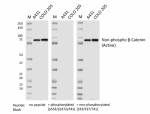
Whole cell extracts (15 µg total protein per lane) from the ... 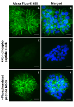
A431 cells were fixed with ice-cold methanol, and then block... 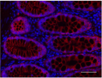
IHC staining using Purified anti-β-catenin non-phospho (acti... 
IHC staining using Purified anti-β-catenin non-phospho (acti...
