- Clone
- 12.1 (See other available formats)
- Regulatory Status
- RUO
- Other Names
- MET, Hepatocyte growth factor receptor, HGF receptor
- Isotype
- Mouse IgG1, κ

-

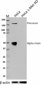
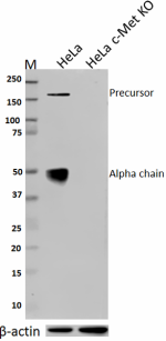
Total lysates (10 µg protein) from HeLa and HeLa c-Met knockout (KO) cells were resolved by electrophoresis (4-20% Tris-glycine gel), transferred to nitrocellulose, and probed with 1:250 (2 µg/ml) purified anti- c-Met antibody , clone 12.1 (upper). Proteins were visualized using chemiluminescence detection by incubation with HRP goat anti-mouse-IgG secondary antibody (cat#405306). Direct-Blot™ HRP anti-&beta:-actin Antibody (Cat#643807) was used as a loading control (lower). Lane M is the Moleculuar weight ladder.
| Cat # | Size | Price | Quantity Check Availability | ||
|---|---|---|---|---|---|
| 689902 | 100 µg | $253.00 | |||
The Mesenchymal Epithelial Transition (MET) receptor belongs to a family of Receptor tyrosine kinases (RTKs). The MET proto-oncogene is located on chromosome 7q21–31 and encodes the receptor tyrosine kinase C-Met. It is a 190 kD glycoprotein heterodimer made up of an extracellular α-chain which is linked by a disulphide bond to a transmembrane β-chain. c-Met is initially synthesised as a 170 kD single polypeptide as precursor that is proteolytically cleaved to form the α-chain and the β-chain. TPR-MET oncogenic fusion protein from human osteosarcoma tumor cells was first discovered in 1984. c-Met is a hepatocyte growth factor (HGF) receptor. The initiation of MET signaling begins with the binding of HGF to the MET receptor at cell membrane that causes C-Met dimerization. Subsequent activation is through a process of trans-phosphorylation of the two tyrosine residues in the catalytical regions Y1234 and Y1235, followed by trans-phosphorylation of two docking tyrosines (Y1349 and Y1356). It enables MET to bind to multiple substrates and activate a variety of signaling pathways, such as MAPK, PI3K-AKT cascades, STAT and NF-κB signaling pathways. This is responsible for driving proliferation, cell survival, migration and invasiveness. c-Met is overexpressed in different human tumors, such as thyroid, gastric, pancreatic, breast, cervical and prostate cancers. Increasing evidence indicated that MET may be a common mechanism of resistance to anticancer treatment such as approved EGFR and VEGFR inhibitors, anti-HER2 therapies and a BRAF inhibitor. Hence, the HGF-MET axis has been targeted in solid tumors to overcome the drug resistance.
Product Details
- Verified Reactivity
- Human
- Antibody Type
- Monoclonal
- Host Species
- Mouse
- Immunogen
- Bacterially expressed human c-Met alpha chain.
- Formulation
- Phosphate-buffered solution, pH 7.2, containing 0.09% sodium azide.
- Preparation
- The antibody was purified by affinity chromatography.
- Concentration
- 0.5 mg/ml
- Storage & Handling
- The antibody solution should be stored undiluted between 2°C and 8°C.
- Application
-
WB - Quality tested
KO/KD-WB - Verified - Recommended Usage
-
Each lot of this antibody is quality control tested by Western blotting. For Western blotting, the suggested use of this reagent is 0.5 - 2.5 µg per ml. It is recommended that the reagent be titrated for optimal performance for each application.
- Application Notes
-
Clone 12.1 does not cross-react with mouse. Clone 12.1 recognizes the precursor and a chain c-Met; the ß chain will not be recognized by this antibody.
- RRID
-
AB_2629791 (BioLegend Cat. No. 689902)
Antigen Details
- Structure
- Heterodimer made of an alpha chain (50 kD) and a beta chain (145 kD) which are disulfide linked.
- Distribution
-
Membrane; single-pass type I membrane protein.
- Function
- Receptor tyrosine kinase that transduces signals from the extracellular matrix into the cytoplasm by binding to hepatocyte growth factor/HGF ligand. Regulates many physiological processes including proliferation, scattering, morphogenesis and survival.
- Interaction
- Interacts with SPSB1, SPSB2, SPSB3, SPSB4, INPP5D/SHIP1, RANBP9, RANBP10, MUC20, GRB10, PTPN1 and PTPN2. When phosphorylated, interacts with STAT3, PIK3R1, SRC, PCLG1, GRB2, GAB1 and INPPL1/SHIP2.
- Ligand/Receptor
- HGF.
- Biology Area
- Cancer Biomarkers, Cell Biology, Cell Proliferation and Viability, Signal Transduction
- Molecular Family
- Protein Kinases/Phosphatase
- Antigen References
-
1. Garajová I, et al. 2015. Transl. Oncogenomics 7:13.
2. Cooper CS, et al. 1984. Nature. 311:29.
3. Furge KA, et al. 2000. Oncogene. 19:5582.
4. Ponzetto C, et al. 1994. Cell. 77(2):261-71. - Gene ID
- 4233 View all products for this Gene ID
- UniProt
- View information about c-Met on UniProt.org
Other Formats
View All c-Met Reagents Request Custom Conjugation| Description | Clone | Applications |
|---|---|---|
| Purified anti-c-Met | 12.1 | WB,KO/KD-WB |
Compare Data Across All Formats
This data display is provided for general comparisons between formats.
Your actual data may vary due to variations in samples, target cells, instruments and their settings, staining conditions, and other factors.
If you need assistance with selecting the best format contact our expert technical support team.
-
Purified anti-c-Met

Total lysates (10 µg protein) from HeLa and HeLa c-Met knock...
