- Clone
- BL28633 (See other available formats)
- Regulatory Status
- RUO
- Other Names
- CAT, Epididymis Secretory Sperm Binding Protein
- Isotype
- Mouse IgG2a, κ
-

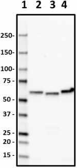
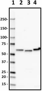
Western blot of purified anti-Catalase antibody (BL28633). Lane 1: Molecular weight marker; Lane 2: 20 µg of human liver lysate; Lane 3: 20 µg of mouse liver lysate; Lane 4: 20 µg of rat liver lysate. The blot was incubated with 0.1 µg/mL of the primary antibody overnight at 4°C, followed by incubation with HRP-labeled goat anti-mouse IgG (Cat. No. 405306). Enhanced chemiluminescence was used as the detection system. -

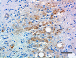
IHC staining of purified anti-Catalase antibody (BL28633) on formalin-fixed paraffin-embedded human liver tissue. Following antigen retrieval using Sodium Citrate H.I.E.R., the tissue was incubated with 0.5 µg/mL of the primary antibody overnight at 4°C. BioLegend’s Ultra Streptavidin (USA) HRP Detection Kit (Multi-Species, DAB, Cat. No. 929901) was used for detection followed by hematoxylin counterstaining, according to the protocol provided. The image was captured with a 40X objective. Scale bar: 50 µm -

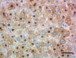
IHC staining of purified anti-Catalase antibody (BL28633) on formalin-fixed paraffin-embedded mouse liver tissue. Following antigen retrieval using Sodium Citrate H.I.E.R., the tissue was incubated with 0.5 µg/mL of the primary antibody overnight at 4°C. BioLegend’s Ultra Streptavidin (USA) HRP Detection Kit (Multi-Species, DAB, Cat. No. 929901) was used for detection followed by hematoxylin counterstaining, according to the protocol provided. The image was captured with a 40X objective. Scale bar: 50 µm -

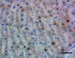
IHC staining of purified anti-Catalase antibody (BL28633) on formalin-fixed paraffin-embedded rat liver tissue. Following antigen retrieval using Sodium Citrate H.I.E.R., the tissue was incubated with 0.5 µg/mL of the primary antibody overnight at 4°C. BioLegend’s Ultra Streptavidin (USA) HRP Detection Kit (Multi-Species, DAB, Cat. No. 929901) was used for detection followed by hematoxylin counterstaining, according to the protocol provided. The image was captured with a 40X objective. Scale bar: 50 µm
| Cat # | Size | Price | Quantity Check Availability | ||
|---|---|---|---|---|---|
| 863301 | 25 µg | $106.00 | |||
Catalase is a heme-containing homotetrameric protein that decomposes hydrogen peroxide to water and oxygen to prevent the generation of hydroxyl radicals in cells. The role of catalase in cellular oxidative stress defense has been widely studied. Overexpression of catalase renders cells more resistant to hydrogen peroxide-induced toxicity and oxidant-mediated injury from hypoxia. Transgenic mice overexpressing catalase are protected against myocardial injury following adriamycin treatment. Although catalase knockout mice develop normally, they show differential sensitivity to oxidant tissue injury. Brain is particularly vulnerable to oxidative stress. Abnormalities and dysfunction of catalase have been indicated in neurodegenerative disorders such as Parkinson’s and Alzheimer’s diseases. Catalase activity was shown to be reduced in Parkinson-derived substantia nigra and putamen tissues. In in vitro cellular models of Alzheimer’s disease, aggregated amyloid beta demonstrates high affinity for catalase leading to the accumulation of hydrogen peroxide and increased oxidative stress.
Product Details
- Verified Reactivity
- Human, Mouse, Rat
- Antibody Type
- Monoclonal
- Host Species
- Mouse
- Immunogen
- Recombinant Catalase
- Formulation
- 0.01 M PBS, pH7.4, containing 0.02% NaN3, 50% glycerol.
- Preparation
- The antibody was purified by affinity chromatography.
- Concentration
- 0.5 mg/ml
- Storage & Handling
- Store at 4°C for up to 3 months. For longer term storage aliquot into small volumes and store at -20°C.
- Application
-
WB - Quality tested
IHC-P - Verified - Recommended Usage
-
Each lot of this antibody is quality control tested by Western blotting. For Western blotting, the suggested use of this reagent is 0.1 - 2.0 µg per ml. For immunohistochemistry on formalin-fixed paraffin-embedded tissue sections, a concentration range of 0.5 - 10 µg/ml is suggested. It is recommended that the reagent be titrated for optimal performance for each application.
- RRID
-
AB_2810758 (BioLegend Cat. No. 863301)
Antigen Details
- Structure
- Human Catalase is a 527 amino acid protein with a molecular mass of 60 kD.
- Distribution
-
Tissue distribution: Ubiquitously expressed; highly expressed in bone marrow and liver.
Cellular distribution: Peroxisome and nucleus - Function
- Catalase is an antioxidant enzyme involved in the body defense against oxidative stress.
- Cell Targets
- Hydrogen peroxide.
- Cell Type
- Astrocytes, Neurons
- Biology Area
- Cell Biology, Cell Death, Neurodegeneration, Neuroscience
- Molecular Family
- Enzymes and Regulators
- Antigen References
-
- Ho YS, et al. 2004. J Biol Chem. 279(31):32804-12.
- Ambani LM, et al. 1975. Arch Neurol. 32(2):114-8.
- Milton NG. 1999. Biochem J. 344(Pt 2): 293–296.
- Habib LK, et al. 2010. J Biol Chem. 285(50):38933-43
- Gene ID
- 547 View all products for this Gene ID
- UniProt
- View information about Catalase on UniProt.org
Other Formats
View All Catalase Reagents Request Custom Conjugation| Description | Clone | Applications |
|---|---|---|
| Purified anti-Catalase | BL28633 | WB,IHC-P |
Compare Data Across All Formats
This data display is provided for general comparisons between formats.
Your actual data may vary due to variations in samples, target cells, instruments and their settings, staining conditions, and other factors.
If you need assistance with selecting the best format contact our expert technical support team.

