- Clone
- W20149K (See other available formats)
- Regulatory Status
- RUO
- Other Names
- CTSB, CPSB, APP secretase, APPS, Cathepsin B1
- Isotype
- Rat IgG2b, κ

-

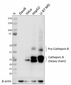
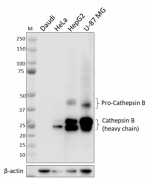
Total cell lysates (15 µg total protein) from Daudi (negative control), HeLa, Hep G2, and U87-MG (positive controls) cells were resolved by 4-12% Bis-Tris gel electrophoresis, transferred to a PVDF membrane, and probed with 0.25 μg/mL (1:2000 dilution) purified anti-Cathepsin B antibody (clone W20149K) overnight at 4°C. Proteins were visualized by chemiluminescence detection using HRP goat anti-rat IgG antibody (Cat. No. 405405) at a 1:3000 dilution. Direct-Blot™ HRP anti-β-actin antibody (Cat. No. 664803) was used as a loading control at a 1:25000 dilution (lower). Western-Ready™ ECL Substrate Premium Kit (Cat. No. 426319) was used as a detection agent. Lane M: Molecular weight marker. -

ICC staining of purified anti-Cathepsin B antibody (clone W20149K) on Daudi (negative control) and Hep G2 (positive control) cells. The cells were fixed and permeabilized with 100% methanol, and blocked with 5% FBS for 1 hour at room temperature. The cells were then stained with 1.0 µg/mL of the primary antibody, followed by incubation with 2.5 µg/mL of Alexa Fluor® 594 goat anti-rat IgG antibody (Cat. No. 405422) for 1 hour at room temperature. Nuclei were counterstained with DAPI, and the image was captured with a 60X objective. -

IHC staining of anti-Cathepsin B antibody (clone W20149K) on formalin-fixed paraffin-embedded human liver (A, positive control) and adipose (B, negative control) tissues. Following antigen retrieval using Sodium Citrate H.I.E.R. (Cat. No. 928502), the tissues were incubated with 1 µg/mL of purified anti-Cathepsin B antibody overnight at 4°C. BioLegend’s Ultra Streptavidin HRP Kit (Multi-Species, DAB, Cat. No. 929501) was used for detection followed by hematoxylin counterstaining, according to the protocol provided. The images were captured with a 40X objective. Scale bar: 50 μm.
| Cat # | Size | Price | Quantity Check Availability | ||
|---|---|---|---|---|---|
| 948001 | 25 µg | $118.00 | |||
| 948002 | 100 µg | $293.00 | |||
Cathepsin B (CTSB) is a lysosomal cysteine protease. While most cathepsins are exclusively endopeptidases, CTSB exhibits both carboxypeptidase and endopeptidase activities. The optimal pH for CTSB activity is between four and six. Cystatin C has been identified as an endogenous CTSB inhibitor. High CTSB protein levels and activity have been found in many tumors including breast, cervix, colon, stomach, glioma, lung, and thyroid. CTSB can be secreted by tumor cells and is associated with the cell membrane of these cells. Membrane associated CTSB promotes extracellular matrix (ECM) degradation, which contributes to cancer motility and invasion. Many ECM proteins, including laminin,
fibronectin, and collagen IV, are substrates of CTSB. CTSB can also activate pro-uPA/PLAU. Activated uPA promotes ECM digestion through serine protease plasminogen. It has been shown that the inhibition of CTSB can limit bone metastasis in breast cancer, which makes it an important anti-cancer drug target. CTSB has been proposed as a new drug target for Alzheimer's disease because of its involvement in the production of neurotoxic β-amyloid (Aβ) peptides. The inhibition of CTSB can reduce the brain Aβ peptides and improve memory that was determined from a mouse model of Alzheimer's disease. CTSB also plays significant roles in immune responses including both T and B cell apoptosis and Th1/Th2 polarization. CTSB is implicated in other pathological conditions including cardiovascular disease, multiple sclerosis, and arthritis. CTSB may also have a role in autophagy, adipogenesis, and cholesterol absorption in the
intestine.
Product Details
- Verified Reactivity
- Human
- Antibody Type
- Monoclonal
- Host Species
- Rat
- Immunogen
- Partial recombinant human cathepsin B protein
- Formulation
- Phosphate-buffered solution, pH 7.2, containing 0.09% sodium azide
- Preparation
- The antibody was purified by affinity chromatography.
- Concentration
- 0.5 mg/mL
- Storage & Handling
- The antibody solution should be stored undiluted between 2°C and 8°C.
- Application
-
WB - Quality tested
ICC, IHC-P- Verified - Recommended Usage
-
Each lot of this antibody is quality control tested by western blotting. For western blotting, the suggested use of this reagent is 0.25 - 1.0 µg/mL. For immunohistochemistry, a concentration range of 1.0 - 5.0 µg/mL is suggested. For immunocytochemistry, a concentration range of 1.0 - 5.0 μg/mL is recommended. It is recommended that the reagent be titrated for optimal performance for each application.
- Application Notes
-
Clone W20149K detects both pro-Cathepsin B and heavy chain cathepsin B by western blot.
Clone W20149K was tested for ICC using the following fixation-permeabilization methods: 4% PFA + Triton X-100, 4% PFA + methanol, and methanol-only. Of these, methanol only was compatible with cathepsin B staining, while 4% PFA + Triton X-100 or methanol resulted in weak/absent staining of cathepsin B.
- RRID
-
AB_2904460 (BioLegend Cat. No. 948001)
AB_2904460 (BioLegend Cat. No. 948002)
Antigen Details
- Structure
- Cathepsin B is a 339 amino acid protein with a predicted molecular weight of 37.8 kD.
- Distribution
-
Ubiquitously expressed/Lysosome
- Function
- Lysosomal cysteine protease involved in intracellular protein turnover
- Interaction
- Interacts with Hepatitis B spliced protein, Bikunin, and TSRC1.
- Biology Area
- Cell Biology, Neurodegeneration, Neuroscience
- Molecular Family
- Lysosomal Markers, Proteases
- Antigen References
-
- Mohamed MM, Sloane BF. 2006. Nat. Rev. Cancer 6:764.
- Wang C, et al. 2012. J. Biol. Chem. 47:39834.
- Grzonka Z, et al. 2001. Acta Biochim. Pol. 48:1.
- Liu J, et al. 2006. FEBS Letters 580:245.
- Brix K, et al. 2008. Biochimie. 90:194.
- Turk V, et al. 2012. Biochim. Biophys. Acta. 1824:68.
- Gene ID
- 1508 View all products for this Gene ID
- UniProt
- View information about Cathepsin B on UniProt.org
Other Formats
View All Cathepsin B Reagents Request Custom Conjugation| Description | Clone | Applications |
|---|---|---|
| Purified anti-Cathepsin B Antibody | W20149K | WB,ICC,IHC-P |
Compare Data Across All Formats
This data display is provided for general comparisons between formats.
Your actual data may vary due to variations in samples, target cells, instruments and their settings, staining conditions, and other factors.
If you need assistance with selecting the best format contact our expert technical support team.


